Quantcast
Channel: Latest Articles of Bio-Synthesis Inc.
Viewing all 591 articles
Browse latest View live

Kaposi’s sarcoma in immunosuppressed patients due to chemotherapy or transplantation, FDA-approved peptide drug to curb HIV-1 infection, and potential peptide drugs to block COVID-19 infection

$
0
0
 In the past, highly prevalent cancers affecting organs such as the lung, colon, breast, or prostate have received a significant level of attention.    Less commonly occurring cancers such as brain cancer (ex. glioblastoma) was covered by the media albeit occasionally—ex. afflicting high-profile figures like the U. S. Senators.  Sarcoma refers to cancerous tumor occurring in connective tissues, which are comprised of bone (ex. osteosarcoma), cartilage (ex. chondrosarcoma), muscle (ex. soft tissue sarcomas in arms or legs), etc.  The advanced-stage sarcoma can spread (metastasize) to distant organs such as the lung.  In the U. S., approximately 15,000 cases of soft tissue sarcoma are diagnosed annually, with 5-year survival rates ranging from ~81% (localized tumors) to ~15% (metastatic cancer).  (https://www.cancer.org/cancer/soft-tissue-sarcoma/detection-diagnosis-staging/survival-rates.html).  Risk factors for sarcomas include exposure to DNA damaging agents, genetic predisposition to develop cancer, [ex. hereditary retinoblastoma (tumors form in the retina due to the inactivation of the tumor suppressor gene RB) for osteosarcoma, Li-Fraumeni syndrome (tumors develop in multiple organs due to the mutation in p53) for rhabdosarcoma] (Shew et al., 1989; Pondrom et al., 2020 ).

The risk factors for sarcoma also include virus infection.    Kaposi’s sarcoma, which affects the skin, mouth, and other tissues, consists of spindle-shaped cells and appears dark purplish due to the leakage of red blood cells from the heavily vascularized (enriched with blood vessels) tumors.  A commonly associated event is the infection by HHV-8 (human gammaherpesvirus 8), a DNA virus with a large genome (~145 kilobases), which encodes the genes involved in cell cycle regulation (ex. cyclin D that complexes with cyclin-dependent kinase to phosphorylate the Rb protein), nucleotide biosynthesis (ex. thymidylate synthetase, dihydrofolate reductase), immune regulation, etc. (Moore et al., 2001;  Gáspár et al., 2002).  It may occur with weakened immunity, ex. transplantation patients taking immunosuppressive drugs to avoid rejection of transplanted organs, and cancer patients undergoing chemotherapy.  Kaposi’s sarcoma has received wide publicity due to its frequent occurrence in HIV-1 (human immunodeficiency virus) infected patients due to the depletion of T helper cells (CD4-positive) and other types of immune cells. 

In the adaptive (as opposed to innate) immune system, T helper cells play a significant role.  Following the encounter with a pathogen (ex. virus, bacteria, fungus), the internalized pathogen is proteolytically processed and selected resultant peptides are presented by class II major histocompatibility complex in antigen processing cells (ex. dendritic cells, macrophages, B lymphocytes).  The presented peptide may then get recognized by naïve ‘T helper cells’ expressing a cognate T cell receptor (specific for the presented peptide) plus the co-receptor CD4 (binds to a distinct site on class II MHC molecule).  [Note: in the case of ‘cytotoxic T cells’, the co-receptor CD8 recognizes class I MHC molecule).  The activated T helper cells contribute to immune function via the secretion of specific cytokines and are involved in the activation of macrophages, B lymphocytes, cytotoxic T cells, etc. 
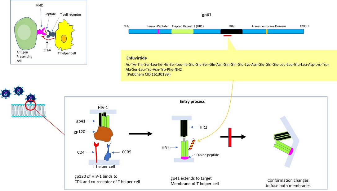


CD4 molecule present on the surface of T helper cells also serves as the receptor for HIV-1.  HIV-1 is a negative-stranded RNA virus that relies on the virally encoded reverse transcriptase to convert its genomic RNA into cDNA, which is converted to double-stranded DNA by using ‘DNA-dependent DNA polymerase’ to synthesize its complementary strand.  During the latent stage, double-stranded HIV-1 genomic DNA is integrated (provirus) into host cells’ chromosomal DNA (via integrase) after entering the nucleus.  The translation of mRNAs transcribed from the provirus produces polypeptides necessary for assembling the HIV-1 virus. 

A high level of errors introduced during the cDNA synthesis by reverse transcriptase, resulting in viruses with highly variant epitopes, has been the key mechanism through which HIV-1 has defied the attempt to suppress the pandemic (caused ~40 million deaths globally) via vaccination.  Its high rate of evolution (ca. million times fast than the human genome) has also contributed to the emergence of resistant strains to antiretroviral drugs (ex. Inhibitors of reverse transcriptase).

This has led to the devising of alternate peptide-based therapeutics to interfere with HIV-1 infection.  Briefly, the entry of HIV-1 is a multi-step process, in which the initial stage (albeit not essential) may consist of the interaction of its Envelope protein (a heterodimer comprised of gp120 and gp41 proteins) with cell surface molecules, i.e. heparan sulfate, alpha4beta7 integrin, or pattern recognition receptor DC-SIGN (dendritic cell–specific intercellular adhesion molecular 3-grabbing non-integrin).  The engagement of CD4 (of T helper cell) by gp120 (of HIV-1) induces a conformational change to expose the domain that interacts with the co-receptor, i.e. chemokine receptor CCR5 or CXCR4 (of T helper cell).  Subsequently, the hydrophobic fusion peptide gp41 inserts into the cell membrane, followed by the folding of hinge regions (N-terminal helix ‘HR-N’ and C-terminal helix ‘HR-C’) to form a 6-helix bundle, fusing viral and cellular membranes to allow the entry of viral capsid (Wilen et al., 2012).  As such, multiple steps could be targeted to disrupt HIV-1 internalization.  The viral entry inhibitor Enfuvirtide (also called Fuzeon; F. Hoffmann-La Roche AG Pharmaceuticals) is a peptide consisting of 36 residues, which was approved by U. S. FDA (Food & Drug Administration) in 2003.  The biomimetic peptide (derived from amino acids 643 -678 of gp41) binds to the prehairpin structure of HR-N to block the 6-helix bundle formation to suppress membrane fusion for HIV-1 entry (Greenberg et al., 2004).  Its limitations include adverse side effects, inability to administer orally, and inflammatory response.

In a similar vein, various peptide drugs have been developed to block the infectivity of SARS-CoV-1, which caused the 2002-2004 SARS outbreak (Madhavan et al., 2021).   A high degree of conservation between SARS-CoV-1 (caused SARS) and SARS-CoV-2 (caused the COVID pandemic in 2019) coronaviruses has inspired the development/testing of similarly designed peptides to suppress the infectivity of the latter albeit mostly in a preclinical setting (Shah et al., 2022).


The key to preventing an epidemic is the ability to diagnose the infected early to preempt further propagation.  For this, Bio-Synthesis, Inc. provides primers and probes (as well as synthetic RNA control) for COVID-19 diagnosis via RT-PCR assay.  It specializes in oligonucleotide modification and provides an extensive array of chemically modified nucleoside analogs (over ~200) including bridged nucleic acid (BNA) in addition to mRNA synthesis.  A number of options are available to label oligonucleotides (DNA or RNA) with fluorophoreseither terminally or internally as well as to conjugate to peptidesor antibodies. It provides custom conjugation of small molecules such as chemical drugs, metabolites and labeled compounds with synthetic or natural polymers (enzymes, peptide, protein, oligonucleotide, antibody, dendrimer, nanoparticle, etc).  It recently acquired a license from BNA Inc. of Osaka, Japan, for the manufacturing and distribution of BNANC, the third generation of BNA oligonucleotides.  To meet the demands of therapeutic application, its oligonucleotide products are approaching GMP grade.  It has recently entered into collaborative agreement with Bind Therapeutics, Inc. to synthesize miR-21 blocker using BNA for triple-negative breast cancer.  The BNA technology provides superior, unequaled advantages in base stacking, binding affinity, aqueous solubility and nuclease resistance.  It also improves the formation of duplexes and triplexes by reducing the repulsion between the negatively charged phosphates of the oligonucleotide backbone.  Its single-mismatch discriminating power is especially useful for diagnosis (ex. FISH using DNA probe).  For clinical application, BNA oligonucleotide exhibits lesser toxicity than other modified nucleotides.   For therapeutic consideration, peptide synthesis or modifications may include labeling, conjugation, cyclization, incorporation of unusual amino acids, and modification of side chain and backbone.


https://www.biosyn.com/oligo-flourescent-labeling.aspx

https://www.biosyn.com/tew/Speed-up-Identification-of-COVID19.aspx

https://www.biosyn.com/covid-19.aspx

https://www.biosyn.com/mrna.aspx

https://www.biosyn.com/bioconjugation.aspx

https://www.biosyn.com/tew/Design-Guidelines-for-BNA-based-Oligonucleotide-Probes.aspx#!

https://www.biosyn.com/tew/cdk-inhibitors-targeting-retinoblastoma-protein-to-block-cell-cycling-emerges-as-the-leading-drug-for-treating-advanced-or-metastatic-breast-cancer.aspx

Peptide Modifications, Modified Peptide Synthesis - Bio-Synthesis (biosyn.com)

https://www.biosyn.com/tew/development-of-a-therapeutic-peptidomimetic-inhibiting-main-protease-to-reduce-the-level-of-rdrp-polymerase-that-replicates-covid-19-coronavirus-genome.aspx

https://www.biosyn.com/tew/peptide-therapeutics-target-dynamic-protein-to-protein-interaction-underlying-human-diseases-such-as-hypertension-cancer-alzheimers-disease-and-potentially-covid-19.aspx


References

Gáspár G, Neyts J, et al. Human herpesvirus 8 gene encodes a functional thymidylate synthase.  J Virol. 76:10530-2 (2002).   PMID: 12239332

Greenberg ML, Cammack N.  Resistance to enfuvirtide, the first HIV fusion inhibitor.  J Antimicrob Chemother. 54:333-40 (2004).   PMID: 15231762

Madhavan M, Mustafa S, et al.  Exploring peptide studies related to SARS-CoV to accelerate the development of novel therapeutic and prophylactic solutions against COVID-19.  J Infect Public Health. 14:1106-1119 (2021).   PMID: 34280732

Moore PS, Chang Y. Molecular virology of Kaposi's sarcoma-associated herpesvirus.  Philos Trans R Soc Lond B Biol Sci.  356:499-516 (2001).   PMID: 11313008

Pondrom M, Brugières L, et al.  Rhabdomyosarcoma associated with germline TP53 alteration in children and adolescents: The French experience.  Pediatr Blood Cancer.   67(9):e28486 (2020).   PMID: 32658383

Shah JN, Dua K, et al.  Peptides-based therapeutics: Emerging potential therapeutic agents for COVID-19.  Therapie.  77:319-328 (2022).   PMID: 34689960

Shew JY, Lee WH, et al.  Antibodies detecting abnormalities of the retinoblastoma susceptibility gene product (pp110RB) in osteosarcomas and synovial sarcomas.  Oncogene Res. 205-214 (1989).  PMID: 2740144

Wilen CB, Doms RW, et al. HIV: cell binding and entry.  Cold Spring Harb Perspect Med. 2:a006866 (2012).  PMID: 22908191

A triplex-forming bridged nucleic acid oligonucleotide peptide conjugate increases transfection efficiency by targeting the nucleus

$
0
0

The mechanism by which molecules move across the cell’s nuclear membrane is called “nuclear transport.” Nuclear pore complexes control the entry and exit of larger molecules.

To move across the nuclear membrane, biomolecules such as plasmid DNA, RNA and proteins need association with transport factors called nuclear transport receptors. Nuclear pore complexes (NPC) form a selectively permeable barrier between the cytoplasm and the nucleus permeable to molecules smaller than ~40 kDa. However, most large molecules cross through the NPC in an energy-dependent process. Soluble transport factors of the importin β-superfamily (also known as β-karyopherins) mediate the transport.

Enhancing drug delivery of therapeutic drugs will increase their efficacy. After delivery, Most drugs end up in endo/lysosomal vesicles, not the nucleus. From there, drugs need to escape from vesicles into the cytoplasm and translocate into the nuclei. Cells have many intracellular resistance mechanisms cytosolic drugs must overcome. As a result, only a small portion of the drug delivered into the cytosol finally reaches the nucleus, particularly in drug-resistant cells. Many research groups now work to enhance the specific delivery of therapeutic drugs to their cellular target.

Signal molecules attached to plasmid DNA (pDNA) enable guided delivery of pDNA to the nucleus. A peptide-bridged nucleic acid (BNA)-pDNA construct is a vehicle for nonviral gene therapy.

Triplex forming bridged nucleic acid (BNA) oligonucleotides conjugated to peptides enable the delivery of plasmid DNA to the nucleus with increased transfection efficiency. This BNA-oligonucleotide-peptide conjugate allows selective intracellular targeting and expression of pDNA-encoded genes.

The conjugation of triplex-forming bridged nucleic acid oligonucleotides to a microtubule interacting peptide enabled the attachment of the peptide conjugate to plasmid DNA (pDNA). The microtubule interacting peptide (MTP) guides the delivery of pDNA to the nucleus along microtubule tracks.


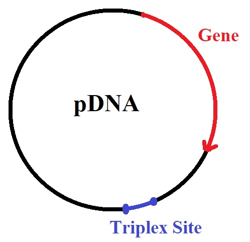
Girardin et al. recently reported a method for the design and synthesis of a BNA-based triplex-forming oligonucleotide conjugate. Using click chemistry, the researchers conjugated the BNA-triplex forming oligonucleotide to a microtubule protein targeting peptide. The peptide-linked BNA oligonucleotide also targets triplex-forming oligonucleotides with oligopurine • oligopyrimidine sites in pDNA. The triplex-forming sequences are inserted into the pDNA outside the luciferase gene sequence.

For the synthesis of the triplex-forming oligonucleotide, the oligonucleotide CTCTCTCTCTC was modified with BNAs to yield the BNA oligonucleotide C+TC+TC+TC+TC+TC in which N+ indicates the position of the BNA placement.


-NNNNN-(TCTCTCTCTCTC)n-NNNNNNNNN---

        ctCtCtCtCtCt-linker-Peptide

 NNNNN-(AGAGAGAGAGAG)N-NNNNNNNNN---


pDNA: Girardin et al. used rpDNA of 5 kbp encoding the luciferase gene and a pDNA of 21 kbp encoding both the luciferase and the full-length dystrophin genes. In vivo transfection was performed by Hydrodynamic Limb Vein (HLV) injection of the large pDNA encoding both the luciferase and the dystrophin genes.

Triplex forming sequences linking the peptide to the pDNA.


BNA-oligonucleotide-peptide conjugate formed by click chemistry.

Linkage of the peptide to pDNA: the linkage occurs via a BNA oligonucleotide-peptide conjugate forming a triple helix with pDNA containing triplex forming sites.

The peptide containing amino acid residues 79 to 98 from the amino acid sequence of the adenovirus E3 early protein of 14.7 kDa (E3-14.7K) specifically interacts with FIP-1 binding to the dynein light chain TCTEL-1. The peptide mediates interaction of pDNA with microtubules. The interaction of the protein complex promotes migration of the pDNA along microtubules toward the nucleus.

N = BNA, n = natural nucleic acid. 


As a side note: There are two known microtuble motor proteins, kinesins and dyneins. Kinesins (with the exception of kinesin 14) move towards the (+) end of MTs to the cellular periphery. Dynein moves towards the (−) end in the direction of the cell nucleus. 


Reference

Girardin, C., Maze, D., Gonçalves, C. et al. Selective attachment of a microtubule interacting peptide to plasmid DNA via a triplex forming oligonucleotide for transfection improvement. Gene Ther (2022).  [nature]

BNAs 

BNAs-as-Tools-for-DNA-or-RNA-Targeting

BNAs as Molecular Tools

BNAs-for-duplex-and-triplex-formation


A-2-4-bridged-nucleic-acid-containing-2-pyridone-as-a-nucleobase-efficient-recognition-of-a-c-g-interruption-by-triplex-formation-with-a-pyrimidine-motif


Triplex-Formation-for-the-Detection-of-microRNAs


Triplex-Medicated-Gene-Modification


Highly-stable-pyrimidine-motif-triplex-formation-at-physiological-ph-values-by-a-bridged-nucleic-acid-analogue


Promotion-of-triplex-formation-by-bna-nc-modification

Molecular Motors in Molecular Biology of the Cell. 4th edition.

---...---
Bio-Synthesis provides a full spectrum of oligonucleotide and peptide synthesis including bio-conjugation services as well as high quality custom oligonucleotide modification services, back-bone modifications, conjugation to fatty acids and lipids, cholesterol, tocopherol, peptides as well as biotinylation by direct solid-phase chemical synthesis or enzyme-assisted approaches to obtain artificially modified oligonucleotides, such as BNA antisense oligonucleotides, mRNAs or siRNAs, containing a natural or modified backbone, as well as base, sugar and internucleotide linkages.
Bio-Synthesis also provides biotinylated mRNA and long circular oligonucleotides.

---...---

Thiamine (Vitamin B1) and RNA capping

$
0
0

RNA molecules like messenger RNA are known to have modifications on their 5’-terminal ends. In recent years, scientists have confirmed many 5’-modified RNA molecules, including RNA modified with adenosine-containing cofactors from the B vitamin group, found in all kingdoms of life.

Thiamine adenosine triphosphate (ThATP) is a vitamin B1 derivative that accumulates in Escherichia coli and other organisms in response to metabolic stress conditions. Thiamine and its phosphorylated derivatives (mono-, di-, and triphosphate) occur naturally in most cells. Frédérich et al., in 2009, reported the presence of the thiamine derivative, adenosine thiamine triphosphate, in Escherichia coli as a response to carbon starvation.

To enable the discovery of 5’-thiamine-capped RNA in organisms, Möhler et al. in 2020 synthesized thiamine adenosine dinucleotides and 5’-thiamine-capped RNA utilizing phosphorimidazolide chemistry. The research group showed that T7 RNA polymerase in the presence of divalent magnesium cations caps RNA with thiamine-ATP and thiamine adenosine diphosphate (thiamine-ADP). This approach allows the capping of 5′-triphosphate RNAs, independent of length, structure, or nucleotide composition, after conversion to the 5′-monophosphate by, e.g., polyphosphatases. In vitro transcription allowed the synthesis of larger capped RNA molecules.

Also, Möhler et al. modified transcripts containing the thiamine modification with biotin, allowing the separation of in vitro transcribed 5’-thiamine RNA from 5’-triphosphate RNA. This new method now provides access to 5’-thiamine-capped RNA useful for developing thiamine-specific RNA capture protocols for discovering and confirming 5’-thiamine-capped RNAs in different organisms.

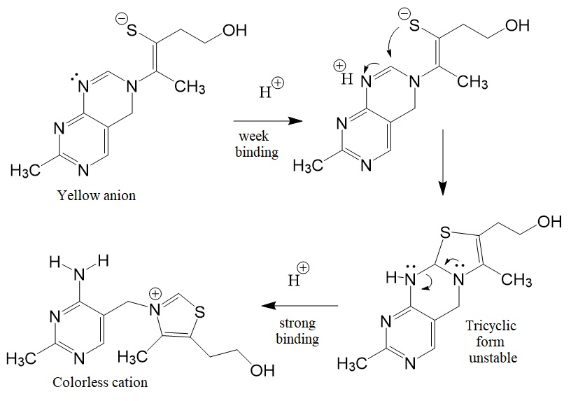
Vitamin B1 is an essential micronutrient the human body cannot make but is found in food, commercially synthesized, and used in supplements and medications. Metabolic reactions in mammals and humans require phosphorylated forms, such as the metabolization of glucose and amino acids. Five natural thiamine phosphates are known: thiamine monophosphate (ThMP), thiamine pyrophosphate (TPP), thiamine triphosphate (ThTP), adenosine thiamine diphosphate (AThDP), and adenosine thiamine triphosphate (AThTP).Vitamin B1 is known as thiamin. The thiamin anion binds protons in a cooperative process. Figure 1 shows the reaction scheme for the cooperative binding of protons by the thiamin anion.

Figure 1: Reaction scheme for the cooperative binding of protons by the thiamine anion (David Metzler. Biochemistry. 2nd Edition).

Thiamine (vitamin B1) functions as a coenzyme, for example, during the fermentation of sugar to ethanol by yeast. Bacteria, fungi, and plants synthesize thiamine from 1-deoxyxylulose 5-phosphate. 1-Deoxyxylose 5-phosphate is an intermediate in the nonmevalonate pathway of polyprenyl synthesis. David Metzler reported that Mizuhare, in 1950, discovered that the hydrogen atom in the two position of the thiazolium ring, between the sulfur and the nitrogen atoms, exchanged quickly with deuterium of 2H2O. The pKa of this proton has been estimated as ~ 18, low enough to permit rapid dissociation and replacement with 2H.


In recent decades, scientists have determined the structures for many thiamine diphosphate-dependent enzymes, including bacterial pyruvate oxidases, yeast, pyruvate decarboxylases, transketolase, and benzoyl formate decarboxylases. The different enzymes have different sequences and catalyze different reactions. However, the thiamin diphosphate is bound similarly in all of them. Figure 2 shows the crystal structure of the thiamine diphosphate-dependent enzyme pyruvate decarboxylase from yeasts.

Figure 2: A; Crystal structure of the thiamine diphosphate dependent enzyme pyruvate decarboxylase from the yeast saccharomyces cerevisiae at 2.3 Angstroms resolution [PDB ID 1PVD]. B and C; Structural models of thiamine diphosphate.

Reference

Arjunan P, Umland T, Dyda F, Swaminathan S, Furey W, Sax M, Farrenkopf B, Gao Y, Zhang D, Jordan F. Crystal structure of the thiamin diphosphate-dependent enzyme pyruvate decarboxylase from the yeast Saccharomyces cerevisiae at 2.3 A resolution. J Mol Biol. 1996 Mar 1;256 (3):590-600. [PubMed]

Frédérich M, Delvaux D, Gigliobianco T, Gangolf M, Dive G, Mazzucchelli G, Elias B, De Pauw E, Angenot L, Wins P, Bettendorff L (2009). Thiaminylated adenine nucleotides. Chemical synthesis, structural characterization and natural occurrence. The FEBS journal 276, 3256-3268. [FEBS]

Metzler, David. Biochemistry. The chemical reactions of living cells. 2nd Edition 2003 [ELSEVIER]

Möhler M, Höfer K, Jäschke A. Synthesis of 5'-Thiamine-Capped RNA. Molecules. 2020 Nov 24;25(23):5492. [PMC]


---...---

" Bio-Synthesis provides a full spectrum of high quality custom oligonucleotide modification services including back-bone modifications, conjugation to fatty acids, biotinylation by direct solid-phase chemical synthesis or enzyme-assisted approaches to obtain artificially modified oligonucleotides, such as BNA antisense oligonucleotidesmRNAs or siRNAs, containing a natural or modified backbone, as well as base, sugar and internucleotide linkages.
Bio-Synthesis also provides biotinylated and capped mRNA as well as long circular oligonucleotides".

---...---


Of Target Effects in small interfering RNA or siRNA

$
0
0

Off-target effects in RNA interference screens occur where small interfering RNAs directly affect the expression of genes other than the targeted gene. Microarray experiments indicated that small interfering RNAs (siRNA) could affect the mRNA levels of many genes.

The recent approval of multiple siRNA therapeutics highlights the potential of siRNA drugs. The success of siRNA drugs was possible due to the availability of chemically modified nucleic acids and their impact on the stability, delivery, potency, and off-target effects of siRNAs.

Therapeutic small interfering RNAs (siRNAs) are double-stranded RNAs that specifically silence genes through the activation of the RNA interference pathway (RNAi). Double-stranded RNAs allow the design of potent siRNAs for specific gene silencing. Since siRNAs are delivered nakedly, they must be modified to resist degradation. Each siRNA strand can downregulate several genes. Also, a saturation of the RNAi machinery leads to the upregulation of miRNA-repressed genes. Often clinical trials face failure because of off-target effects such as off-target gene dysregulation.

Eliminating sense strand uptake reduces off-target gene silencing and limits the disruption to endogenous regulatory mechanisms. Therefore, optimal strand selection impacts the success of future siRNA therapeutics. Extensive modification and encapsulation in protective carrier molecules can prevent the rapid degradation by blood serum nucleases of siRNAs. The formulation in lipid nanoparticles (LNPs) facilitates the delivery of siRNAs to tissues such as the liver. The chemical conjugation of double-stranded siRNAs to biological molecules such as GalNac enables their cellular delivery.

Cuccato et al. estimated that 103 to 104 RISC molecules are present in the average mammalian cell, corresponding to an intracellular concentration of approximately 3 to 5 nM.

Each siRNA strand can have a range of distinct gene-silencing effects. Limiting sense strand selection allows the management of off-target effects. Minimizing off-target effects is possible using chemical modification and sequence design strategies which reduce seed-region binding and possible matches in the 3’-untranslated region (3′UTR).

Conjugates can inhibit their corresponding strands resulting in the development of conjugates added to the sense strand.

siRNAs modified with hydrophobic conjugates and adding 5’-E-vinylphosphonate (5′-E-VP) improves tissue retention, silencing activity, and duration of the silencing effect.

Nomenclature

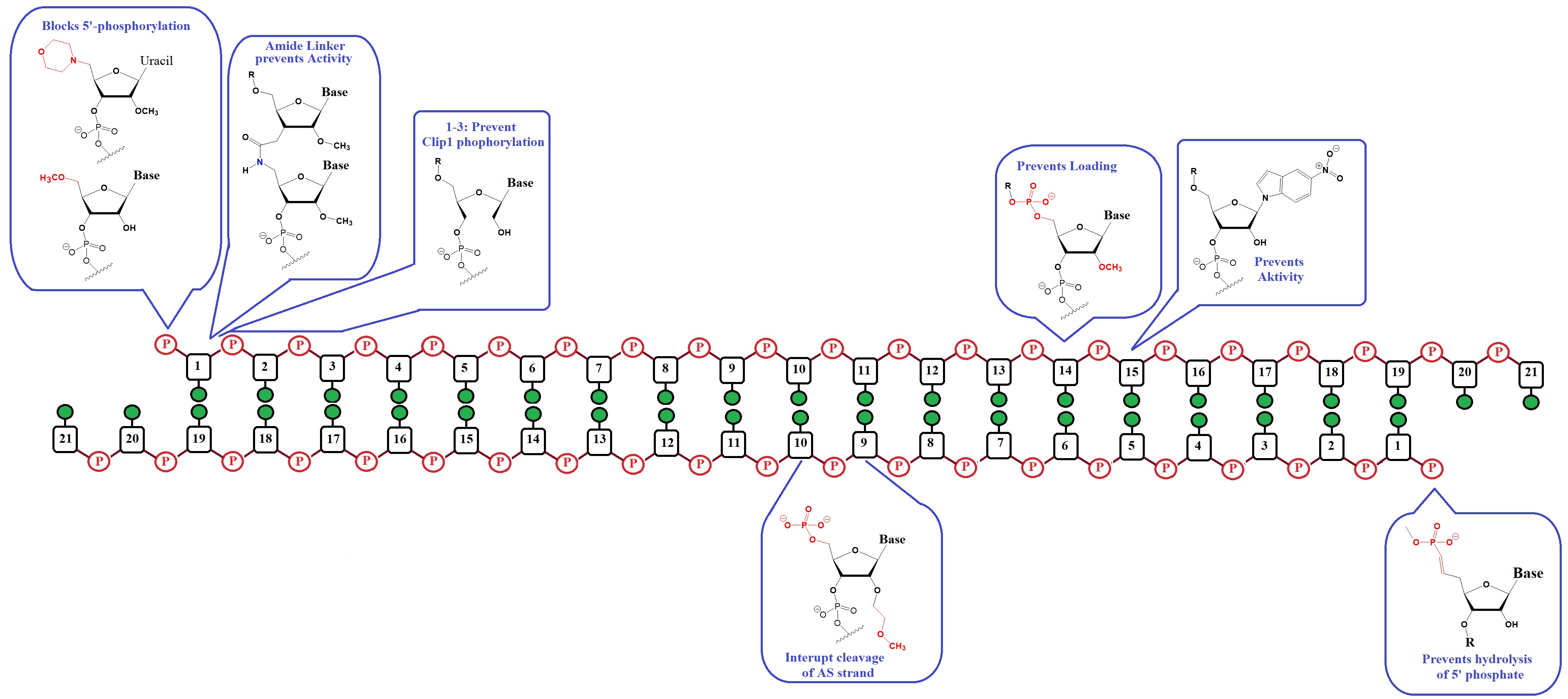
In the siRNA nomenclature as proposed by Varley & Desaulniers in 2020 [AS/SS:##] indicates the duplex molecules where AS refers to the antisense strand, SS refers to the sense strand, and ## indicates the nucleotide number from the 5′ end of the corresponding strand. 
For example, SS:14 refers to the 14th nucleotide from the 5′ end of the sense strand.

Chemical modifications of siRNA impacting strand selection.


Figure 1: Strategies to mitigate sense strand uptake into RISC (Adapted from Varley & Desaulniers).

On-target and off-target effects:

[1]  The 5’-phosphate is the most critical region for the uptake by AGO2. 

[2]  A phosphate group at the 5’-end of a strand is essential for AGO2 uptake. Clp1 kinase and phosphatase(s), which balance 5’-phosphorylated and free hydroxyl ends in cells, recognize siRNAs.

[3]  The efficiency of these enzymes is influenced by modifications of the 5'-ends of these enzymes resulting in a shift in phosphorylation which affects the likelihood of uptake of the associated strand.  

Effectively stabilized 5’-phosphate mimics must facilitate RNAi activity by overcoming three hurdles:

[1] They must sterically fit within the deep binding pocket of AGO2;

[2] Permit interactions with AGO2 side chains without significant distortion to the attached nucleoside; and

[3] Prevent hydrolysis by phosphatases within the cell.

These strict requirements resulted in relatively few effective 5’-phosphate mimics.

Stabilized 5’-phophate mimics for the antisense strand can carry modifications within the first three nucleotides of the 5’-end. These modifications can reduce Clp1 kinase activity, preventing intracellular phosphorylation of the strand.

Successful chemical strategies for improving strand selection for siRNAs are:

[1] Control of the 5’-phosphorylation status.

[2] Alter internal structure to prevent cleavage of the antisense loaded in the passenger strand.

[3] Alter siRNA structure that prevents sense strand capture.

All improvements will support the development of effective, safe, and potent therapeutics.


Kobayashi et al. recently reported rules for siRNA sequences allowing them to be functional in mammalian cells: 

[1] Place A/U at the 5′ end of the guide strand, G/C at the 5′ end of the passenger strand. Place more than 4 A/Us in the 5′-terminus 7-nucleotide of the guide strand, and no GC stretch longer than 8 nucleotides. 

[2] siRNA sequences with weak off-target activity: siRNAs with low Tm value in the seed region (nucleotides 2–8) exhibit weak off-target effect.

[3] Nucleotides in the seed region can be functionally divided into two domains in response to 2′-OMe modifications: 

[a] 2′-OMe modifications of nucleotides 2–5 inhibit the off-target effect of siRNAs

[b] 2′-OMe modifications of nucleotides 6–8 promote both on-target and off-target effects.

Chemical modifications of the 5’-terminus of siRNA influencing strand selection and antisense activity.

Many researchers have investigated 5’-terminal modifications in siRNA and ss-siRNA. 5’-Phosphate Modifications include methylene- and vinyl-phosphonate, 5′-Hydrogen substitutions, and 5′-alternative structures. The structures of these modifications are shown below (Varley & Desaulniers, 2021).


  Natural 5’-Phosphate


  5-Methylene Phosphonate NA.

  5-(E)-Vinyl-Phosphonate NA.

 5′-(E)-VP phosphate mimic is the preferred choice for phosphate stabilization. 
 Addition to a GalNac3 conjugated siRNA improves the IC50 3 to 20 fold.

 5-(R)-Me-Phosphonate NA.

 Improves the IC50 ~ 2 to 3-fold.

 5-(S)-Me-Phosphonate NA.

 No impact on IC50 of siRNA.


 5-R-MeOCH3-Phosphonate NA.

 Improves activity 2 to 20-fold.

 5’-(S)-FMe-P NA.

 Improved IC50 ~ 2-fold.


 5’-Deoxy-5’-Morpholino-2’-O-Methyl Uridine.

 Reduced activity ~3-fold. 
 May prevent uptake of sense strand.

 5’-O-Methyl NA.

 Disrupts activity.


Chemical internal modifications of siRNA influencing strand selection and antisense activity.


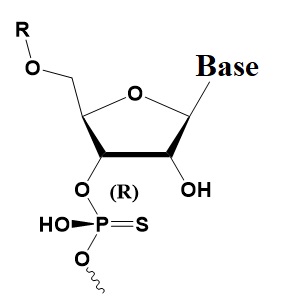
Rp Phosphorothioate

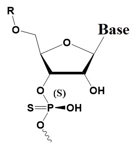
Sp Phosphorothioate

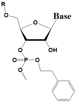
Phenylethyl Phosphate

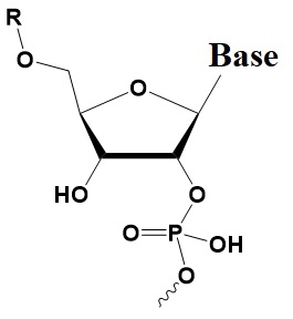
2’-5’-Phosphate linkage

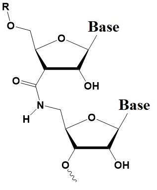
Amide linkage


Reference

Birmingham A, Anderson EM, Reynolds A, Ilsley-Tyree D, Leake D, Fedorov Y, Baskerville S, Maksimova E, Robinson K, Karpilow J, Marshall WS, Khvorova A: 3' UTR seed matches, but not overall identity, are associated with RNAi off-targets. Nat Methods. 2006, 3 (3): 199-204. [Nature Methods]

Chernikov IV, Gladkikh DV, Meschaninova MI, Ven'yaminova AG, Zenkova MA, Vlassov VV, Chernolovskaya EL. Cholesterol-Containing Nuclease-Resistant siRNA Accumulates in Tumors in a Carrier-free Mode and Silences MDR1 Gene. Mol Ther Nucleic Acids. 2017 Mar 17;6:209-220. [PMC]


Cuccato G, Polynikis A, Siciliano V, Graziano M, di Bernardo M, di Bernardo D. Modeling RNA interference in mammalian cells. BMC Syst Biol. 2011 Jan 27;5:19. [PMC]


Dong Y, Siegwart DJ, Anderson DG. Strategies, design, and chemistry in siRNA delivery systems. Adv Drug Deliv Rev. 2019 Apr;144:133-147 [PMC]


Fedorov Y, Anderson EM, Birmingham A, Reynolds A, Karpilow J, Robinson K, Leake D, Marshall WS, Khvorova A. Off-target effects by siRNA can induce toxic phenotype. RNA. 2006;12(7):1188–1196. [PMC]


Jackson AL, Burchard J, Schelter J, Chau BN, Cleary M, Lim L, Linsley PS: Widespread siRNA "off-target" transcript silencing mediated by seed region sequence complementarity. RNA. 2006, 12 (7): 1179-1187. [PMC]


Varley AJ, Desaulniers JP. Chemical strategies for strand selection in short-interfering RNAs. RSC Adv. 2021 Jan 11;11(4):2415-2426. [PMC]


---...---

" Bio-Synthesis provides a full spectrum of high quality custom oligonucleotide modification services including back-bone modifications, conjugation to fatty acids, biotinylation by direct solid-phase chemical synthesis or enzyme-assisted approaches to obtain artificially modified oligonucleotides, such as BNA antisense oligonucleotidesmRNAs or siRNAs, containing a natural or modified backbone, as well as base, sugar and internucleotide linkages.
Bio-Synthesis also provides biotinylated and capped mRNA as well as long circular oligonucleotides".

---...---


Custom synthesis of N-acetylgalactosamine (GalNac) siRNA

$
0
0

When conjugated to oligonucleotide drugs, Triantennary N-Acetylgalactosamine (GalNAc) enhances their specific delivery to targets in liver cells. Already, the FDA has approved several GalNAc-conjugated oligonucleotides for clinical use.

The ligand of the asialoglycoprotein receptor (ASGPR), triantennary N-acetylgalactosamine (tri-GalNAc), when conjugated to oligonucleotides, improves their cellular uptake and tissue-specific delivery. ASGPR binds glycoproteins with terminal galactose residues in liver cells and removes targeted glycoproteins from circulation. The half-life of therapeutic GalNAc-RNAi drugs is relatively long, allowing monthly to half-yearly dosing regiments.

The conjugation of siRNAs to multivalent GalNAc linkers enhances their delivery and gene silencing in liver cells. GalNAc linkers attached to the 3'-end of siRNA sense strands allow delivery into cells without additional delivery reagents. GalNac-conjugates oligonucleotides exhibit improved tissue-specific delivery for antisense oligonucleotides (ASOs) and siRNAs.

---...---

" Bio-Synthesis provides custom synthesized GalNAc-conjugated oligonucleotides including GalNAc-siRNAs. 

Bio-Synthesis also provides a full spectrum of high quality custom oligonucleotide modification services including back-bone modifications, conjugation to fatty acids, biotinylation by direct solid-phase chemical synthesis or enzyme-assisted approaches to obtain artificially modified oligonucleotides, such as BNA antisense oligonucleotidesmRNAs or siRNAs, containing a natural or modified backbone, as well as base, sugar and internucleotide linkages.
Bio-Synthesis also provides 
biotinylated and capped mRNA as well as long circular oligonucleotides".

---...---


Affinity-controlled release of 5-fluorouracil by synthetic peptide D-Leu-Phe-Phe-derived hydrogels for the potential treatment of skin cancer

$
0
0
 Molecular advances in the arena of biotechnology are increasingly making an impact on future biomedicine.  As opposed to directly utilizing natural product-derived medicine, understanding its underlying pharmacological mechanism allows one to design more efficacious therapeutics based on the mechanism.  The design could utilize readily accessible molecular tools such as synthetic peptides or oligonucleotides.  By doing so, the costly endeavor required to extract medicinal agents from naturally occurring herbs, plants, or other biological materials could be bypassed.   Alternatively, such tools could be exploited to deliver naturally occurring therapeutics to their intended targets to reduce side effects.

In the last several decades, the potential utility of synthetic peptides has been greatly expanded.  Exploiting the intrinsic properties of peptides to adopt myriad conformations, pharmaceutical industries as well as academic institutions have been increasingly focusing on applying synthetic peptides to address previously unsolved medical needs.  The advantages of utilizing peptides are multiple, including greater tissue penetration (ex. for solid tumor therapy) (Hong et al., 2000), the ability to modulate stability or overall charge through chemical modification (ex. peptide mimetics for oral delivery) (Cooper et al., 2021), the relative ease for interrogating the structure/conformation of shorter peptides than complex proteins composed of longer polypeptides via molecular modeling, etc.  The recently developed FDA-approved peptidomimetic drug for the COVID-19 coronavirus (Paxlovid) represents one such example.

More importantly, the ability to synthesize peptides through organic chemistry at an industrial scale has facilitated greater than 6400 clinical trials.  The scope of its potential being explored through clinical studies includes antiviral therapy, cancer radiotherapy, cancer immunotherapy, and treating complex disorders involving multiple genetic mutations.  As some of these illnesses are becoming quite common, the demand for peptide therapeutics is likely to grow with an increasing number of elderly patients approaching or exceeding 90-100 years of age globally (ex. >3.4 million centenarians expected by 2050)  (Cho et al., 2020).

On another front, the potential of utilizing synthetic peptides to address the problem of targeted drug delivery has been actively explored.  Critical to this undertaking is the ability to chemically modify amino acids comprising the peptide during the synthesis at specific residues.  These include phosphorylation, acetylation, amidation, citrullination, succinylation, PEGylation, etc.  Equally important is the ability to conjugate peptides to a variety of agents to improve their pharmacological properties.  Multiple candidate synthetic peptides have been discovered in the past utilizing phage-display technology to target normal tissues or cancerous tissues (ex.  RGD, NGR, HN-1 PEP-1, etc.)

With the refinement of the delivery system, further improvements to meet specific pharmacokinetic and/or pharmacodynamic requirements for treating complex disorders are being sought.   For treating diseases such as diabetes, persistent dosing of drugs may be necessary to achieve the desired therapeutic outcome.  Here, the traditional schedule consisting of a singular injection (resulting in an early burst of drug dose) may not be optimal due to the eventual depletion of drugs in circulation.  To meet such requirements, pharmaceutical researchers have been bioengineering ways through which a sustained release of drugs could be attained following the injection.  In principle, the ‘long-acting drugs could be achieved by delaying the access of injected drugs to blood vessels from the site of injection (ex. subcutaneous tissue)—through the formation of insoluble aggregates, for instance.


An alternate method to attain persistent dosing is to compartmentalize drugs in a vehicle that allows a controlled release of the enclosed drugs while in circulation.  The rapid elimination could potentially be avoided by designing vehicles whose hydrodynamic diameter exceeds the threshold for renal clearance.  One such example is hydrogel comprised of synthetic peptides.  Hydrogels are artificial 3-dimensional structures formed by synthetic or natural biopolymers that retain a large amount of water.  It has been used for a variety of biomedical applications, including tissue implants, tissue engineering/regeneration, biosensor, etc. (Chyzy et al., 2022).  To date, ~30 hydrogels have been approved by U.S. FDA for clinical use (Mandal et al., 2020).

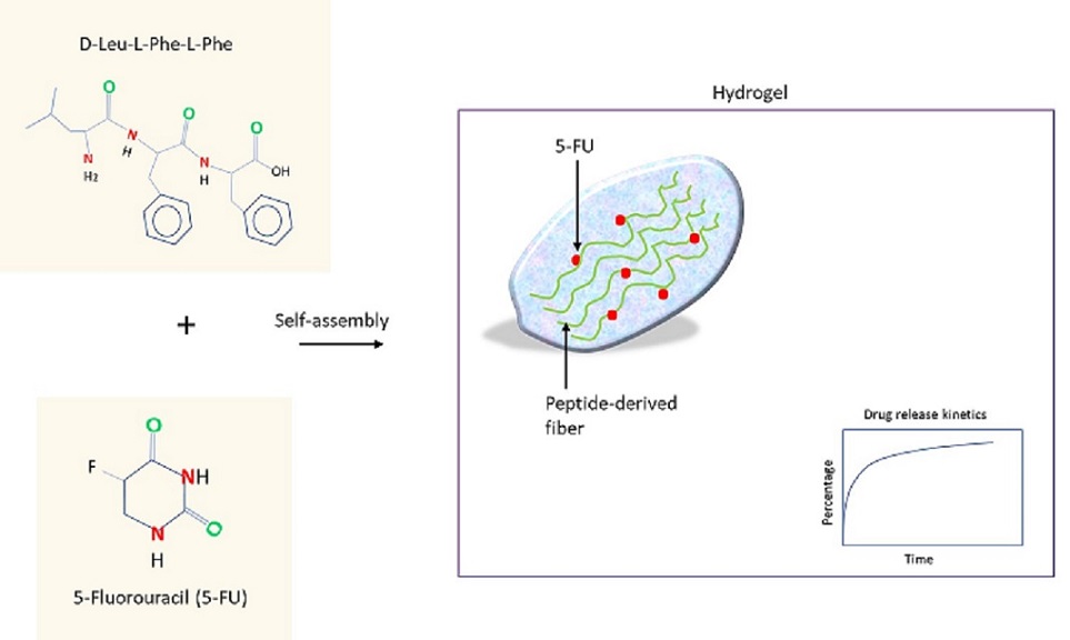
Hydrogels self-assembled from synthetic peptides have also been utilized for drug delivery (Nguyen et al, 2011).  The basic principle encompasses the formation of fibers from peptides, followed by their cross-linking to achieve gelation, the extent of which can be regulated (ex. by light).  The gelation process is sensitive to multiple parameters, ex. pH, chirality, pie-pie stacking (interaction of aromatic groups), peptide sequence, etc.  Hydrogels with a defined mesh size will allow a slower release of larger drugs than smaller-sized drugs.  However, the process of manufacturing hydrogels with a defined mesh size via the self-assembly of peptides has been difficult to control (Namblar et al., 2022).

An innovative method is to utilize an affinity-based method to control the release of drugs.  For peptide-based systems, commonly used interactions include electrostatic, hydrophobic, reversible covalent bonds (ex. disulfide bonds), etc.  Upon identifying the proper binding entity for the drug, it can be engineered into the amino acids used for peptide synthesis.  To release drugs, the electrostatic interaction of drugs with charged residues (ex. glutamic acid, aspartic acid, arginine) could be disrupted by altering pH or ionic strength.

Skin cancer is the most commonly diagnosed cancer in the U.S.; among the subtypes, invasive melanoma accounts for the majority of incurred deaths (~7650 death expected in 2022).  (Cancer Facts & Figures 2022 )   5-fluorouracil (5-FU) is an antimetabolite drug used to treat skin cancer and it functions by inhibiting thymidylate synthetase, which converts dUMP to dTMP to support DNA replication (Longley et al., 2003).  To resolve multiple outstanding issues such as shorter half-life or dosage variability, investigators at the University of Trieste (Italy) have developed a supramolecular hydrogel comprised of a tripeptide and the drug (Parisi et al., 2019).  Intriguingly, the self-assembly by the tripeptide D-Leu-Phe-Phe occurred in the presence of 5-FU.  Molecular dynamics simulation study revealed that the pie-pie stacking between the ring structure of 5-FU and the phenylalanine may underlie the drug-to-peptide interaction, which lasted transiently (~100 nanoseconds).  The majority of the contained drug was released within the first hour.


The key to preventing an epidemic is the ability to diagnose the infected early to preempt further propagation.  For this, Bio-Synthesis, Inc. provides primers and probes (as well as synthetic RNA control) for COVID-19 diagnosis via RT-PCR assay.  It specializes in oligonucleotide modification and provides an extensive array of chemically modified nucleoside analogs (over ~200) including bridged nucleic acid (BNA) in addition to mRNA synthesis.  A number of options are available to label oligonucleotides (DNA or RNA) with fluorophoreseither terminally or internally as well as to conjugate to peptidesor antibodies. It provides custom conjugation of small molecules such as chemical drugs, metabolites and labeled compounds with synthetic or natural polymers (enzymes, peptide, protein, oligonucleotide, antibody, dendrimer, nanoparticle, etc).  It recently acquired a license from BNA Inc. of Osaka, Japan, for the manufacturing and distribution of BNANC, the third generation of BNA oligonucleotides.  To meet the demands of therapeutic application, its oligonucleotide products are approaching GMP grade.  It has recently entered into collaborative agreement with Bind Therapeutics, Inc. to synthesize miR-21 blocker using BNA for triple-negative breast cancer.  The BNA technology provides superior, unequaled advantages in base stacking, binding affinity, aqueous solubility and nuclease resistance.  It also improves the formation of duplexes and triplexes by reducing the repulsion between the negatively charged phosphates of the oligonucleotide backbone.  Its single-mismatch discriminating power is especially useful for diagnosis (ex. FISH using DNA probe).  For clinical application, BNA oligonucleotide exhibits lesser toxicity than other modified nucleotides.   For therapeutic consideration, peptide synthesis or modifications may include labeling, conjugation, cyclization, incorporation of unusual amino acids, and modification of side chain and backbone.


https://www.biosyn.com/oligo-flourescent-labeling.aspx

https://www.biosyn.com/tew/Speed-up-Identification-of-COVID19.aspx

https://www.biosyn.com/covid-19.aspx

https://www.biosyn.com/mrna.aspx

https://www.biosyn.com/bioconjugation.aspx

https://www.biosyn.com/tew/Design-Guidelines-for-BNA-based-Oligonucleotide-Probes.aspx#!

Peptide Modifications, Modified Peptide Synthesis - Bio-Synthesis (biosyn.com)

https://www.biosyn.com/tew/development-of-a-therapeutic-peptidomimetic-inhibiting-main-protease-to-reduce-the-level-of-rdrp-polymerase-that-replicates-covid-19-coronavirus-genome.aspx

https://www.biosyn.com/tew/peptide-therapeutics-target-dynamic-protein-to-protein-interaction-underlying-human-diseases-such-as-hypertension-cancer-alzheimers-disease-and-potentially-covid-19.aspx


References

Cho J, Hirose N, et al. Caregiving centenarians: Cross-national comparison in Caregiver-Burden between the United States and Japan.  Aging Ment Health. 24: 774-783 (2020).  PMID: 30596257

Chyzy A, Plonska-Brzezinska ME, et al. Microwave-Assisted Synthesis of Modified Glycidyl Methacrylate-Ethyl Methacrylate Oligomers, Their Physico-Chemical and Biological Characteristics.  Molecules. 27:337 (2022).  PMID: 35056652

Cooper BM, Spring DR, et al.  Peptides as a platform for targeted therapeutics for cancer: peptide-drug conjugates (PDCs).  Chem Soc Rev.  50:1480-1494 (2021).  PMID: 33346298

Hong FD, Clayman GL. Isolation of a peptide for targeted drug delivery into human head and neck solid tumors.  Cancer Res. 60:6551-6 (2000).  PMID: 11118031

Longley DB, Johnston PG, et al.  5-fluorouracil: mechanisms of action and clinical strategies.  Nat Rev Cancer.  3:330-8 (2003).  PMID: 12724731

Mandal A, Mitragotri S., et al. Hydrogels in the clinic.  Bioeng Transl Med.  5:e10158 (2020).  PMID: 32440563

Nambiar M, Schneider JP. Peptide hydrogels for affinity-controlled release of therapeutic cargo: Current and potential strategies.  J Pept Sci. 28: e3377 (2022).  PMID: 34747114

Nguyen LH, Linse KD, et al.  Unique biomaterial compositions direct bone marrow stem cells into specific chondrocytic phenotypes corresponding to the various zones of articular cartilage.  Biomaterials. 32:1327-38 (2011).  PMID: 21067807

Parisi E, Marchesan S., et al. Supramolecular Tripeptide Hydrogel Assembly with 5-Fluorouracil.  Gels. 5 :5 (2019).  PMID: 30691142


Antisense Oligonucleotides as Tracers for PET Imaging

$
0
0

Oligonucleotides hybridize with high selectivity to RNA sequences allowing monitoring of gene expression or its inhibition in experimental and therapeutic applications. Custom-synthesized antisense oligonucleotides enable the design of molecular imaging technics needed for medical diagnostics. Molecular imaging technics allow visualization and quantification of molecular events in cellular contexts. In nuclear medicine, positron emission tomography (PET) and single-photon emission tomography (SPECT) are widely used in clinical therapeutic diagnostics, also called theranostics. PET-based imaging is fast enough to determine the pharmacokinetics of radiotracer uptake and distribution. PET typically utilizes radiolabeled molecules with positron-emitting nucleotides such as 15O, 13N, 11C, and 18F with relatively short half-lives. However, Gallium-68 and technetium-99m now also see increased use.

For the production of isotope-labeled probes, modified custom antisense oligonucleotides are provided with an amino group, a chelator, or any other functional group needed for labeling with the radioemitter located on either the 5’- or 3’-end ready for labeling with the radioemitter label.


In recent decades scientists developed several methods for labeling antisense oligonucleotides with positron-emitting isotopes. A selection of methods enables the labeling of antisense oligonucleotides with positron-emitting isotopes. Examples are positron-emitting isotopes 11C, 18F, or 76Br. Several molecular imaging trials with experimental animals have also been described. A recent example is the PET radioligand utilized for imaging 
Tau protein in the brain of patients with tauopathies.

Also, radiolabeled amino acid PET tracers targeting specific tumor-expressed receptors offer improved accuracy in defining the tumor-to-background contrast and in more particular treatments. Therefore, Gallium-68 (68Ga) has recently become an alternative positron emitter to the most commonly used 18F-2-fluoro-2-deoxy-D-glucose (18F-FDG).

Pan et al., in 1998, utilized 5'-deoxy-5'-fluoro-O4-methylthymidine to develop radiofluorinated antisense oligodeoxynucleotide probes useful for PET.

In 1999, Kobori et al. imaged mRNAs in rat glioma tumors with antisense phosphorothioate oligodeoxynucleotides. The antisense oligonucleotides targeting the glial fibrillary acidic protein (GFAP) contained the positron emitter 11C as a label.

Roivainen et al., in 2004, reported that labeling oligonucleotides with 68Ga is a convenient approach for in vivo imaging and quantifying oligonucleotide biokinetics in living animals with PET.

Gallium-68 has a short half-life of 68 minutes. A germanium-68/gallium-68 generator allows its convenient production using a germanium-68/gallium-68 generator. These features enabled its increased clinical use in recent years.

Roivainen et al. labeled synthetic oligonucleotides with 68Ga and DOTA chelate conjugated to the oligonucleotides. Intravenously injected 68Ga-oligonucleotides of 17mer length generated high-quality PET images, allowing quantification of the biokinetics in major organs and tumors.

Liu et al., in 2007, showed that 99mTechnetium (99mTc)-labeled antisense oligonucleotide probes allowed imaging of human telomerase reverse transcriptase (hTERT) messenger RNA in malignant tumors. 99mTc is a metastable nuclear isomer of technetium-99. This isotope (Mw: 98.9063 Da) has a half-life of 6.0067 hours. The isotope enables a variety of imaging and diagnostic methods.

Cole et al., in 2014, showed that the short-lived fluorine-18 atom (t1/2 = 109.77 min) could be incorporated into molecular imaging probes late in the probe’s synthetic pathway. This approach enables the development of rapid and efficient late-stage fluorination methodologies.

Jacobsen et al., in 2015, reviewed labeling strategies and synthetic routes for Fluorine-18 radiochemistry. Typically, fluorine-18, with a nuclear characteristic of beta decay and a half-life of 109.7 minutes, is added to a functional group present at the reporter molecule (18F; 97% β+ decay, 109.7 min half-life, 635 keV).

---...---

Controlled Release Linkers For New Therapeutic Drugs

$
0
0

New drugs, such as oligonucleotide-based antisense drugs, peptide-drug conjugates, or lipid-drug conjugates, can potentially treat or cure many diseases. However, the drugs must reach their specific target and have improved pharmacokinetics to function as desired. The conjugation of drugs to macromolecular carriers improves the pharmacokinetics of the drug. However, stable conjugates can have several limitations:

(1) loss of the potency of the drug,

(2) confining the drug to the extracellular space, and

(3) requirement for circulating conjugates.

To improve the pharmacokinetics of drug conjugates, Santi et al. described conjugation linkers with highly tunable cleavage rates. The research group reviewed a set of tunable release linkers with half-lives ranging from hours to up to one year. Santi et al. suggest that these releasable linkers provide several benefits, including lower Cmax, the highest concentration of a drug in the blood, cerebrospinal fluid, or target organ after a dose is given, and the pharmacokinetic coordination of drug combinations. The multiple developed linkers have cleavage rates predetermined by the acidity of a C-H bond on the linker. A hydroxide-catalyzed β-elimination reaction releases the native, active drug.

Electron-withdrawing groups attached to the ionizable C-H group control the acidity of the linker. The linkers described by Santi et al. enable the construction of drug conjugates with predictable, tunable release kinetics over long periods useful in vitro and in vivo environments. Figure 1 shows a chemically releasable carbamate linker.


Figure 1: A chemically releasable carbamate linker. A macromolecular carrier is attached to a linker which is attached to the drug or prodrug via a carbamate group. The β-carbon has an acidic C-H group and contains an electron-withdrawing “modulator” that controls the pKa of that C-H group. Removing a proton initiates rapid β-elimination, cleaving the C-O of the linker–carbamate bond. A further loss of CO2 provides the free drug or prodrug and a substituted alkene. The drug release rate is proportional to the acidity (pKa) of the proton adjacent to the modulator. The chemical nature of the modulator controls the pKa and the release of the drug.

As a next step, the research group tested a carbamate containing Ne-2,4-dinitrophenyl-l-lysine [Lys(DNP)OH] and various electron-withdrawing pKa modulators. Figure 2 illustrates the releasing reaction. 

Figure 2: Releasing reaction of a linker scaffold containing electron-withdrawing pKa modulators linked via carbamate bonds to Ne-2,4-dinitrophenyl-l-lysine [Lys(DNP)OH]. Modulators utilized aromatic groups, ketones, nitriles, and sulfones. 

Figure 3: Modulator groups tested. [A] Half-lives for β-elimination varied between 14 hours to 2 weeks. [B to E] These less electron-withdrawing modulators extended the half-lives for up to 12 months (Santi et al. 2012).

Furthermore, the research group reported the preparation of bifunctional linkers containing succinimidyl carbonate on one end and an azide moiety connected to the other. After reacting the succinimidyl carbonate with the amino group of a carrier or a similar molecule, copper-free click chemistry allows conjugation to other desired functional groups. The reported heterobifunctional linkers allow attachment to the amine group of a drug or prodrug as a carbamate on one end and any macromolecular carrier on the other end.

The acidity of the ionizable C-H group is controlled by electron-withdrawing pKa modulators. After proton abstraction, the linkers undergo β-elimination to release the native, amine-containing drug or prodrug from the carrier molecules. Controlling the acidity of the ionizable C-H group by the pKa modulator allows for controlling the drug release rate.

Reference

Santi DV, Schneider EL, Reid R, Robinson L, Ashley GW. Predictable and tunable half-life extension of therapeutic agents by controlled chemical release from macromolecular conjugates. Proc Natl Acad Sci U S A. 2012 Apr 17;109(16):6211-6. [PMC]

Links

https://www.biosyn.com/oligo-modified-linker-attachment-chemsitry.aspx#!

https://www.biosyn.com/tew/oligonucleotide-linkers-for-conjugation.aspx#!

https://www.biosyn.com/oligonucleotideproduct/photocleavable-pc-linker.aspx

https://www.biosyn.com/tew/affinity-controlled-release-of-5-fluorouracil-by-synthetic-peptide-d-leu-phe-phe-derived-hydrogels-for-the-potential-treatment-of-skin-cancer.aspx

Pharmacokinetics NIH Library 

---...---

" Bio-Synthesis provides custom synthesized GalNAc-conjugated oligonucleotides including GalNAc-siRNAs. 

Bio-Synthesis also provides a full spectrum of high quality custom oligonucleotide modification services including back-bone modifications, conjugation to fatty acids, biotinylation by direct solid-phase chemical synthesis or enzyme-assisted approaches to obtain artificially modified oligonucleotides, such as BNA antisense oligonucleotidesmRNAs or siRNAs, containing a natural or modified backbone, as well as base, sugar and internucleotide linkages.

Bio-Synthesis also provides 
biotinylated and capped mRNA as well as long circular oligonucleotides".

---...---


MITO-Porters Enable Delivery of Antisense Drugs to Mitochondria

$
0
0

Antisense RNA oligonucleotides (ASOs) potentially allow gene silencing by mitochondrial delivery to target mtDNA-encoded mRNA. MITO-Porters are liposomal nanocarrier systems designed for mitochondrial delivery. Well-designed MITO-Porters may enable their use as therapeutics to regulate mitochondrial function. The efficient packaging of ASOs in a MITO-Porter via a nanoparticle packaging method has a 10-fold higher packaging efficiency than the conventional method. The delivery of the constructed carrier resulted in a decrease in the target mRNA levels and ATP production.

Mitochondrial diseases include multisystem disorders involving metabolic errors. The dysfunction of mitochondria appears to cause various diseases, including cancer, Alzheimer’s disease, Parkinson’s disease, diabetes mellitus, and others. Mitochondrial diseases most often affect the brain, retina, and skeletal muscles. However, multisystem damage can also involve the liver, gastrointestinal tract, pancreas, kidneys, etc.

Mitochondria are energy-generating cellular organelles with their own coding DNA, a circular mtDNA of approximately 16,000 base pairs. More than 1,000 genes from nuclear DNA (nDNA) and 37 genes from mitochondrial DNA (mtDNA) control the mitochondrial proteome. Mutations in more than 350 genes in both genomes appear to cause different mitochondrial diseases.

Figure 1: Mitochondrial electon transport chain (Adapted from Wiki commens. METC).

Gene therapy promises the correction of mitochondrial disorders. However, to be successful, the dynamics of mitochondrial genetics will need to be better understood. Current research focuses on increasing transfection efficiency while lowering cytotoxicity.

Historically, drug delivery into cells utilizes recombinant adeno-associated viruses as viral vectors. However, to selectively treat mitochondria dysfunctions, the targeted delivery of engineered genes or gene products to the nucleus of mitochondria is essential. Because large molecules, including plasmid DNA (pDNA), antisense oligonucleotides, and folded proteins, do not readily pass through the mitochondrial membrane, delivery into mitochondria is difficult. To address these limitations, Yamada et al. developed a liposome-based carrier for delivering cargo molecules called a “MITO-Porter.”

Two independent processes, “cytoplasmic delivery through the cell membrane” and “mitochondrial delivery through the mitochondrial membrane,” are required for efficient drug delivery to mitochondria.

MITO-Porters

Yamada et al., in 2008, described a liposome-based carrier for delivering macromolecular cargos to the mitochondrial interior via membrane fusion. The liposome particles utilized are called MITO-Porters. The nanoparticles carry octa-arginine (R8) surface modifications to enable their entry into cells as intact vesicles. The research team identified lipid compositions that promote the fusion of the nanoparticles with the mitochondrial membrane and the release of its cargo to the intra-mitochondrial compartment in living cells. This uptake process, called “macropinocytosis,” is a non-selective liquid-phase endocytic pathway to uptake extracellular substances.

High-density octa arginine-modified liposomes (R8-LPs) stimulate micropinocytosis by enabling intracellular trafficking. R8 is a synthetic peptide mimicking the trans-activating transcriptional activator derived from the human immunodeficiency virus. R8-LPs can escape from macropinosomes into the cytosol by keeping the encapsulated compounds intact. Low-density R8-LPs are taken up via clathrin-mediated endocytosis and degraded by lysosomal enzymes. MITO-Porter delivered to the cytosol binds to mitochondria via electrostatic interactions with R8. Encapsulated cargo is delivered to the intra-mitochondrial compartment via membrane fusion with the help of sphingomyelin or phosphatidic acid, the lipids that fuse with the mitochondrial membrane. Upon release from the macropinosomes, the MITO-Porter binds to the mitochondrial membrane via electrostatic interactions, inducing fusion between the MITO-Porter and mitochondrion.

The research team screened for liposomes fused with isolated rat liver mitochondria to find the best MITO-Porter liposome components. The variation of the lipid composition using a panel of liposomes and monitoring membrane fusion via fluorescence resonance energy transfer (FRET) analysis allowed the identification of two highly fusogenic lipid compositions, which form the basis of the MITO-Porter.

Green fluorescence protein (GFP) was used as a model macromolecule to validate the MITO-Porter. The use of confocal laser scanning microscopic analysis validated its delivery to mitochondria. Also, FRET analysis allowed the evaluation of membrane fusion between the MITO-Porter and mitochondria in living cells.


In 2019, Kawamura et el. reported that the MITO-Porter is a practical delivery vehicle for antisense oligonucleotides (ASOs) regulating mitochondrial function. The MITO-Porter showed a 10-fold higher packaging efficiency than conventional delivery methods. The use of ASO carriers resulted in a decrease in the targeted mRNA and ATP production.

In 2020, Gao et al. reported that transfected siRNAs could enter the mitochondrial matrix and allow targeted mitochondrial transcripts to be silenced. The study investigated whether siRNAs and small hairpin RNAs (shRNAs) can target mitochondria DNA (mtDNA) encoded transcripts.

DF-MITO-Porter

In 2011, Yamada et al. reported the development of a dual-function MITO-Porter called DF-MITO-Porter. The DF-MITO-Porter is a result of integrating R8-modified liposomes with the MITO-Porter. The research team showed that the DF-MITO-Porter delivers exogenous macro-biomolecules into the mitochondrial matrix. The DF-MITO-porter was adapted to contain an outer endosome-fusogenic envelope facilitating a more efficient escape from the endosome via membrane fusion.


As an example, the construction of a DF-MITO-Porter encapsulating DNase I requires the following three steps: 

(i) the construction of nanoparticles containing DNase I; 

(ii) coating the nanoparticles with a mitochondria-fusing envelope; 

(iii) another coating endosome-fusogenic envelope step-wise, based on previous reports regarding gene packaging with two different types of lipid layers.

More recently, Chernega et al., in 2022, reviewed mitochondrion-targeted RNA therapies as a potential treatment strategy for mitochondrial diseases. The research team pointed out that a careful and thorough examination of possibly disease-associated mtDNA variants and heteroplasmic load is needed to identify genetic causes in patients with possible mitochondrial diseases.

The currently utilized RNA-based therapeutic agents include antisense oligonucleotides (ASOs), small interfering RNAs (siRNAs), and mRNA therapeutic agents. ASOs are single-stranded oligonucleotides that complementary bind to a target mRNA or premature mRNA, which induce degradation, altered splicing, or inhibited translation upon binding.

However, until this day, RNA-based therapeutic agents have yet to be approved for treating mitochondrial diseases. Targeting mitochondrial RNAs could be valuable therapeutic based on mitochondrial biology and conditions.

Also recently, Xu et al., in 2022, reviewed how to design mitochondria-targeted drugs for neurodegenerative diseases, the rescue mechanism of the drugs, and how to assess their therapeutic effect, including structures of small molecules and peptides targeting mitochondria.

References

Chernega T, Choi J, Salmena L, Andreazza AC. Mitochondrion-targeted RNA therapies as a potential treatment strategy for mitochondrial diseases. Mol Ther Nucleic Acids. 2022 Oct 27;30:359-377. [PMC]

Chial, H.: mtDNA and Mitochondrial Diseases [Nature]

Gao K, Cheng M, Zuo X, Lin J, Hoogewijs K, Murphy MP, Fu XD, Zhang X. Active RNA interference in mitochondria. Cell Res. 2021 Feb;31(2):219-228. [PMC]

Kawamura E, Hibino M, Harashima H, Yamada Y. Targeted mitochondrial delivery of antisense RNA-containing nanoparticles by a MITO-Porter for safe and efficient mitochondrial gene silencing. Mitochondrion. 2019 Nov;49:178-188. [PubMed]

Rath S, Sharma R, Gupta R, Ast T, Chan C, Durham TJ, Goodman RP, Grabarek Z, Haas ME, Hung WHW, Joshi PR, Jourdain AA, Kim SH, Kotrys AV, Lam SS, McCoy JG, Meisel JD, Miranda M, Panda A, Patgiri A, Rogers R, Sadre S, Shah H, Skinner OS, To TL, Walker MA, Wang H, Ward PS, Wengrod J, Yuan CC, Calvo SE, Mootha VK. MitoCarta3.0: an updated mitochondrial proteome now with sub-organelle localization and pathway annotations. Nucleic Acids Res. 2021 Jan 8;49(D1):D1541-D1547.  [PMC]

Soldatov VO, Kubekina MV, Skorkina MY, Belykh AE, Egorova TV, Korokin MV, Pokrovskiy MV, Deykin AV, Angelova PR. Current advances in gene therapy of mitochondrial diseases. J Transl Med. 2022 Dec 5;20(1):562. doi: 10.1186/s12967-022-03685-0. Erratum in: J Transl Med. 2023 Feb 8;21(1):96.  [
PMC]

Song S, Zhang Y, Ding T, Ji N, Zhao H. The Dual Role of Macropinocytosis in Cancers: Promoting Growth and Inducing Methuosis to Participate in Anticancer Therapies as Targets. Front Oncol. 2021 Jan 19;10:570108. [
PMC]

Taanman, Jan-Willem; The mitochondrial genome: structure, transcription, translation and replication. Biochimica et Biophysica Acta (BBA) - Bioenergetics, Volume 1410, Issue 2, 1999, Pages 103-123. [Sciencedirect]

Xu J, Du W, Zhao Y, Lim K, Lu L, Zhang C, Li L. Mitochondria targeting drugs for neurodegenerative diseases-Design, mechanism and application. Acta Pharm Sin B. 2022 Jun;12(6):2778-2789. [
PMC]

Yamada Y, Akita H, Kamiya H, Kogure K, Yamamoto T, Shinohara Y, Yamashita K, Kobayashi H, Kikuchi H, Harashima H. MITO-Porter: A liposome-based carrier system for delivery of macromolecules into mitochondria via membrane fusion. Biochim Biophys Acta. 2008 Feb;1778(2):423-32. [sciencedirect]


Yamada Y, Furukawa R, Yasuzaki Y, Harashima H. Dual function MITO-Porter, a nano carrier integrating both efficient cytoplasmic delivery and mitochondrial macromolecule delivery. Mol Ther. 2011 Aug;19(8):1449-56. [
PMC] 

---...---

" Bio-Synthesis provides custom synthesized GalNAc-conjugated oligonucleotides including GalNAc-siRNAs and antisense oligonucleotides (ASOs). 

Bio-Synthesis also provides a full spectrum of high quality custom oligonucleotide modification services including back-bone modifications, conjugation to fatty acids, biotinylation by direct solid-phase chemical synthesis or enzyme-assisted approaches to obtain artificially modified oligonucleotides, such as BNA antisense oligonucleotidesmRNAs or siRNAs, containing a natural or modified backbone, as well as base, sugar and internucleotide linkages.

Bio-Synthesis also provides 
biotinylated and capped mRNA as well as long circular oligonucleotides".

---...---

Custom single- and double-stranded RNA to study uridylation

$
0
0

RNA uridylation has now emerged as a widespread posttranscriptional regulator of gene expression. Single- and double-stranded RNAs available as custom RNAs allow the study of uridylation mechanisms. The availability of novel high throughput sequencing protocols revealed the pervasiveness of messenger RNA (mRNA) uridylation, ranging from plants to humans.

mRNAs in both prokaryotes and eukaryotes need to be resistant to decay to be translated but must eventually undergo degradation to allow appropriate regulation of gene expression.

A vital part of gene expression control is the degradation of messenger RNA (mRNA) involving the removal of a poly(A) tail in both prokaryotes and eukaryotes.


In 2008, Mullen and Marzluff discovered a new mechanism of mRNA decay. Histone mRNAs, which are never polyadenylated in mammalian cells, degrade by a cell cycle-regulated mechanism that involves addition of a short oligo (U) tail at the 3 end which is recognized by the Lsm1–7 complex, guiding the transcript into the standard mRNA decay pathways. The two researchers observed that the degradation of histone mRNAs requires the stem–loop sequence, which binds the stem–loop-binding protein (SLBP), active translation of the histone mRNA, and the location of the stem–loop close to the termination codon. The initial step in histone mRNA degradation is the addition of uridines to the 3′ end of the histone mRNA, both after inhibition of DNA replication and at the end of S phase.

Figure 1: Histone mRNA decay. Histone mRNAs undergo oligouridylation by a cytoplasmic terminal uridyl transferase at the end of S phase leading to the association with Lsm1–7 and recruitment of the decapping and 5’–3’ decay machinery. Decay also occurs 3’–5’ by the exosome. How Lsm1–7 association influences exosome activity is unclear, although there is evidence for an inhibitory role (Adapted from Wilusz & Wilusz).

Uridylation refers to adding one or more uridine molecules to target molecules. RNA uridylation has been detected in many eukaryotes, including trypanosomes, animals, plants, and fungi, but not in budding yeast. All classes of eukaryotic RNAs can be uridylated.


Uridylation can tag viral RNAs. The untemplated addition of a few uridines at the 3' end of a transcript can determine the fate of these RNA. In rare instances, uridylation is an intrinsic step in the maturation of noncoding RNAs, such as the U6 spliceosomal RNA or mitochondrial guide RNAs in trypanosomes.

Uridylation can also switch specific miRNA precursors from a degradative to a processing mode. The switch depends on the number of uridines added, regulated by the cellular context. Typically, the uridylation of mature noncoding RNAs or their precursors accelerates their decay.

Uridylation also tags mRNAs.

High throughput sequencing protocols have recently revealed the pervasiveness of mRNA uridylation from plants to humans.

For noncoding RNAs, the primary function to date for mRNA uridylation is to promote degradation.

3' uridylation is an essential modification associated with coding and noncoding RNA degradation in eukaryotes.

Additional roles of U-tailing begin to emerge, such as the control of mRNA deadenylation, translation control, and possibly storage.


Scientists are just beginning to appreciate RNA uridylation's diverse roles and its full temporal and spatial implications in regulating gene expression.

In the adult human nervous system, uridine is the primary source of pyrimidine nucleosides. When taken up by the brain, uridine is phosphorylated to nucleotides. Phosphorylated uridine is a source for DNA and RNA synthesis, membrane parts, and glycosylation synthesis. Neural and glial cells can release uridine nucleotides and uridine diphosphate-sugars (UDP-sugars).

Recently Ye et al. suggested that uridine, when taken as a supplement, potentially prevents osteoarthritis (OA) induced by aging. The research group found that uridine can alleviate chondrocytes and mesenchymal stem cells (MSCs) in vivo. Their study indicated that uridine could relieve OA in vivo. These findings suggest that uridine, used as a functional food, could treat and prevent early aging and OA. 

In 2012, a study by Heo et al. revealed the functional duality of uridylation. The research group identified the terminal uridylyl transferases TUT7/4/2 as parts of the miRNA biogenesis pathway. Terminal uridylyl transferases are responsible for pre-miRNA mono-uridylation. The TUTs act specifically on dsRNAs with a 1 nucleotide 3′-overhang, creating a 2 nt 3′-overhang. The depletion of TUTs reduces let-7 levels and disrupts let-7 function. Heo et al. noticed that group II pre-miRNAs acquire a shorter (1 nt) 3′ overhang during Drosha processing, requiring a 3′-end mono-uridylation for Dicer processing.

In 2014, Lee et al. reviewed the domain organization of five human terminal uridylyl transferases, also called “writers,” that uridylate RNAs. These writers of uridylation belong to a family of ribonucleotidyl transferases containing a catalytic domain with sequence homology to DNA polymerase β. Some ribonucleotidyl transferases have uridylation activity. Noncanonical poly(A) polymerases (PAPs) are also called terminal uridylyl transferases (TUTs) or poly(U) polymerases (PUPs). Humans have seven proteins with potential TUT activity potentially uridylating mRNAs or microRNAs. 

In 2015, Kim et al. showed that terminal uridylyl transferases (TUTs) function as integral microRNA (miRNA) biogenesis regulators. The researchers utilized biochemistry, single-molecule, and deep sequencing techniques to investigate how human TUT7 (also known as ZCCHC6) recognizes and uridylates precursor miRNAs (pre-miRNAs) in the absence of Lin28. The study found that the overhang of a pre-miRNA is the key structural element recognized by TUT7 and its paralogues, TUT4 (ZCCHC11) and TUT2 (GLD2/PAPD4). 
This study revealed dual roles and mechanisms of uridylation in repairing and removing defective pre-miRNAs.

TUT7 catalyzed mono-uridylation restores the canonical end structure (2-nt 3′-overhang) of group II pre-miRNAs with a 1-nt 3′-overhang. This modification promotes miRNA biogenesis.

For pre-miRNAs where the 3′-end is further recessed into the stem (as in 3′-trimmed pre-miRNAs), TUT7 generates an oligo-U tail that leads to degradation.

TUT7 uses processive Lin28-stimulated oligo-uridylation for both mono- and oligo-uridylation in the absence of Lin28. The overhang length dictates the frequency (but not duration) of the TUT7-RNA interaction, explaining how TUT7 differentiates pre-miRNA species with different overhangs. 

Also, in 2015, Song et al. showed that the posttranscriptional addition of nontemplated nucleotides to the 3′-ends of RNA molecules could significantly impact their stability and biological function. The researchers reported that the nontemplated addition of uridine or adenosine to the 3′-ends of RNAs occurs in different organisms ranging from algae to humans and on different kinds of RNAs, including histone mRNAs, mRNA fragments, U6 snRNA, mature small RNAs and their precursors as well as others.

These modifications lead to different outcomes, such as increasing RNA decay, promoting or inhibiting RNA processing, or changing RNA activity.

Modifications can be RNA sequence-specific and subjected to temporal or spatial regulation in development. RNA tailing and its outcomes have been associated with human diseases.

In 2016, Lee et al. reported that higher animals have multiple isoforms of let-7 miRNAs. The isoforms share a consensus sequence called the ‘seed sequence’ categorized as the let-7 miRNA family. The expression of the let-7 family is required for developmental timing and tumor suppressor function. However, for the self-renewal of stem cells, the let-7 expression must be suppressed. Because dysregulation of let-7 processing is deleterious, the biogenesis of let-7 is tightly regulated by cellular factors, such as by the RNA binding proteins LIN28A/B and DIS3L2.

In 2018, De Almeida et al. reviewed RNA uridylation as a potent and widespread posttranscriptional regulator of gene expression. The advent of novel high-throughput sequencing protocols has recently revealed the pervasiveness of mRNA uridylation in many organisms. 

Reference

Bernstein, David L., Xinpei Jiang, and Slava Rom. 2021. "let-7 microRNAs: Their Role in Cerebral and Cardiovascular Diseases, Inflammation, Cancer, and Their Regulation" Biomedicines 9, no. 6: 606. [biomedicinesmdpi]

De Almeida C, Scheer H, Zuber H, Gagliardi D. RNA uridylation: a key posttranscriptional modification shaping the coding and noncoding transcriptome. Wiley Interdiscip Rev RNA. 2018 Jan;9(1). doi: 10.1002/wrna.1440. Epub 2017 Oct 5. PMID: 28984054. [
pubmed]

Dobolyi A, Juhász G, Kovács Z, Kardos J. Uridine function in the central nervous system. Curr Top Med Chem. 2011;11(8):1058-67. doi: 10.2174/156802611795347618. PMID: 21401495. [pdf]


Glogovitis I, Yahubyan G, Würdinger T, Koppers-Lalic D, Baev V. isomiRs-Hidden Soldiers in the miRNA Regulatory Army, and How to Find Them? Biomolecules. 2020 Dec 30;11(1):41. doi: 10.3390/biom11010041. PMID: 33396892; PMCID: PMC7823672. [
PMC]

Heo I, Ha M, Lim J, Yoon MJ, Park JE, Kwon SC, Chang H, Kim VN. Mono-uridylation of pre-microRNA as a key step in the biogenesis of group II let-7 microRNAs. Cell. 2012 Oct 26;151(3):521-32. doi: 10.1016/j.cell.2012.09.022. Epub 2012 Oct 11. PMID: 23063654. [
cell]

Kim B, Ha M, Loeff L, Chang H, Simanshu DK, Li S, Fareh M, Patel DJ, Joo C, Kim VN. TUT7 controls the fate of precursor microRNAs by using three different uridylation mechanisms. EMBO J. 2015 Jul 2;34(13):1801-15. [
PMC]

Lee H, Han S, Kwon CS, Lee D. Biogenesis and regulation of the let-7 miRNAs and their functional implications. Protein Cell. 2016 Feb;7(2):100-13. [
PMC]

Medhi R, Price J, Furlan G, Gorges B, Sapetschnig A, Miska EA. RNA uridyl transferases TUT4/7 differentially regulate miRNA variants depending on the cancer cell type. RNA. 2022 Mar;28(3):353-370. [
PMC]

Menezes MR, Balzeau J, Hagan JP. 3' RNA Uridylation in Epitranscriptomics, Gene Regulation, and Disease. Front Mol Biosci. 2018 Jul 13;5:61. [
PMC]

Mihye Lee, Boseon Kim, V. Narry Kim; Emerging Roles of RNA Modification: m6A and U-Tail. Cell, Volume 158, Issue 5, 2014, Pages 980-987. [
cell,sciencedirect]

Mullen, T.E. and Marzluff, W.F. 2008. Degradation of histone mRNA requires oligouridylation followed by decapping and simultaneous degradation of the mRNA both 5 to 3 and 3 to 5. Genes & Dev. 2008, 22(1): 50-65.


Qian Hu, Huiru Yang, Mingwei Li, Lingru Zhu, Mengqi Lv, Fudong Li, Zhiyong Zhang, Guodong Ren, Qingguo Gong, Molecular mechanism underlying the di-uridylation activity of Arabidopsis TUTase URT1, Nucleic Acids Research, Volume 50, Issue 18, 14 October 2022, Pages 10614–10625. [
NAR]

Song J, Song J, Mo B, Chen X. Uridylation and adenylation of RNAs. Sci China Life Sci. 2015 Nov;58(11):1057-66. [
PMC]

Wang, Y., Weng, C., Chen, X. et al. CDE-1 suppresses the production of risiRNA by coupling polyuridylation and degradation of rRNA. BMC Biol 18, 115 (2020). [
BMC,biomedcentral]

Warkocki Z, Krawczyk PS, Adamska D, Bijata K, Garcia-Perez JL, Dziembowski A. Uridylation by TUT4/7 Restricts Retrotransposition of Human LINE-1s. Cell. 2018 Sep 6;174(6):1537-1548. [
PMC]

Wilusz, C.J., and Wilusz J.; New ways to meet your (3’) end—oligouridylation as a step on the path to destruction. GENES & DEVELOPMENT 22:1–7, 2008. [PMC]


Yamashita, S., Nagaike, T. & Tomita, K. Crystal structure of the Lin28-interacting module of human terminal uridylyltransferase that regulates let-7 expression. Nat Commun 10, 1960 (2019). [
nature]

Ye J, Jin Z, Chen S, Guo W. Uridine relieves MSCs and chondrocyte senescence in vitvo and exhibits the potential to treat osteoarthritis in vivo. Cell Cycle. 2022 Jan;21(1):33-48. [
PMC]

Zhu L, Hu Q, Cheng L, Jiang Y, Lv M, Liu Y, Li F, Shi Y, Gong Q. Crystal structure of Arabidopsis terminal uridylyl transferase URT1. Biochem Biophys Res Commun. 2020 Apr 2;524(2):490-496. [
PubMed]

Zigáčková D, Vaňáčová Š. The role of 3' end uridylation in RNA metabolism and cellular physiology. Philos Trans R Soc Lond B Biol Sci. 2018 Nov 5;373(1762):20180171. [
PMC]

---...---

UV Absorption and Extinction Coefficients of DNA and RNA

$
0
0

The extinction coefficient of DNA and RNA refers to the ability of these molecules to absorb ultraviolet (UV) light at a specific wavelength. The extinction coefficient allows measuring the concentration of nucleic acids in a sample, as the amount of UV absorption is directly proportional to the concentration of nucleic acid molecules in the sample.

The extinction coefficient of DNA and RNA depends on the nucleotide composition and the wavelength of the UV light used for measurement. The extinction coefficient is generally expressed in absorbance units per unit concentration, typically in liters per mole per centimeter (L/mol/cm). The most used wavelength for measuring the extinction coefficient of nucleic acids is 260 nm.


Also, specific functional groups in the nucleotide bases, such as the amino and keto groups, influence the extinction coefficient of DNA and RNA. In general, purine bases (adenine and guanine) absorb more UV light than pyrimidine bases (cytosine, thymine, and uracil) due to an additional double bond in their ring structure. As a result, DNA and RNA molecules containing more purine bases have higher extinction coefficients than those containing more pyrimidine bases.


The extinction coefficient of DNA is typically higher than that of RNA due to the presence of an additional hydroxyl group in RNA's ribose sugar. This hydroxyl group can interfere with UV absorption and reduce the extinction coefficient of RNA compared to DNA.


In addition to measuring the concentration of nucleic acids in a sample, the extinction coefficient also allows the determination of the purity of a nucleic acid preparation. Pure DNA or RNA will have a high extinction coefficient at 260 nm and a low extinction coefficient at 280 nm, while impurities such as proteins will absorb more UV light at 280 nm.


The extinction coefficient of DNA and RNA is a valuable parameter for measuring the concentration and purity of nucleic acid samples. The nucleotide composition and the wavelength of the UV light used for measurement influence the calculated extinction coefficient. The extinction coefficient is typically higher for DNA than for RNA due to differences in their chemical structure.


Nucleic acids, both DNA and RNA, contain conjugated double bonds in their purine and pyrimidine rings with a specific absorption peak at 260 nm. According to the Beer-Lambert law, the amount of energy absorbed to a particular wavelength is a function of the concentration of the absorbing material.

The extinction coefficient of double-stranded DNA is less than the sum of the extinction coefficients of the individual strands. This property is known as hypochromicity caused by base stacking in dsDNA.

The Beer-Lambert law is expressed as 

  I = I010-ɛdc,  

Where I is the intensity of transmitted light; I0 is the intensity of the incident light; ɛ is the molecular extinction coefficient (also known as the molecular absorption coefficient); d is the optical path length (in cm); c is the concentration of the absorbing material (in moles/liter); and ɛ is numerically equal to the absorbance of a 1 M solution in 1-cm light path expressed as M-1cm-1.

Absorbance data collected are generally reported as absorbance [log(I/I0)]. Where d = 1 cm, A is called the optical density or OD at a particular wavelength:

ODl = ɛc

The Beer-Lambert law is valid for at least up to an OD = 2. 
The molecular extinction coefficient (ɛ) for nucleic acids decreases as adjacent purines and pyrimidines' ring system becomes stacked in a polynucleotide chain. The value for ɛ falls in the following order: 

Free base > small oligonucleotides > single-stranded nucleic acids > double-stranded nucleic acids.

Absorbance measurements at 260 nm permit the direct calculation of nucleic acid concentration in a sample:

RNA:μg/ml = A260× dilution × 40.0

Where A260 = absorbance (in optical densities) at 260 nm, dilution = dilution factor (usually 200–500), 40.0 = average extinction coefficient of RNA.

A similar approach allows for determining the concentration of a DNA sample:


DNA:μg/ml = A260× dilution × 50.0

Where A260 = absorbance (in optical densities) at 260 nm, dilution = dilution factor (usually 200–500), 50.0 = average extinction coefficient of double-stranded DNA.

It is important to note that concentrations calculated from UV 260 nm absorbance are only accurate for purified DNA and RNA molecules. The solution's ionic strength and pH affect the extinction coefficients of nucleic acids. Controlling the pH of the solution helps to achieve accurate results. Also, the ionic strength of the solution needs to be low (<0.1 M).

dsDNA: The molar extinction coefficient of double-stranded DNA at 260 nm is 6.6.

ssDNA and ssRNA: The molar extinction coefficient of single-stranded DNA and RNA is ~7.4.

dsDNA: For double-stranded DNA, the average coefficient is 50 (mg/mL)-1cm-1.  

ssDNA or ssRNA: The average coefficient is 38 (mg/mL)-1cm-1.  

ddDNA: 1 OD260 unit = 50 mg/ml.

ssDNA and ssRNA: 1 OD260 unit = 38 mg/ml.

Cavaluzzi and Borer, in 2004, noticed that nearly all of the previously published extinction coefficients for the nucleoside-5′-monophosphates are too large, with an error of as much as 7%. The researchers noted that the accuracy of the results is potentially limited by uncertainties in the material's extinction coefficient, ε, in the Beer–Lambert law: A = ε·C·l, where l is the pathlength of the cuvette (ε = A/C·l).

Due to turbidity, these uncertainties may arise from UV-absorbing impurities, pH effects, and light scattering. Other possible causes for deviations from a linear Beer's law behavior are reorientation of the chromophores due to base pairing, stacking, and other conformational changes such as aggregation and formation of complexes with ligands.

A recent study by Nwoekeoji et al. determined the extinction coefficient (ɛ) for dsRNA to be between 46.18 and 47.29 μg mL−1/A260 by measuring the change in absorbance of samples upon thermal denaturation in the presence of DMSO. The research group determined the hypochromicity of the oligonucleotide or complex nucleic acid structure to allow for accurate quantification. Using the chemical denaturant dimethyl sulfoxide and a short thermal denaturation step prevented the renaturation of the duplex nucleic acids (dsDNA/RNA).

More recently, Strezsak et al. developed a nucleic acid digestion method to digest double- and single-stranded RNA and DNA into nucleosides. A reversed-phase HPLC/UV method allowed the separation and quantitation of the monomeric nucleosides.

This method allowed the researchers to calculate the absorptivity coefficient (a proxy for the extinction coefficient) for dsRNA to be 45.9 ± 0.52 μg mL-1/A260.

However, the scientists noticed that the sequence design could dramatically change the extinction coefficient of the molecule. A 5% reduction in the calculated extinction coefficient was observed for molecules with ssRNA overhangs.

Reference

Cavaluzzi MJ, Borer PN. Revised UV extinction coefficients for nucleoside-5'-monophosphates and unpaired DNA and RNA. Nucleic Acids Res. 2004 Jan 13;32(1):e13. [PMC]

Green & Sambrook; Molecular Cloning. A Laboratory Manual. 4th Edition. Page 78 to 79. Cold Spring Harbor, NY. (www.molecularcloning.org)

Nwokeoji AO, Kilby PM, Portwood DE, Dickman MJ. Accurate Quantification of Nucleic Acids Using Hypochromicity Measurements in Conjunction with UV Spectrophotometry. Anal Chem. 2017 Dec 19;89(24):13567-13574. [
ACS]

Shen, Chang-Hui; Detection and Analysis of Nucleic Acids. 2019 in Diagnostic Molecular Biology (Book).[
sciencedirect.com]

Strezsak SR, Beuning PJ, Skizim NJ. Complete enzymatic digestion of double-stranded RNA to nucleosides enables accurate quantification of dsRNA. Anal Methods. 2021 Jan 21;13(2):179-185. [RSC]


---...---

The efficacy of FDA-approved drug inhibiting the ‘cap snatching’ endonuclease of RdRP polymerase is undermined by the emergence of resistant influenza virus strains

$
0
0
 With the gradual decline in the number of COVID-19 infections (total of ~103 million cases in the U. S. alone during the 3-year pandemic), the attention is returning to flu, which is projected to incur 25-51 million cases in the U. S. during 2022-2023 (https://www.cdc.gov/flu/about/burden/preliminary-in-season-estimates.htm ).  Globally, the influenza virus, which causes flu, infects 2–10% of the entire population, causing 250,000-500,000 deaths annually (El Ramahi et al., 2019).  Unlike COVID-19, flu can impact the younger and healthier populations.  The disease can deteriorate further by contracting pneumonia caused by the virus or bacterial infection, which may lead to death.  Since 1900, the influenza virus has caused pandemics 5 times, with the most notable one being  ‘The Spanish flu (1918)’, which may have led to as many as 50 million deaths globally.


For cancer patients, the death rate due to flu is significantly higher than the general public, i.e. 17.4% versus 1.3% (per 100,000 person-years), respectively.  ‘Person-year’ is defined as the number of individuals multiplied by the study duration in years (El Ramahi et al., 2019).  Over 70% of cancer patients, who contracted flu, received chemotherapy within the preceding 1 month (possibly immunocompromised) during the influenza pandemic in 2009.   Those with lung cancer suffered the worst outcome.


There are 4 types of influenza virus: A, B, C, and D.  Of these, influenza A and B are capable of infecting humans.  To infect, the virus uses its glycoprotein (hemagglutinin) to bind to the sialic acid moiety of saccharides attached to the cell surface.  To access the receptor, virally encoded neuraminidase degrades mucus; it may also assist in the release of viruses from a cell.   For influenza virus type A, 20 subtypes (H1-H18) of hemagglutinin are known.  Neuraminidases are classified into subtypes N1 through N11.   Spanish flu was caused by the H1N1 subtype.


Each year, the Center for Disease Control prepares distinct vaccines to prevent seasonal flu in the U. S.  The vaccine primarily targets the hemagglutinin antigen of the influenza virus.  The need for constantly updating flu vaccine lies in ‘antigenic drift’, which refers to amino acid changes caused by errors introduced during transcription by the virally encoded RNA polymerase (RdRP) (Taubenberger et al., 2010).

Unlike the genome of the coronavirus that caused COVID-19, the genome of the influenza virus consists of 8 segments.  The segmented genome allows shuffling of the genomic segments through a process called ‘reassortment’.  This could happen if the same cell is infected by more than 1 influenza virus.  Through the ‘antigenic shift’, drastically altered antigens can be generated, which could cause a pandemic.


The genome of the influenza virus is composed of negative-sense single-stranded RNA, which is transcribed to generate viral mRNA (to translate into proteins) or cRNA (complementary RNA) to generate vRNA (virus RNA).  The positive-sense cRNA template is used to produce the negative-sense genomic RNA of the virus.  The synthesis of viral mRNA utilizes the ‘cap-snatching’ mechanism, in which the capped nucleotide primer is derived from the pre-mRNA of the host cell.  RNA-dependent RNA polymerase of influenza virus is comprised of 3 subunits:  PA, PB1, and PB2.  Upon binding of the PB2 subunit to m7G (N7-methyl guanosine)-capped 5’ end of cellular mRNA, the endonuclease activity of the PA subunit cleaves the host pre-mRNA at a position 10-13 nucleotides downstream of the cap structure (De Vlugt et al., 2018).  Within the PB1 subunit, the last nucleotide (usually guanidine) of capped host RNA primer is annealed to cytosine of template virus RNA and then extended by RdRP to synthesize mRNA (Li et al., 2020).  Recent works indicate that ~55% of the host RNA primers were derived from noncoding RNAs (ex. snRNA U1 and U2) (Li et al., 2020).


Previously, Tamiflu (Gilead Sciences) was developed to inhibit the neuraminidase activity of influenza viruses A and B.  More recently, Baloxavir marboxil (Xofluza), a chemical drug that inhibits the cap-dependent endonuclease activity responsible for the cap snatching process was developed.  The prodrug is hydrolyzed by arylacetamide deacetylase to release the active agent, baloxavir acid.  It was approved by U. S. FDA in 2018 as an antiviral drug to manage the influenza virus.  Nevertheless, strains resistant to the drug were detected by 2019 (Yoshino et al., 2019).


The key to preventing an epidemic is the ability to diagnose the infected early to preempt further propagation.  For this, Bio-Synthesis, Inc. provides primers and probes (as well as synthetic RNA control) for COVID-19 diagnosis via RT-PCR assay.  It specializes in oligonucleotide modification and provides an extensive array of chemically modified nucleoside analogs (over ~200) including bridged nucleic acid (BNA) in addition to mRNA synthesis.  A number of options are available to label oligonucleotides (DNA or RNA) with fluorophoreseither terminally or internally as well as to conjugate to peptidesor antibodies. It provides custom conjugation of small molecules such as chemical drugs, metabolites and labeled compounds with synthetic or natural polymers (enzymes, peptide, protein, oligonucleotide, antibody, dendrimer, nanoparticle, etc).  It recently acquired a license from BNA Inc. of Osaka, Japan, for the manufacturing and distribution of BNANC, the third generation of BNA oligonucleotides.  To meet the demands of therapeutic application, its oligonucleotide products are approaching GMP grade.  It has recently entered into collaborative agreement with Bind Therapeutics, Inc. to synthesize miR-21 blocker using BNA for triple-negative breast cancer.  The BNA technology provides superior, unequaled advantages in base stacking, binding affinity, aqueous solubility and nuclease resistance.  It also improves the formation of duplexes and triplexes by reducing the repulsion between the negatively charged phosphates of the oligonucleotide backbone.  Its single-mismatch discriminating power is especially useful for diagnosis (ex. FISH using DNA probe).  For clinical application, BNA oligonucleotide exhibits lesser toxicity than other modified nucleotides.  For therapeutic consideration, peptide synthesis or modifications may include labeling, conjugation, cyclization, incorporation of unusual amino acids, and modification of side chain and backbone.


https://www.biosyn.com/oligo-flourescent-labeling.aspx

https://www.biosyn.com/tew/Speed-up-Identification-of-COVID19.aspx

https://www.biosyn.com/covid-19.aspx

https://www.biosyn.com/mrna.aspx

https://www.biosyn.com/bioconjugation.aspx

https://www.biosyn.com/tew/Design-Guidelines-for-BNA-based-Oligonucleotide-Probes.aspx#!

Peptide Modifications, Modified Peptide Synthesis - Bio-Synthesis (biosyn.com)


References

De Vlugt C, Sikora D, Pelchat M. Insight into Influenza: A Virus Cap-Snatching.  Viruses. 10:641 (2018).  PMID: 30453478

El Ramahi R, Freifeld A.  Epidemiology, Diagnosis, Treatment, and Prevention of Influenza Infection in Oncology Patients.  J Oncol Pract. 15:177-184 (2019).   PMID: 30970229

Li L, Dai H, Nguyen AP, Hai R, Gu W. Influenza A virus utilizes noncanonical cap-snatching to diversify its mRNA/ncRNA.  RNA. 26:1170-1183 (2020).  PMID: 32444459

Taubenberger JK, Kash JC.  Influenza virus evolution, host adaptation, and pandemic formation.  Cell Host Microbe. 7:440-51 (2010).  PMID: 20542248

Yoshino R, Yasuo N, Sekijima M. Molecular Dynamics Simulation reveals the mechanism by which the Influenza Cap-dependent Endonuclease acquires resistance against Baloxavir marboxil.  Sci Rep. 9:17464 (2019).  PMID: 31767949


Methods to calculate nucleic acid mass concentration using absorbance maxima at 260 nm.

$
0
0

Utilizing UV absorbance spectrophotometry allows for the rapid quantification of nuclei acids. This type of analysis is the most popular analytical method used. UV absorbance spectrophotometry enables quantifying nucleic acids, such as DNA and RNA. The process allows quantifying nucleic acids based on their ability to absorb UV light, which is directly proportional to their concentration.

Nucleic acids absorb UV light because they contain aromatic bases in their structure. Adenine, guanine, cytosine, and thymine (in DNA) or uracil (in RNA) have a characteristic absorption spectrum in the UV region, with a maximum absorbance at around 260 nm.

The amount of UV light absorbed at this wavelength is directly proportional to the concentration of nucleic acids in the sample and the light path length. The linearity of the measured absorbance value allows for calculating nucleic acid concentration in the sample.

However, sample impurities such as proteins and other organic molecules can interfere with the measurement.

Method 1 (Cavaluzzi & Borer, 2004):  

This method ignores base composition and assumes that the average molar mass and extinction coefficient of nucleotides is 330 g/mol and 10 mmol-1cm-1, respectively.

For an A260 nm absorbance of 1, a concentration of 33 μg/ml is obtained for a single-stranded oligonucleotide using the Lambert-Beer equation.

Method 2 (Kallansrud & Ward 1996):

This method assumes that ɛ is the sum of nucleotide extinction coefficients weighted by the number of times each base appears in the sequence.

Method 1 and 2 do not account for potential hypochromicity in the measured oligonucleotide or complex nucleic acid structures. 
However, for accurate quantification, the hypochromicity of the oligonucleotides needs to be considered.  

Method 3:

This method uses a near-neighbor calculation to account for hypochromicity. Based on published data, this approach yields extinction coefficients within 20% of experimentally measured extinction coefficients.

Hypochromic measurements are made by comparing the absorbance of non-denatured and denatured nucleic acids and determining the melting profile using UV spectrophotometry. Unfortunately, at high temperatures, partial hydrolysis of RNA can occur. At moderate temperatures, complete denaturation is not guaranteed. Also, large dsRNA requires high temperatures for denaturation, and the extinction coefficients are affected by temperature.

Method 4 (Wilson et al., 2014):

Determination of RNA concentration by thermal hydrolysis.

Using the absorbance values for hydrolyzed oligonucleotides allows for a more accurate calculation of concentrations present in a sample.

For this method, measure the UV absorbance at 260 nm with a Nanodrop microvolume UV/VIS spectrometer.

[1] Start with an aliquot of RNA sample (~2 μl).

[2] Dilute the sample to a starting A260 of ~10 AU in hydrolysis buffer. Mix equal amounts of sample solution (2 μl) with 500 mM Na2CO3, and 100 mM EDTA at pH 7-9 (2μl) and add sterile water (16 μl) in a reaction vial with a safety lock lid (Total volume 20 μl).

[3] Incubate at 95ºC for 90 minutes in a dry-heat block.

[4] Cool the reaction mixture and briefly centrifuge prior to opening.

[5] Neutralize the hydrolysis solution using 0.1 M acetic acid if the hydrolysis was performed at a higher pH (pH 8 to 9).

[6] Measure UV absorbance at 260 nm using 2 μl aliquots. To minimize errors, measure three different aliquots.

[7] Calculate the concentration of the RNA sample.

[8] Calculations:

The concentration of RNA sample = A260 value for hydrolyzed divided by the product of the sum of the number of nucleotides present in the sequence and published extinction coefficients for the 5’-mononucleotides. 

 ,

Where b is the path length, is the nucleotide identity (A, C, U, or G), ni is the number of nucleotides i present in the sequence, and ɛi is the extinction coefficient for the given mononucleotide found in the literature. Use the following extinction coefficients for the 5′ mononucleotides to approximate the extinction coefficients for the hydrolysis products: pA = 15,020 M−1cm−1, pC = 7,070 M−1cm−1, pG = 12,080 M−1cm−1, pU = 9,660 M−1cm−1 (Cavaluzzi & Borer, 2004).

Method 5 (Nwokeoji et al., 2023):

This method utilized the chemical denaturant dimethyl sulfoxide (DMSO) and a short thermal denaturation step. DMSO prevents renaturation of the duplex nucleic acids (dsDNA/RNA). The absorbance of unstructured and structured nucleic acids is accurately measured to determine their hypochromicity and extinction coefficients.

Using this method, Nwokeoji et al. determined extinction coefficient values of 46.18 - 47.29 μg/mL/A260 for dsRNA.

References

Adler AJ, Greenfield NJ, Fasman GD. Circular dichroism and optical rotatory dispersion of proteins and polypeptides. Methods Enzymol. 1973;27:675-735. [sciencedirect]

Cavaluzzi MJ, Borer PN. Revised UV extinction coefficients for nucleoside-5'-monophosphates and unpaired DNA and RNA. Nucleic Acids Res. 2004 Jan 13;32(1):e13. [
PMC]

Handbook of Biochemistry and Molecular Biology (R. Lundblad and F. Macdonald (eds.), 4th Edn., CRC Press, Taylor & Francis Group, Boca Raton-London-New York, 2010.

Kallansrud G, Ward B. A comparison of measured and calculated single- and double-stranded oligodeoxynucleotide extinction coefficients. Anal Biochem. 1996 Apr 5;236(1):134-8. 

Lee J, Vogt CE, McBrairty M, Al-Hashimi HM. Influence of dimethylsulfoxide on RNA structure and ligand binding. Anal Chem. 2013 Oct 15;85(20):9692-8. [
PMC]

Mergny JL, Lacroix L. Analysis of thermal melting curves. Oligonucleotides. 2003;13(6):515-37. [liebertpub]

Nwokeoji AO, Kilby PM, Portwood DE, Dickman MJ. Accurate Quantification of Nucleic Acids Using Hypochromicity Measurements in Conjunction with UV Spectrophotometry. Anal Chem. 2017 Dec 19;89(24):13567-13574. [
ACS]

Strauss, J. H., Jr., Kelly, R. B., Sinsheimer, R. L.: Denaturation of RNA with dimethyl sulfoxide. Biopolymers 6, 793–807 (1968). [
onlinelibrary]

Wilson SC, Cohen DT, Wang XC, Hammond MC. A neutral pH thermal hydrolysis method for quantification of structured RNAs. RNA. 2014 Jul;20(7):1153-60. [PMC]


---...---


Bio-Orthogonal Conjugation, the Staudinger Reaction, and the 2022 Nobel Prize in Chemistry

$
0
0

Bio-orthogonal reactions allow the tagging of proteins, carbohydrates, nucleic acids, and oligonucleotides (DNA and RNA) with reporter molecules, the conjugation of molecular building blocks to each other, and other selected biomolecules of interest as well.

The Staudinger ligation is a bio-orthogonal reaction helpful in studying biomolecules. Staudinger ligation allows the formation of a native amide bond between a tagging group and a biomolecule. Staudinger ligation enables coupling azides to phosphines to form an amide bond. The German chemists Hermann Staudinger and Jules Meyer first described the Staudinger reaction in the early 20th century, which has since become a standard tool in chemical biology.


The general form of the reaction is shown here:

Figure 1: Classical Staudinger Reaction.

During the Staudinger reaction, triphenylphosphines react with azides forming an intermediate iminophosphorane with the release of nitrogen gas. In the presence of water molecules, the intermediate breaks down into a triphenylphosphine oxide and a primary amine.

In 2000, Saxon and Bertozzi introduced a modified Staudinger reaction that enabled a coupling reaction with biomolecules, now known as Staudinger ligation enabling the conjugation of biomolecules in living animals.


Figure 2: Modified Staudinger reaction (Saxon & Bertozzi. Science 2000, 287, 2007-2010; Hermanson G.T., Bioconjugate Techniques. 3rd Editon. Academic Press 2013).

Twenty-two years later, in 2022, the Nobel Prize in chemistry was awarded to Carolyn R. Bertozzi, Morten Meldal and K. Barry Sharpless for their development of click chemistry and bio-orthogonal chemistry.


Staudinger ligation is particularly useful in chemical biology because it allows site-specific modifications of biological molecules, such as oligonucleotides, proteins, peptides and nucleic acids.

The Staudinger ligation reaction utilizes the introducing of an azide group into a target molecule by genetic or chemical means and then reacting it with a phosphine-containing reagent to form a covalent bond between the target molecule and the reagent. The reaction allows labeling or modifying specific sites within a complex biological system for the study of biological processes in situ. The azide group can be incorporated into amino acids, lipids, oligonucleotides (DNA and RNA), and sugars to label these molecules in vivo or in vivo using phosphine probes.

Figure 3: Example of a Staudinger ligation reaction utilizing a biotin-phosphine tag for specific labeling of azido-sialic acid derivatives in vitro and in vivo.

In the traceless variant of the Staudinger ligation, a modified phosphine reagent containing a cleavable linker allows the removal of the phosphine after the reaction.

The traceless Staudinger ligation eliminates the need for a separate step to remove the phosphine from the product, resulting in a more efficient and practical valuable approach for biological applications.


Reference

Click chemistry and biorthogonal chemistry. Scientific background. [pdf]

Hermanson G.T., Bioconjugate Techniques. 3rd Editon. Academic Press 2013.

Kitoun C, Fonvielle M, Sakkas N, Lefresne M, Djago F, Blancart Remaury Q, Poinot P, Arthur M, Etheve-Quelquejeu M, Iannazzo L. Phosphine-Mediated Bioconjugation of the 3'-End of RNA. Org Lett. 2020 Oct 16;22(20):8034-8038. [ACS]

Kitoun C, Fonvielle M, Arthur M, Etheve-Quelquejeu M, Iannazzo L. Traceless Staudinger Ligation for Bioconjugation of RNA. Curr Protoc. 2021 Feb;1(2):e42. [CURRENT PROTOCOLS]

Nobel Prize in Chemistry 2022

Saxon & Bertozzi. Cell surface engineering by a modified Staudinger reaction Science 2000, 287, 2007-2010. [Science]

Sletten EM, Bertozzi CR. From mechanism to mouse: a tale of two bioorthogonal reactions. Acc Chem Res. 2011 Sep 20;44(9):666-76. [PMC]

Staudinger Reaction [Wiki]
 

---...---

Bio-Synthesis provides a full spectrum of bio-conjugation services including high quality custom oligonucleotide modification services, back-bone modifications, conjugation to fatty acids and lipids, cholesterol, tocopherol, peptides as well as biotinylation by direct solid-phase chemical synthesis or enzyme-assisted approaches to obtain artificially modified oligonucleotides, such as BNA antisense oligonucleotides, mRNAs or siRNAs, containing a natural or modified backbone, as well as base, sugar and internucleotide linkages.

Bio-Synthesis also provides biotinylated mRNA and long circular oligonucleotides.

---...---

Dideoxynucleotide chain termination oligonucleotides and their application

$
0
0

Because of their unique properties, several molecular biology methods utilize dideoxynucleotides. For example, the dideoxynucleotide chain-terminating method uses deoxyribonucleotides lacking a hydroxyl group (OH) at the 3′-positions of the ribose sugar. Oligonucleotides modified with chain terminator or end blocker dideoxynucleotides at the 3’-end block ligation or prevent polymerase extension from the 3’-terminus. For oligonucleotides modified with a deoxyribonucleotide, a phosphodiester bond cannot form with a 5′-hydrogen resulting in a chain elongation stop.

The lack of a 3'-OH group on the ribose sugar makes dideoxynucleotides a valuable tool for the following applications:


1. DNA sequencing: The Sanger sequencing method, a widely used technique to determine the sequence of DNA molecules, uses dideoxynucleotides.


2. Site-directed mutagenesis: Dideoxynucleotides help to create mutations in specific regions of DNA. This technique incorporates a dideoxynucleotide into the growing DNA strand during replication, which terminates the chain and introduces a mutation.


3. In vitro transcription: Dideoxynucleotides can terminate RNA synthesis during in vitro transcription producing RNA molecules that have a defined 5'- and 3'-end.


4. Primer extension: In primer extension assays, dideoxynucleotides allow the determination of the position of specific nucleotides in a DNA or RNA molecule. In this technique, a primer is annealed to the target molecule, and DNA polymerase extends the primer in the presence of dideoxynucleotides. Incorporating a dideoxynucleotide at a specific position terminates the extension reaction, indicating the position of the nucleotide of interest.


The Sanger DNA sequencing method uses dideoxynucleotides as chain-elongating inhibitors or chain terminators of DNA polymerase. The abbreviation of dideoxynucleotides is ddNTPs (ddGTP, ddATP, ddTTP, or ddCTP). Because the ribose's 2'- and 3’-position do not contain hydroxyl groups, dideoxynucleotides are also known as 2', 3’-dideoxynucleotides.


Dideoxynucleotides can be labeled with a radioactive or nonradioactive tag to visualize fragments containing ddNTPs.


The earlier Taq polymerases used were deficient in two respects:

(i) During sequencing, the enzymes incorporate each of the four dideoxynucleoside 5′ triphosphates (ddNTPs) at widely different rates (ddGTP, for example, was incorporated ten times faster than the other three ddNTPs), and 

(ii) The enzymes exhibited uneven band-intensity or peak-height patterns in radio-labeled or dye-labeled DNA sequence profiles; therefore, Li et al., in 1999, created Taq polymerase variants with improved biotechnological specificities converting these polymerases to functional tools:


[1] With a better extension of guanine (G) bases, and


[2] a more consistent band-intensity pattern allowing for more accurate sequencing results.


As a result, during genome sequencing, using Taq DNA polymerases mutated at position 660 helped limit errors and reduce the requirement for redundancy, thereby decreasing cost and labor.

Reference

Li Y, Mitaxov V, Waksman G. Structure-based design of Taq DNA polymerases with improved properties of dideoxynucleotide incorporation. Proc Natl Acad Sci U S A. 1999 Aug 17;96(17):9491-6. [PMC]

Sanger, F.
 (8 December 1980). "Determination of Nucleotide Sequences in DNA (Nobel lecture)".

---...---


Synthetic 5’-Triphosphate Oligonucleotides

$
0
0

Synthetic oligonucleotide-based therapeutics, including antisense oligonucleotides (ASOs), small interfering RNAs (siRNAs), and modified oligonucleotides, require high-quality products at high quantities and free of impurities.

Phosphorylated oligonucleotides such as 5′-triphosphates are necessary biochemical and therapeutic tools. The 5’-triphosphate modification is an essential nucleic acid modification found in all living organisms taking part in metabolic processes providing energy to drive many processes in living cells.

5'-triphosphate oligonucleotides can act as antiviral and anticancer inhibitors, as substrates for polymerase chain reactions, and nucleic acids ligation reactions, allowing structural and mechanistic studies. Also, 5'-triphosphate oligonucleotides can stimulate an immune response stimulation and are intermediates in the enzymatic synthesis of m7G-5′-ppp capped RNAs.

Oligonucleotides containing a 5'-triphosphate group are short chains of nucleotides modified with a triphosphate group at their 5' end. Various applications in molecular biology and biotechnology utilize 5'-triphosphate oligonucleotides in molecular biology and biotechnology, particularly in nucleic acid-based therapeutics.


Some key features and applications of 5'-TP oligonucleotides are:


1. Immunostimulatory Properties: 5'-triphosphate (5'-TP) oligonucleotides can activate the immune system by interacting with pattern recognition receptors, for example, Toll-like receptors (TLRs), specifically TLR3, TLR7, TLR8, and TLR9. This activation leads to the induction of cytokines and other immune response mediators, making them useful in immunotherapy and vaccine development.


2. Antiviral Activity: Certain 5'-TP oligos containing specific sequences of nucleotides can exert antiviral effects by targeting viral nucleic acids or proteins. They can interfere with viral replication and trigger immune responses against viral infections.


3. RNA Interference (RNAi): 5'-TP oligonucleotides as part of small interfering RNA (siRNA) molecules trigger RNA interference. RNAi is a process that silences or knocks down specific genes. The presence of the triphosphate group at the 5' end enhances the efficiency of siRNA uptake and subsequent gene silencing.


4. Gene Editing and Genome Engineering: In the gene-editing technique CRISPR-Cas9, 5'-TP oligonucleotides can guide the Cas9 nuclease to specific genomic targets. 5'-TP oligonucleotides serving as the template for repairing or modifying DNA sequences enable precise gene editing and genome engineering.


5. Diagnostic and Therapeutic Applications: 5'-TP oligonucleotides are used in diagnostics and therapeutics, for example, as probes in molecular diagnostic techniques such as polymerase chain reaction (PCR) or situ hybridization (ISH).

5'-TP oligonucleotides can be modified with various functional groups or conjugated to other molecules (for example, targeting ligands or therapeutic agents) for enhanced specificity and efficacy in therapeutic applications.

The cytosolic pattern recognition receptor RIG-I detects negative-stranded RNA viruses that do not have double-stranded RNA but contain panhandle blunt short double-stranded 5′-triphosphate RNA in their single-stranded genome. In 2009, Schlee et al. reported that synthetic single-stranded 5′-triphosphate oligoribonucleotides could not bind and activate RIG-I. However, the addition of the synthetic complementary strand resulted in optimal binding and activation of RIG-I.


Reference

ATP [ NIH , wiki ]

Bare, G. A. L., Horning, D. P. Chemical Triphosphorylation of Oligonucleotides. J. Vis. Exp. (184), e63877, doi:10.3791/63877 (2022). [ jove ]


Cytosolic pattern recognition receptor Rig-I [wiki/RIG-I ]

Schlee M, Roth A, Hornung V, Hagmann CA, Wimmenauer V, Barchet W, Coch C, Janke M, Mihailovic A, Wardle G, Juranek S, Kato H, Kawai T, Poeck H, Fitzgerald KA, Takeuchi O, Akira S, Tuschl T, Latz E, Ludwig J, Hartmann G. Recognition of 5' triphosphate by RIG-I helicase requires short blunt double-stranded RNA as contained in panhandle of negative-strand virus. Immunity. 2009 Jul 17;31(1):25-34.[ 
PMC ]

Zlatev I, Lackey JG, Zhang L, Dell A, McRae K, Shaikh S, Duncan RG, Rajeev KG, Manoharan M. Automated parallel synthesis of 5'-triphosphate oligonucleotides and preparation of chemically modified 5'-triphosphate small interfering RNA. Bioorg Med Chem. 2013 Feb 1;21(3):722-32. [ 
sciencedirect ]

---...---


Bio-Synthesis provides a full spectrum of bio-conjugation services including high quality custom oligonucleotide modification services, back-bone modifications, conjugation to fatty acids and lipids, cholesterol, tocopherol, peptides as well as biotinylation by direct solid-phase chemical synthesis or enzyme-assisted approaches to obtain artificially modified oligonucleotides, such as BNA antisense oligonucleotides, mRNAs or siRNAs, containing a natural or modified backbone, as well as base, sugar and internucleotide linkages.

Bio-Synthesis also provides biotinylated mRNA and long circular oligonucleotides.

---...---

Targeted delivery of siRNA

$
0
0

Targeted delivery of small interfering RNA (siRNA) allows precise delivery of siRNA molecules to a particular tissue, cell type, or cellular compartment within an organism. siRNAs are a class of small RNA molecules that can silence or downregulate specific genes by targeting complementary mRNA molecules for degradation. For siRNAs to reach their site of action in the cytosol, siRNA therapeutics need to overcome several barriers standing in their way.

The targeted delivery of siRNA is crucial for therapeutic applications because it allows for precise gene regulation while minimizing off-target effects. Without targeted delivery, siRNA molecules will quickly degrade in the bloodstream, fail to reach the intended target cells or organelle, or accumulate in non-specific tissues, leading to potential side effects.


Various approaches to achieve targeted delivery of siRNA are now available:


1.   Chemical modifications

Chemical modifications of siRNAs enhance their stability, reduce immune responses, and improve their ability to enter cells. Also, these modifications can increase siRNA's resistance to degradation and improve its pharmacokinetic properties.


2.   Nanoparticles

Nanoparticles used as carriers protect and deliver siRNA molecules. These nanoparticles can be engineered with specific properties to facilitate targeted delivery. For example, lipid-based, polymer-based, or inorganic nanoparticles can deliver siRNA to specific cells or tissues when formulated to encapsulate siRNA.


3.   Antibody conjugates

When conjugated to siRNA molecules, antibodies bind to cell surface receptors expressed on target cells. This approach takes advantage of antibodies' high specificity and affinity to deliver siRNA to specific cell types.


4.   Ligand-mediated targeting

Attaching ligands, such as peptides or small molecules, to siRNA molecules or nanoparticle carriers enable targeting specific receptors or transporters on the surface of target cells. These ligands can facilitate receptor-mediated endocytosis and enhance siRNA delivery to the desired cells.


5.   Cell-specific promoters

siRNA expressed from a DNA vector under the control of a cell-specific promoter allows the delivery to target cells, where it is transcribed and processed into siRNA molecules, enabling specific gene silencing within those cells.


These approaches, individually or in combination, offer strategies for achieving targeted delivery of siRNA to specific cells or tissues, thereby enhancing the therapeutic potential of siRNA-based therapies while minimizing off-target effects. Targeted delivery is critical when developing siRNA-based therapeutics for various diseases, including cancer, genetic disorders, and viral infections.


Table 1: Targeted siRNA delivery systems for brain, leukocytes, and tumors. Receptor-specific ligands are paired with the respective molecular targets and siRNA-formulations such as nanoparticles or bioconjugates (Source: Lorenzer et al. 2015).

Tissue

Ligands

 Target

Formulation

Blood–brain-barrier

(BBB)

Transferrin antibody

Transferrin receptor

Liposomes

I. van Rooy, E. Mastrobattista, G. Storm,W.E. Hennink, R.M. Schiffelers, Comparison  of five different targeting ligands to enhance accumulation of liposomes into the brain, J. Control. Release 150 (2011) 30–36. [Pubmed] 

BBB

Transferrin-targeted fusion peptide


Transferrin receptor

Peptide/siRNA complexes

P. Youn, Y. Chen, D.Y. Furgeson, A myristoylated cell-penetrating peptide bearing a transferrin receptor-targeting sequence for neuro-targeted siRNA delivery, Mol. Pharm. 11 (2014) 486–495. [PMC]


BBB


Rabies virus glycoprotein (RVG)


Not identified

Peptide/siRNA complexes

P. Kumar, H.Wu, J.L. McBride, K.-E. Jung, M. Hee Kim, B.L. Davidson, S. Kyung Lee, P. Shankar, N. Manjunath, Transvascular delivery of small interfering RNA to the central nervous system, Nature 448 (2007) 39–43. [nature]


BBB


Rabies virus glycoprotein (RVG)


nAChR, NCAM,

and p75NTR are possible targets

Polyethyleneimine complexes

Hwang DW, Son S, Jang J, Youn H, Lee S, Lee D, Lee YS, Jeong JM, Kim WJ, Lee DS. A brain-targeted rabies virus glycoprotein-disulfide linked PEI nanocarrier for delivery of neurogenic microRNA. Biomaterials. 2011 Jul;32(21):4968-75. doi: 10.1016/j.biomaterials.2011.03.047. [sciencedirect.com]

Liposomes

Y. Tao, J. Han, H. Dou; Brain-targeting gene delivery using a rabies virus glycoprotein peptide modulated hollow liposome: bio-behavioral study. J. Mater. Chem., 22 (2012), pp. 11808-11815. [RSC]

PAMAM nanoparticles

Y. Liu, R. Huang, L. Han, W. Ke, K. Shao, L. Ye, J. Lou, C. Jiang; Brain-targeting gene delivery and cellular internalization mechanisms for modified rabies virus glycoprotein RVG29 nanoparticles. Biomaterials, 30 (2009), pp. 4195-4202.
[sciencedirect]

Targeted exosomes

Alvarez-Erviti, L., Seow, Y., Yin, H. et al. Delivery of siRNA to the mouse brain by systemic injection of targeted exosomes. Nat Biotechnol 29, 341–345 (2011).
[nature]


Leukocytes


Integrin antibody


Integrins

Protamine-antibody/siRNA complexes

D. Peer, P. Zhu, C.V. Carman, J. Lieberman, M. Shimaoka; Selective gene silencing in activated leukocytes by targeting siRNAs to the integrin lymphocyte function-associated antigen-1. Proc. Natl. Acad. Sci. U. S. A., 104 (2007), pp. 4095-4100.

[PNAS]

Liposomes

D. Peer, E.J. Park, Y. Morishita, C.V. Carman, M. Shimaoka; Systemic leukocyte-directed siRNA delivery revealing cyclin D1 as an anti-inflammatory target. Science, 319 (2008), pp. 627-630. [Science]


Tumor tissues


Folate


Folate-receptor

Polythyleneimine complexes

C. Dohmen, T. Frohlich, U. Lachelt, I. Rohl, H.-P. Vornlocher, P. Hadwiger, E. Wagner Defined folate-PEG-siRNA conjugates for receptor-specific gene silencing. Mol. Ther. Nucleic Acids, 1 (2012), p. e7 [sciencedirect]


H. Lee, A.K.R. Lytton-Jean, Y. Chen, K.T. Love, A.I. Park, E.D. Karagiannis, A. Sehgal, W. Querbes, C.S. Zurenko, M. Jayaraman, C.G. Peng, K. Charisse, A. Borodovsky, M. Manoharan, J.S. Donahoe, J. Truelove, M. Nahrendorf, R. Langer, D.G. Anderson Molecularly self-assembled nucleic acid nanoparticles for targeted in vivo siRNA delivery. Nat. Nanotechnol., 7 (2012), pp. 389-393. [Nature]


J.S. Kim, M.H. Oh, J.Y. Park, T.G. Park, Y.S. Nam; Protein-resistant, reductively dissociable polyplexes for in vivo systemic delivery and tumor-targeting of siRNA. Biomaterials, 34 (2013), pp. 2370-2379. [Sciencedirect]


PEG–siRNA conjugate

Li JM, Wang YY, Zhang W, Su H, Ji LN, Mao ZW. Low-weight polyethylenimine cross-linked 2-hydroxypopyl-β-cyclodextrin and folic acid as an efficient and nontoxic siRNA carrier for gene silencing and tumor inhibition by VEGF siRNA. Int J Nanomedicine. 2013;8:2101-17. [PMC]


Self-assembled nanoparticles

Julian Willibald, Johannes Harder, Konstantin Sparrer, Karl-Klaus Conzelmann, and Thomas Carell; Click-Modified Anandamide siRNA Enables Delivery and Gene Silencing in Neuronal and Immune Cells. Journal of the American Chemical Society 2012 134 (30), 12330-12333. [ACS]


Tumor tissues


Hyaluronic acid


CD44

Hyaluronic acid-graft-poly(dimethyl-aminoethyl methacrylate) (HPD) conjugate complexes

Ahrens T, Assmann V, Fieber C, Termeer C, Herrlich P, Hofmann M, Simon JC. CD44 is the principal mediator of hyaluronic-acid-induced melanoma cell proliferation. J Invest Dermatol. 2001 Jan;116(1):93-101. doi: 10.1046/j.1523-1747.2001.00236.x. PMID: 11168803. [jidonline]


Tumor tissues


Antibody affibody


Her-2 receptor

Chitosan/quantum dot nanoparticles

Tan WB, Jiang S, Zhang Y. Quantum-dot based nanoparticles for targeted silencing of HER2/neu gene via RNA interference. Biomaterials. 2007 Mar;28(8):1565-71. doi: 10.1016/j.biomaterials.2006.11.018. Epub 2006 Dec 11. PMID: 17161865. [Science]

Bionanocapsule/liposome complexes

Yoon HY, Kim HR, Saravanakumar G, Heo R, Chae SY, Um W, Kim K, Kwon IC, Lee JY, Lee DS, Park JC, Park JH. Bioreducible hyaluronic acid conjugates as siRNA carrier for tumor targeting. J Control Release. 2013 Dec 28;172(3):653-61. [Sciencedirect]


Tumor tissues


Anisamide


Sigma receptor

Conjugate

Nishimura Y, Mieda H, Ishii J, Ogino C, Fujiwara T, Kondo A. Targeting cancer cell-specific RNA interference by siRNA delivery using a complex carrier of affibody-displaying bio-nanocapsules and liposomes. J Nanobiotechnology. 2013 Jun 24;11:19. doi: 10.1186/1477-3155-11-19. [PMC]

Lipid/protamine nanoparticles

Li SD, Chen YC, Hackett MJ, Huang L. Tumor-targeted delivery of siRNA by self-assembled nanoparticles. Mol Ther. 2008 Jan;16(1):163-9. doi: 10.1038/sj.mt.6300323. Epub 2007 Oct 9. PMID: 17923843; PMCID: PMC2739987. [PMC]


Tumor tissues


Designed ankyrin repeat protein


Epithelial cell adhesion molecule

Nanocomplexes

Winkler J, Martin-Killias P, Plückthun A, Zangemeister-Wittke U. EpCAM-targeted delivery of nanocomplexed siRNA to tumor cells with designed ankyrin repeat proteins. Mol Cancer Ther. 2009 Sep;8(9):2674-83. [PMC]


Tumor tissues


EGFR-targeted peptide


EGF-receptor

Chitosan nanoparticles

Nascimento AV, Singh A, Bousbaa H, Ferreira D, Sarmento B, Amiji MM. Mad2 checkpoint gene silencing using epidermal growth factor receptor-targeted chitosan nanoparticles in non-small cell lung cancer model. Mol Pharm. 2014 Oct 6;11(10):3515-27. doi: 10.1021/mp5002894. Epub 2014 Sep 26. PMID: 25256346; PMCID: PMC4186685. [PMC]


Tumor tissues


Bombesin-like histidine-rich peptide


BB2

Conjugate

Nakagawa O, Ming X, Carver K, Juliano R. Conjugation with receptor-targeted histidine-rich peptides enhances the pharmacological effectiveness of antisense oligonucleotides. Bioconjug Chem. 2014 Jan 15;25(1):165-70.  [PMC]


Tumor tissues


EGFR-, vimentin-targeted peptides


EGF-receptor

Proteinticle

Lee, E. J., Lee, S. J., Kang, Y. S., Ryu, J. H., Kwon, K. C., Jo, E., Yhee, J. Y., Kwon, I. C., Kim, K., & Lee, J. (2015). Engineered proteinticles for targeted delivery of siRNA to cancer cells. Advanced Functional Materials25(8), 1279-1286. 

https://experts.umn.edu/en/publications/engineered-proteinticles-for-targeted-delivery-of-sirna-to-cancer-3

A proteinticle is a nanoscale particle constructed of proteins that self-assembles inside cells to form a constant structure and surface topology.


Reference

Lorenzer C, Dirin M, Winkler AM, Baumann V, Winkler J. Going beyond the liver: progress and challenges of targeted delivery of siRNA therapeutics. J Control Release. 2015 Apr 10; 203:1-15. https://www.sciencedirect.com/science/article/pii/S0168365915000930?via%3Dihub

https://www.biosyn.com/tew/Delivery-Of-Active-Silencing-RNA-(siRNA)-Into-Mitochondria-Is-Possible.aspx

https://www.biosyn.com/tew/Efficient-delivery-of-therapeutic-RNA.aspx

https://www.biosyn.com/tew/N-Acetylgalactosamine-(GalNAc)-Conjugated-Oligonucleotides-for-Cell-Delivery.aspx

https://www.biosyn.com/tew/A-specific-intracellular-peptide-delivery-system-for-targeting-cellular-compartments.aspx

https://www.biosyn.com/tew/In-vivo-delivery-of-lipophilic-siRNA.aspx

https://www.biosyn.com/tew/Protease-Resistant-Peptides-for-Targeted-Cell-Delivery.aspx

https://www.biosyn.com/tew/Efficient-intracellular-delivery-of-prodrugs-with-ProTides.aspx

https://www.biosyn.com/tew/MITO-Porters-Enable-Delivery-of-Antisense-Drugs-to-Mitochondria.aspx

https://www.biosyn.com/tew/ongoing-pharmaceutical-endeavor-to-develop-orally-administrable-drugs-including-oligonucleotide-therapeutics-for-inflammation-cancer-and-other-disorders.aspx

---...---

Phosphoryl guanidine oligo-2′-O-methylribonucleotides in neutral therapeutic oligonucleotides enable enhanced cell penetration!

$
0
0

Phosphoryl guanidine oligo-2’-O-methylribonucleotides (2’-OMe PGOs) are a newer version of uncharged or neutral RNA analogs with high RNA affinity. 2’-OMe PGOs penetrate through bacterial cell walls more efficiently.

Neutral oligonucleotides are short sequences of nucleotides that do not carry any net positive or negative charge. In their natural form, nucleic acids have a negative charge due to phosphate groups in their backbone. However, certain modifications of the nucleotide structure allow removing the backbone charge. These DNA or RNA oligonucleotides typically contain neutral functional groups at their inter-nucleoside linkages.

One common modification used in therapeutic oligonucleotides is the phosphorothioate linkage. In this modification, a sulfur atom replaces one of the non-bridging oxygen atoms in the phosphate group. This substitution renders the internucleotide linkage resistant to nuclease degradation but does not totally remove the charges carried by phosphate groups. For therapeutic uses, phosphorothioate and phosphorodithioate modifications are often chemically introduced into synthetic nucleic acids. The enhanced affinity of racemic phosphorothioate DNA with target oligonucleotides arises from diastereomer-specific hydrogen bonds and hydrophobic contacts.


Oligonucleotides with neutral backbones have now found applications in various research and therapeutic applications. Examples are their use as control sequences in experiments studying the effects of charges on nucleic acid interactions or to determine the specific contribution of the charged backbone to a biological process.


Two types of nucleic analogs that render the backbone of oligonucleotides neutral are peptide nucleic acids (PNAs) and phosphorodiamidate morpholino oligomers (PMO). Both types have been well studied over the past 20 years and can sequence-specifically bind natural DNA and RNA. To find better therapeutic solutions, the search for oligonucleotide therapeutics able to efficiently penetrate cells without transfection agents continues to this day.

Figure 1: Structures of neutral nucleic acid analogues used in therapeutic oligonucleotides (ODNs).

PMO: Phosphorodiamidate morpholino oligonucleotides. PAN ODN: Peptide nucleic acid oligonucleotides.  
Morpholinos or PMOs; PNAs }

Recently, scientists investigated new types of neutral oligonucleotides for their use in therapeutic applications, such as antisense oligonucleotide-based drugs, where the neutral charge can help improve their stability, biodistribution, and cellular uptake. Figure 2 shows the structures for phosphorothioates oligodeoxynucleotides (PS ODNs), mesyl phosphoramidate oligodeoxynucleotides (μ-ODNBs), tetramethyl phosphoryl guanidine oligonucleotides (2’-OMe PGO ODNs), and phosphoryl guanidine oligo-2′-O-methylribonucleotides (2’-OMe PGO ODN).

  PS ODN: 

 Oligodeoxynucleotide
  phosphorothioate

  μ-ODNB: 

 oligodeoxynucleotide
 mesyl phosphoramidate

  TMG ODN:
tetramethyl phosphoryl guanidine

  2’-OMe PGO ODN: phosphoryl guanidine oligo-2′-O-methylribonucleotide


ASOs, siRNAs,
therapeutic oligonucleotides, and others.


Su et al. 2019.

Parallel G-quadruplexes.


Kupryushkin et al. 2014.
Pavlova et al. 2021.
Sue t al. 2019.

ASOs, siRNAs,
Parallel G-quadruplexes.


Su et al. 2019.
Pavlova et al. 2021.

ASOs, siRNAs,
Parallel G-quadruplexes.


Figure 2: Structures of nucleic acid analogs used in oligonucleotides (ODNs) containing inter-nucleoside modifications.

PS ODN: Oligodeoxynucleotide phosphorothioate or PS oligodeoxynucleotide, negatively charged.

μ-ODNB: Oligodeoxynucleotide mesyl phosphoramidate, negatively charged.

TMG ODN: Oligonucleotide tetramethyl phosphoryl guanidine, neutral.

2’-OMe PGO ODN: Oligonucleotide phosphoryl guanidine oligo-2′-O-methylribonucleotide, neutral.

In 2014, Kupryushkin et al. reported the synthesis of a new neutral nucleic acid analog containing a phosphoryl guanidine group. The oxidation of a polymer-supported dinucleotide 2-cyanoethyl phosphite by iodine in the presence of 1,1,3,3-tetramethyl guanidine yielded a dinucleotide with an internucleoside tetramethyl phosphoryl guanidine (Tmg) group as the main product. The Tmg group is stable under conditions of solid-phase DNA synthesis and subsequent cleavage and deprotection with ammonia.

The research group showed that the Tmg functional group has hydrophobic properties characterized by a longer retention time (τR) of the Tmg group-containing oligonucleotides compared with that of the unmodified oligonucleotide. Several modified oligothymidylates up to 20 nucleotides long have been synthesized, with one or two Tmg groups at various positions in the oligonucleotide chain. MALDI- TOF mass spectrometry allows confirmation of the presence of tetramethyl phosphoryl guanidine groups in the synthetic oligonucleotides. All oligonucleotides studied having one or more Tmg groups bind their complementary DNA or RNA with affinities similar to natural oligodeoxyribonucleotides. Further, 
Kupryushkin et al. pointed out that, unlike the oligonucleotide analogs PNA or PMO, phosphoryl guanidine derivatives can be synthesized by conventional phosphoramidite chemistry using a standard DNA synthesizer.

In 2018, Pavlova et al. showed that SDS-PAGE is suitable for the analysis of the un-charged oligonuclotides such as morpholino oligonucleotides (PMOs) and peptide nucleic acids (PNAs). The research group demonstrated that sodium dodecyl sulfate (SDS) establish hydrophobic interactions with these nucleic acids.  SDS adds a net negative charge to the polymers making these molecules mobile in polyacrylamide slab gels under the influence of an electric field.

In 2019, Su et al. evaluated structural, thermodynamic, and kinetic properties of the parallel G-quadruplexes formed by oligodeoxynucleotides d(G4T), d(TG4T), and d(TG5T), in which all phosphates were replaced with N-methanesulfonyl (mesyl) phosphoramidate or phosphoryl guanidine groups resulting in either negatively charged or neutral DNA sequences, respectively. The research study established that all modified sequences formed G-quadruplexes of parallel topology. The presence of modifications caused a decrease in thermal stability relative to unmodified G4s.

Compared to negatively charged G4s, the assembly of neutral G4 DNA species was faster in the presence of sodium ions than potassium ions but was independent of the salt concentration used. The formation of mixed G4s composed of both native and neutral G-rich strands was confirmed using native gel electrophoresis, size-exclusion chromatography, and ESI-MS. The study showed that nucleic acids modified with N-methanesulfonyl (mesyl) phosphoramidate or phosphoryl guanidine groups are compatible with G-quadruplex formation.

Also in 2019, Lomzov et al. reported the physicochemical properties of diastereomers of a mono-substituted phosphoryl guanidine trideoxyribonucleotide d(TpCp*A), including information on isolation, identification, treatment with snake venom phosphodiesterase, structural analysis by 1D and 2D NMR spectroscopy as well as restrained molecular dynamics analysis.

Also in 2019, Skvortsova et al. reported that phosphoryl guanidine oligo-2′-O-methylribonucleotides (2′-OMe PGOs specific for the alanine dehydrogenase-encoding ald gene inhibited the growth of Mycobacterium smegmatis and downregulated ald expression at both the transcriptional and translational levels through an RNase H-independent mechanism with a higher biological activity than its phosphorothioate oligonucleotide. The observed antisense activity and efficient uptake of the new RNA analog, 2′-OMe PGO, by intracellular microorganisms could promote the development of novel therapeutic strategies to treat tuberculosis and prevent the emergence of drug-resistant mycobacterial strains.

In 2021, Pavlova et al. investigated silencing effects in cell cultures of siRNAs modified with phosphoryl guanidine (PGs) groups. PG modifications make oligonucleotides resistant to snake venom phosphodiesterase.

Adding the PG group to siRNAs decreased their thermodynamic stability but resulted in increased resistance to RNase A. A gene silencing experiment showed that the PG-modified siRNA retained activity if the passenger strand contained PG modifications. However, the PG group introduces steric hindrances limiting access of nucleases to neighboring sites. Modifying the guide strand with PG groups abrogates the silencing effect. Pavlova et al. suggested that adding nucleic acids containing the PG group to the passenger strand could eliminate off-target effects due to the passenger strand unintentionally entering RISC.

In 2022, Dyudeeva and Pyshnaya demonstrated that PGOs could act as primers in the presence of a fragment of human ribosomal RNA with a complex spatial structure. This study showed that the proportion (in %) of abortive elongation products of a PGO primer depends on the ionic strength of the buffer, the nucleotide sequence of the primer, and the presence and location of phosphoryl guanidine groups in the primer. The results indicate the suitability of PGOs as primers for reverse-transcription PCR.


Reference

Antisense Oligonucleotides in Alzheimer’s Research [ R&D World ]

Dyudeeva ES, Pyshnaya IA. Phosphoryl guanidine oligonucleotides as primers for RNA-dependent DNA synthesis using murine leukemia virus reverse transcriptase. Vavilovskii Zhurnal Genet Selektsii. 2022 Feb;26(1):5-13. [PMC]

Kupryushkin MS, Pyshnyi DV, Stetsenko DA. Phosphoryl guanidines: a new type of nucleic Acid analogues. Acta Naturae. 2014 Oct;6(4):116-8. [PMC]

Lomzov AA, Kupryushkin MS, Shernyukov AV, Nekrasov MD, Dovydenko IS, Stetsenko DA, Pyshnyi DV. Data for isolation and properties analysis of diastereomers of a mono-substituted phosphoryl guanidine trideoxyribonucleotide. Data Brief. 2019 Jun 20;25:104148. [
PMC]

Pavlova AS, Dyudeeva ES, Kupryushkin MS, Amirkhanov NV, Pyshnyi DV, Pyshnaya IA. SDS-PAGE procedure: Application for characterization of new entirely uncharged nucleic acids analogs. Electrophoresis. 2018 Feb;39(4):670-674. [Wiley]

Pavlova AS, Yakovleva KI, Epanchitseva AV, Kupryushkin MS, Pyshnaya IA, Pyshnyi DV, Ryabchikova EI, Dovydenko IS. An Influence of Modification with Phosphoryl Guanidine Combined with a 2'-O-Methyl or 2'-Fluoro Group on the Small-Interfering-RNA Effect. Int J Mol Sci. 2021 Sep 10;22(18):9784. [
PMC]

Skvortsova YV, Salina EG, Burakova EA, Bychenko OS, Stetsenko DA, Azhikina TL. A New Antisense Phosphoryl Guanidine Oligo-2'-O-Methylribonucleotide Penetrates Into Intracellular Mycobacteria and Suppresses Target Gene Expression. Front Pharmacol. 2019 Sep 19;10:1049. [PMC]

Su Y, Fujii H, Burakova EA, Chelobanov BP, Fujii M, Stetsenko DA, Filichev VV. Neutral and Negatively Charged Phosphate Modifications Altering Thermal Stability, Kinetics of Formation and Monovalent Ion Dependence of DNA G-Quadruplexes. Chem Asian J. 2019 Apr 15;14(8):1212-1220. [Pubmed]

Yamasaki K, Akutsu Y, Yamasaki T, Miyagishi M, Kubota T. Enhanced affinity of racemic phosphorothioate DNA with transcription factor SATB1 arising from diastereomer-specific hydrogen bonds and hydrophobic contacts. Nucleic Acids Res. 2020 May 7;48(8):4551-4561. [PMC]

---...---
Bio-Synthesis provides a full spectrum of bio-conjugation services including high quality custom oligonucleotide modification services, back-bone modifications, conjugation to fatty acids and lipids, cholesterol, tocopherol, peptides as well as biotinylation by direct solid-phase chemical synthesis or enzyme-assisted approaches to obtain artificially modified oligonucleotides, such as BNA antisense oligonucleotides, mRNAs or siRNAs, containing a natural or modified backbone, as well as base, sugar and internucleotide linkages.

Bio-Synthesis also provides biotinylated mRNA and long circular oligonucleotides.
---...---

The photolabile 2-(2-nitrophenyl) propoxy-carbonyl (NPPOC) group allows for orthogonal protection of oligonucleotides

$
0
0

The development of the 2-(2-nitrophenyl) propoxy-carbonyl (NPPOC) group permitted the manufacture of microarrays of long oligonucleotides. The NPPOC photolabile protecting group essentially allows quantitative deprotection with a high photolysis quantum yield during the manufacture of microarrays of long oligonucleotides.

Figure 1: Structure of the photolabile NPPOC protecting group and its photocleavage products.

Figure 2: Proposed photo-deprotection mechanism of NPPOC. 

According to Hasan et al. (1997), Buehler et al. (2004), and Chen et al. (2022), the photocyclization of NPPOC to the N-hydroxy indole ketone involves the initial formation of nitrostyrene products that could cyclize with the cleavage of the Cβ-O bond because several of the side products are inactive. This reaction is advantageous in the synthesis of oligonucleotides and cyclic peptides.

NPPOC or 2-(2-nitrophenyl)-propoxy-carbonyl oligonucleotides are photolabile oligonucleotides useful in molecular biology and genetics research. NPPOC oligonucleotides contain the photolabile nitrophenyl)-propoxy-carbonyl group at the 2'-position of the ribose sugar. BzNPPOC oligonucleotides have the more labile 2-(2-nitrophenyl)-propoxy-carbonyl group attached at the 2'-position. NPPOC oligonucleotides are chemically modified nucleic acids that offer selective protection, reversible deprotection, and stability. Their versatility makes them valuable tools for various molecular biology applications, including oligonucleotide synthesis, library construction, and antisense technologies.


The availability of commercial NPPOC phosphoramidites now enables automated custom solid phase synthesis of NPPOC modified oligonucleotides.

Light as an external trigger signal allows for controlled direct chemical methods with high spatial and temporal accuracy. Photolabile protecting groups can be removed with light allowing for a high degree of chemo-selectivity. The nitrobenzyl-modified photolabile protecting groups are the most used.

Photolabile groups extend available orthogonal protecting strategies to enable technics such as photopolymerization, cross-linking, and functionalization in polymer chemistry for 3D patterning and fabrication, as well as creating biologically inactivated (caged) molecules. After the introduction into cells, a beam of light can activate caged molecules. Caged adenosine triphosphate (ATP) photolysis is an excellent example of a controlled photo-releasable reaction. Caged ATP [NPE-caged ATP; P3-(1-(2-nitrophenyl)-ethyl-adenosine 5’-triphosphate] is a nucleotide analog containing a blocking or protecting group at the terminal phosphate group, the γ-phosphate. The presence of the blocking group renders the molecule biologically inactive. Flash photolysis of the blocking or caging group with UV light illumination at around 360 nm rapidly releases the caging group, releasing the free nucleotide locally.

Photolabile groups enable high-resolution spatial control of reactions when optical imaging systems deliver the light. Spatial control is beneficial for the combinatorial synthesis of biopolymer microarrays. This approach can produce microarrays with >106 unique sequences per square centimeter.

Hasan et al., in 1997, reported the development of photosensitive protecting groups for nucleoside 5’-hydroxyl group containing 2-(2-nitrophenyl)-ethoxy-carbonyl groups. The researchers reported the synthesis of a series of thymidine derivatives with 5’-photolabile protecting groups. During this study, the research group identified the 2-(2-nitrophenyl)-propoxy-carbonyl (NPPOC) group as a particularly promising candidate.

Giegrich et al., in 1998, showed that the NPPOC group can be readily removed from 5’-O-protected thymidine derivatives by irradiation at 365 nm.


In 1997, Dell’Aquilla et al. reported the use of the o-nitro-phenyl-1,3-propanediol group for the synthesis of a solid-phase support for automated oligonucleotide synthesis.
The research group covalently linked o-nitrophenyl-1,3-propanediol to long-chain alkyl amine-controlled pore glass (LCAA-CPG) beads through a stable phosphoramidate diester link. This solid support allowed the automated synthesis of 3′-phosphate unmodified and base-sensitive oligonucleotides. Photolytic cleavage released the final oligonucleotides from the solid support with high efficiency.


Figure 3: Photolabile thymidines investigated by Hasan et al. in 1997.

Figure 4: o-Nitrophenyl-1,3-propandial long chain alkyl amine controlled pore glass.


The NPPOC group is now widely used for synthesizing genomic DNA microarrays, the synthesis of aptamers, gene assembly, RNAs, and peptide microarrays, in carbohydrate chemistry, as cleavable linkers, and for caging. The photolysis quantum yield of NPPOC is relatively high (0.41 in MeOH). Still, its low absorptivity (ε365nm/MeOH≈ 230 M−1 cm−1) has led to both the search for derivatives with higher absorptivity and the development of photosensitization techniques based on intra- and intermolecular energy transfer from a triplet sensitizer. A triplet sensitizer absorbs a low-energy photon and generates a singlet exciton that is rapidly converted into a triplet exciton via intersystem crossing to transfer the triplet exciton to an emitter material with low-lying triplet and high-lying singlet state energies.

Introducing the 2-(2-nitrophenyl)-propoxy-carbonyl (NPPOC) or benzoyl-2-(2-nitrophenyl)-propoxy-carbonyl (Bz-NPPOC) group into oligonucleotides offers several advantages for various applications. The NPOC modification allows selective protection of the 2'-hydroxyl group, which is crucial for controlling the reactivity and stability of oligonucleotides. Masking the 2'-hydroxyl group with the NPPOC moiety prevents unwanted chemical reactions or enzymatic degradation at that position.

The synthesis of NPPOC oligonucleotides involves coupling a 2-(2-nitrophenyl)-propoxy-carbonyl phosphoramidite building block to the growing oligonucleotide chain during solid-phase synthesis. This step introduces the NPPOC modification at the desired position, and subsequent deprotection and purification steps yield the final NPPOC oligonucleotide product.


The NPPOC modification can be selectively removed under mild conditions, allowing the recovery of the unmodified 2'-hydroxyl group for further functionalization or conjugation reactions. This flexibility makes NPPOC oligonucleotides highly versatile in a range of applications. For example, deprotection of NPPOC enables enzymatic ligation, which is particularly useful in assembling longer oligonucleotides or synthesizing nucleic acid constructs allowing for various
 solid-phase synthesis strategies. The reversible nature of the NPPOC modification enables iterative coupling and deprotection steps, useful for the synthesis of complex oligonucleotide libraries or combinatorial structures.

The NPPOC modification enhances the stability and resistance of oligonucleotides against nuclease degradation, ensuring their prolonged activity in biological systems.


Examples of applications and studies

Beier and Hoheisel, in 2000, reported the photolithographic synthesis of DNA chips utilizing 5’-[2-(2-nitrophenyl)-propyloxycarbonyl]-2’-deoxynucleoside (NPPOC) phosphoramidites. The efficient quantitative photo-deprotection step influenced the quality of the resulting DNA chips. This approach resulted in an increase of synthesis yields by more than 10-fold for 20mer oligonucleotides. The researchers pointed out that DNA chips enable hybridization applications.

Pirrung et al., 2001, developed pyrimidine building blocks for 5’-3’ DNA synthesis utilizing the NPPOC protecting groups to allow for automated photochemical DNA synthesis in a synthesizer.

Blair et al., in 2006, reported the synthesis of DNA oligonucleotide strands in capillaries utilizing photolabile NPPOC-chemistry and ultraviolet-light emitting diodes (UV-LEDs). The researchers synthesized multiple oligonucleotides in single capillaries and characterized the final products via hybridization, sequencing, and gene synthesis. The capillary-based DNA synthesis produced functional oligonucleotides with up to 44% perfect sequences. According to the researchers, the capillary-based synthesis system offers a novel, scalable approach for synthesizing high-quality oligonucleotides useful for biological applications but may need improvements to allow the synthesis of longer oligonucleotides
.

Wöll et al., in 2007, studied various covalently linked thioxanthone (TX)−linker−2-(2-nitro-phenyl)-propoxy-carbonyl (NPPOC)−substrate conjugates where the TX chromophore functioned as an intramolecular sensitizer to the NPPOC moiety. Quantitative stationary fluorescence spectroscopy allowed the researchers to determine the rate of electronic energy transfer between TX and NPPOC. The study observed a dual mechanism of triplet−triplet energy transfer encompassing a slower mechanism involving the T1(
ππ*) state of TX with linker-length-dependent time constants longer than 20 ns and a fast mechanism with linker-length-dependent time constants shorter than three ns. The latter mechanism involved the energy transfer from the T2(nπ*) state, which is in fast equilibrium with the fluorescent S1(ππ*) state. The spectroscopic results revealed the presence of one united chromophore, which shows the typical NPPOC cleavage reaction triggered by intramolecular hydrogen atom transfer to the nitro group.

Agbavwe et al., in 2011, studied efficiencies, errors, and yields for the light-directed maskless synthesis (MAS) of DNA microarrays. During MAS array synthesis, the phosphoramidite chemistry achieved coupling efficiencies comparable to solid-phase oligonucleotide synthesis of ~99%. The reported increased coupling efficiency allowed the synthesis of 60mers.

Light-directed synthesis of microarrays is a photolithographic technology borrowed from the semiconductor industry combined with combinatorial chemistry of phosphoramidites utilizing a photolabile 5’-hydroxyl protecting group. In 1991, Fodor et al. showed that solid-phase chemistry, photolabile protecting groups, and photolithography could be combined to achieve light-directed, spatially addressable parallel chemical synthesis to produce a highly diverse set of chemical products.

The MAS method is similar to conventional solid-phase synthesis of oligonucleotides, however, the synthesis of microarrays is more complex. The unique synthesis kinetics on the glass substrate requires careful tuning of parameters and modifications to the synthesis cycle to achieve optimal deprotection and phosphoramidite coupling.

Franssen et al., in 2013, demonstrated that light-activatable aptamers enable the production of microarrays. Franssen et al. utilized in situ synthesis to synthesize oligonucleotide microarrays and suggested increasing the spacer length and maximizing oligonucleotide sequence fidelity can significantly improve aptamer microarray detection.

According to the study, aptamer microarrays are less sensitive than hybridization microarrays to molecular crowding. However, the functionalization chemistry of the glass substrate affected the aptamer binding signal, either by modifying the oligonucleotide surface density or via electrostatic or hydrophobic interactions with the aptamers or target protein. According to Franssen et al., aptamer microarrays are a promising high-throughput method for ultrasensitive detection of multiple analytes.

More recently, Kretchy et al., in 2015, introduced two more o-nitrobenzyl derivatives of the NPPOC group with improved photo-deprotection efficiencies. The structures of oligonucleotides modified with these photo-cleavable groups are illustrated in figure 4.


Figure 5: Structures and photocleavage products of DNA oligonucleotides modified with NPPOC, Bz-NPPOC, and SPh-NPPOC 5'-OH protecting groups on the 5’-terminal ends.

Hoelz et al., in 2018, optimized a photolithographic in situ maskless array synthesizer system for the fabrication of 5′→3′ oligomers. Regular in situ synthesized DNA arrays is done in the traditional 3′→5′ direction. However, recently emerged new applications, such as spatial transcriptomics and enzymatic synthesis of RNA, require a reverse, 5′→3ʹ oriented array synthesis or combined 5′→3′ and 3′→5′ synthesis. 


Reference


Agbavwe C, Kim C, Hong D, Heinrich K, Wang T, Somoza MM. Efficiency, error and yield in light-directed maskless synthesis of DNA microarrays. J Nanobiotechnology. 2011 Dec 8;9:57. doi: 10.1186/1477-3155-9-57. [PMC]

Beier M, Hoheisel JD. Production by quantitative photolithographic synthesis of individually quality checked DNA microarrays. Nucleic Acids Res. 2000 Feb 15;28(4):E11. [
PMC]

Blair S, Richmond K, Rodesch M, Bassetti M, Cerrina F. A scalable method for multiplex LED-controlled synthesis of DNA in capillaries. Nucleic Acids Res. 2006;34(16):e110. doi: 10.1093/nar/gkl641. Epub 2006 Sep 8. Erratum in: Nucleic Acids Res. 2007;35(2):703. [
PMC]

Bühler S., Lagoja I., Giegrich H., Stengele K.P., Pfleiderer W. New Types of Very Efficient Photolabile Protecting Groups Based upon the [2-(2-Nitrophenyl)Propoxy]Carbonyl (NPPOC) Moiety. Helv. Chim. Acta. 2004; 87:620–659. [Helvetica]

Chen T, Wang G, Tang L, Yang H, Xu J, Wen X, Sun Y, Liu S, Peng T, Zhang S, Wang L. Synthesis of Cyclic Peptides in SPPS with Npb-OH Photolabile Protecting Group. Molecules. 2022 Mar 29;27(7):2231. doi: 10.3390/molecules27072231. [
PMC]

Dell'Aquila, Ch., Imbach, J.-L., Rayner, B.; Photolabile linker for the solid-phase synthesis of base-sensitive oligonucleotides. Tetrahedron Letters, 38, 30, 1997, 5289-5292. [
Tetrahedron Letters]

Franssen-van Hal NL, van der Putte P, Hellmuth K, Matysiak S, Kretschy N, Somoza MM. Optimized light-directed synthesis of aptamer microarrays. Anal Chem. 2013 Jun 18;85(12):5950-7. [
PMC]

Fodor S, Read J, Pirrung M, Stryer L, Lu A, Solas D. Light-directed, spatially addressable parallel chemical synthesis.  Science. 1991; 251:767–773. [
PubMed]

Giegrich H., Eisele-Bühler,S., Hermann,C., Kwasyuk,E., Charubala,R. and Pfleiderer,W. (1998) New Photolabile Protecting Groups in Nucleoside and Nucleotide Chemistry—Synthesis, Cleavage Mechanisms and Applications. Nucleosides, Nucleotides & Nucleic Acids.  Nucl. Nucl., 17, 1987–1996. [
NN&N]

Hasan A, Stengele KP, Giegrich H, Cornwell P, Isham KR, Sachleben RA, Pfleiderer W, Foote RS. Photolabile protecting groups for nucleosides: Synthesis and photodeprotection rates. (1997) Tetrahedron. 53: 4247-4264. [
Tetrahedron

Hölz, K., Hoi, J., Schaudy, E. et al. High-Efficiency Reverse (5′→3′) Synthesis of Complex DNA Microarrays. Sci Rep 8, 15099 (2018). [
Nature]

Jemt, A. et al. An automated approach to prepare tissue-derived spatially barcoded RNA-sequencing libraries. Sci Rep 6, 37137, (2016). [Scientific Reports] Derived spatially barcoded RNA-sequencing libraries.


Kretschy N, Holik AK, Somoza V, Stengele KP, Somoza MM. Next-Generation o-Nitrobenzyl Photolabile Groups for Light-Directed Chemistry and Microarray Synthesis. Angew Chem Int Ed Engl. 2015 Jul 13;54(29):8555-9. [
PMC]

Pirrung MC, Wang L, Montague-Smith MP. 3'-nitrophenylpropyloxycarbonyl (NPPOC) protecting groups for high-fidelity automated 5' --> 3' photochemical DNA synthesis. Org Lett. 2001 Apr 19;3(8):1105-8. [
PubMed] -> 5'->3'-Synthesis.

Ståhl PL, Salmén F, Vickovic S, Lundmark A, Navarro JF, Magnusson J, Giacomello S, Asp M, Westholm JO, Huss M, Mollbrink A, Linnarsson S, Codeluppi S, Borg Å, Pontén F, Costea PI, Sahlén P, Mulder J, Bergmann O, Lundeberg J, Frisén J. Visualization and analysis of gene expression in tissue sections by spatial transcriptomics. Science. 2016 Jul 1;353(6294):78-82. [PubMed] -> Spatial transcriptomics. 

Vickovic, S. et al. Massive and parallel expression profiling using microarrayed single-cell sequencing. Nat Commun 7, 13182, (2016).  [Nature Communications] -> Microarrayed single-cell sequencing.

Wöll D, Laimgruber S, Galetskaya M, Smirnova J, Pfleiderer W, Heinz B, Gilch P, Steiner UE. On the mechanism of intramolecular sensitization of photocleavage of the 2-(2-nitrophenyl)propoxycarbonyl (NPPOC) protecting group. J Am Chem Soc. 2007 Oct 10;129(40):12148-58. [
JACS]

Wu, C.-H., Holden, M. T. & Smith, L. M. Enzymatic Fabrication of High-Density RNA Arrays. Angew Chem, Int Ed 53, 13514–13517.  (2014). [PMC] -> Enzymatic Fabrication.


--...--

The protein musclin promotes exercise-induced cardiac conditioning

$
0
0

The protein musclin, known as osteocrin, regulates muscle metabolism and growth and is essential for exercise-induced cardiac protection. Inducing musclin signaling might serve as a novel therapeutic strategy for cardioprotection.

Exercise is the best way to promote physical and metabolic well-being. Exercise triggers cardiac conditioning, which is beneficial for healthy and diseased hearts. Muscles produce and secrete myokines. Myokines mediate local and systemic "crosstalk" during exercise to promote exercise tolerance and overall health, including cardiac conditioning. However, molecular mechanisms of exercise tolerance and its plasticity are only partially understood.

The myokine musclin acts as a ligand for natriuretic peptidereceptor NPR3/NPR-C and promotes bone growth and physical endurance in muscles.

In mammals, osteocrin regulates osteoblast differentiation and bone growth by binding to natriuretic peptide receptor NPR3/NPR-C, thereby preventing binding between NPR3/NPR-C and natriuretic peptides. In humans, osteocrin is a regulator of dendritic growth in the developing cerebral cortex in response to sensory inputs. Brain membrane depolarization induces the hormone. It inhibits dendritic branching in neurons of the developing cortex by binding to natriuretic peptide receptor NPR3/NPR-C, thereby preventing binding between NPR3/NPR-C and natriuretic peptides, leading to an increase in cyclic guanidine monophosphate (cGMP) production required to enhance physical endurance. Musclin (osteocrin) also may act as an autocrine and paracrine factor linked to glucose metabolism in skeletal muscle.

Using a mouse model, Harris et al., 2023, recently investigated the role and mechanisms by which the myokine musclin promotes exercise-induced cardiac conditioning. The infusion of the synthetic musclin peptide reproduced the cardioprotective benefits of exercise in sedentary wild-type (WT) and osteocrin knockout (Ostn-KO) mice.

The musclin peptide used for the study was the peptide with the sequence SFSGFGSPLDRLSAGSVEHRGKQRKAVDHSKKR.

Figure 1 shows the alignment of musclin protein sequences from rat, mouse, and human, with the sequence of the musclin peptide and sequences of several natriuretic peptides from atrial natriuretic peptide receptor-peptide complexes.


Figure 1: Alignment of sequences from rat, mouse, and human musclin with the sequences of natriuretic peptide A and B.

Relatively recently discovered, the musclin protein has been primarily studied in the context of skeletal muscle physiology. Musclin is a secreted protein predominantly expressed in skeletal muscle tissues. It belongs to the family of proteins known as "immunoglobulin-like domain-containing proteins." It is also called "IGFBP-rP10" (Insulin-like Growth Factor-Binding Protein-related Protein 10) due to its structural similarities to other proteins in the IGFBP family.

Thomas et al., 2003, utilized a viral-based signal-trap strategy to identify a novel gene the scientists called "osteocrin." A 1280-bp mRNA encoding osteocrin, producing a mature protein of 103 amino acids with a molecular mass of 11.4 kDa. The study identified two proteins in the medium of cells overexpressing osteocrin, a full-length 11.4 kDa protein and a processed approximately 5 kDa protein. Mutation of the 76KKKR79 dibasic cleavage site abolished the appearance of this smaller osteocrin fragment.

Nishizawa et al. 2004 identified musclin using a signal sequence trap of mouse skeletal muscle cDNAs. Musclin's copy or complementary DNA (cDNA) encoded 130 amino acids, including an NH2-terminal 30-amino acid signal sequence. The musclin protein contained a region homologous to the natriuretic peptide family and the amino acid sequence KKKR, a putative serine protease cleavage site similar to the natriuretic peptide family. Musclin cDNA-transfected mammalian cell cultures secreted the full-length musclin protein and a KKKR-dependent cleaved fragment. The musclin mRNA was expressed almost exclusively in the skeletal muscle of mice.

Recent research suggests that musclin has multiple functions in muscle metabolism. It enhances glucose uptake in muscle cells, promoting glucose utilization and improving insulin sensitivity. Musclin also appears to have an anti-inflammatory effect, as it can inhibit the production of specific pro-inflammatory cytokines. Additionally, studies have indicated that musclin may have anabolic effects on skeletal muscle. Musclin stimulates muscle protein synthesis and promotes muscle hypertrophy, potentially by activating signaling pathways involved in muscle growth.

The overexpression of musclin in muscle reduces cardiac dysfunction and myocardial fibrosis during pressure overload. Mechanistically, Musclin enhances the abundance of C-type natriuretic peptide (CNP), thereby promoting cardiomyocyte contractility through protein kinase A and inhibiting fibroblast activation through protein kinase G signaling.

However, more research is needed to fully understand its exact role in muscle physiology and its potential implications for various conditions such as metabolic disorders and muscle wasting diseases.

Atrial natriuretic peptide receptor/peptide complexes

Natriuretic peptides are a family of three structurally related hormones or paracrine factors. The cardiac atria, the two top chambers of the heart, and the ventricles, the two bottom chambers of the heart involved in pumping blood, secrete the atrial natriuretic peptide (ANP) and B-type natriuretic peptide (BNP). ANP signals in an endocrine and paracrine manner to decrease blood pressure and cardiac hypertrophy. BNP acts locally to reduce ventricular fibrosis. The C-type natriuretic peptide (CNP) primarily stimulates long bone growth but may also have other functions.


ANP and BNP activate the transmembrane guanylyl cyclase, natriuretic peptide receptor-A (NPR-A). CNP activates a related cyclase, natriuretic peptide receptor-B (NPR-B). Both receptors catalyze cGMP synthesis, which mediates the known effects of natriuretic peptides. A third natriuretic peptide receptor, natriuretic peptide receptor-C (NPR-C), clears natriuretic peptides from circulation through receptor-mediated internalization and degradation.

As of July 2023, several 3D structures of the atrial natriuretic peptide receptor are available in the PDB database, including 7BRK, 1YK1, 1YK0, 1JDP, and 1JDN.

The structural models of two selected receptor-peptide complexes are shown below.

7BRK: Atrial Natriuretic Peptide Receptor complexed with deletion mutant of human Atrial Natriuretic Peptide [5-27]

Structural models of the human Atrial Natriuretic Peptide [5-27] as bound to the receptor.


>pdb|7BRK|L Chain L, Natriuretic peptides A

SSCFGGRMDRIGAQSGLGCNSFR

1YK1: Structure of Natriuretic Peptide Receptor-C complexed with Brain Natriuretic Peptide

Brain natriuretic peptide


>pdb|1YK1|E Chain E, Natriuretic peptides B

GCFGRKMDRISSSSGLGCKVL 


Reference

Harris, Matthew P., Shemin Zeng, Zhiyong Zhu, Vitor A. Lira, Liping Yu, Denice M. Hodgson-Zingman, and Leonid V. Zingman.; 2023. "Myokine Musclin Is Critical for Exercise-Induced Cardiac Conditioning." International Journal of Molecular Sciences 24, no. 7: 6525. [IJMS]

He XL, Dukkipati A, Garcia KC.; Structural determinants of natriuretic peptide receptor specificity and degeneracy. J Mol Biol. 2006 Aug 25;361(4):698-714. [ScienceDirect]

Nishizawa H, Matsuda M, Yamada Y, Kawai K, Suzuki E, Makishima M, Kitamura T, Shimomura I.; Musclin, a novel skeletal muscle-derived secretory factor. J Biol Chem. 2004 May 7;279(19):19391-5.  [
PubMed

Potter LR, Yoder AR, Flora DR, Antos LK, Dickey DM.; Natriuretic peptides: their structures, receptors, physiologic functions and therapeutic applications. Handb Exp Pharmacol. 2009;(191):341-66. [PMC]

Szaroszyk M, Kattih B, Martin-Garrido A, Trogisch FA, Dittrich GM, Grund A, Abouissa A, Derlin K, Meier M, Holler T, Korf-Klingebiel M, Völker K, Garfias Macedo T, Pablo Tortola C, Boschmann M, Huang N, Froese N, Zwadlo C, Malek Mohammadi M, Luo X, Wagner M, Cordero J, Geffers R, Batkai S, Thum T, Bork N, Nikolaev VO, Müller OJ, Katus HA, El-Armouche A, Kraft T, Springer J, Dobreva G, Wollert KC, Fielitz J, von Haehling S, Kuhn M, Bauersachs J, Heineke J.; Skeletal muscle derived Musclin protects the heart during pathological overload. Nat Commun. 2022 Jan 10;13(1):149. [PMC]

Thomas G, Moffatt P, Salois P, Gaumond MH, Gingras R, Godin E, Miao D, Goltzman D, Lanctôt C.; Osteocrin, a novel bone-specific secreted protein that modulates the osteoblast phenotype. J Biol Chem. 2003 Dec 12;278(50):50563-71. [
PubMed]

Zunner BEM, Wachsmuth NB, Eckstein ML, Scherl L, Schierbauer JR, Haupt S, Stumpf C, Reusch L, Moser O.; Myokines and Resistance Training: A Narrative Review. Int J Mol Sci. 2022 Mar 23;23(7):3501. [PMC]

---...---

Viewing all 591 articles
Browse latest View live