Quantcast
Channel: Latest Articles of Bio-Synthesis Inc.
Viewing all 591 articles
Browse latest View live

Exploring the efficacy of combination therapy for DNA crosslinking drug mitomycin C (or antimetabolite drug 5-fluorouracil) and the synthetic strategies for crosslinked oligonucleotide duplex

$
0
0
 The recent surge in interest in herbal medicine stems from an increased curiosity in determining whether the efficacy of anticancer drugs could be augmented via additional ingredients.  In the case of digestion, effective processing of nutrients obtained through the diet may require additional components (ex. vitamin C), some of which serve as co-factors for enzymatic catalysis.  Given the high rate of cancer-associated mortality that remains unabated over the years (~608,000 deaths in the U.S. alone in 2021), additional means of improving current therapeutic regimens are being sought (Siegel et al, 2021).   Worldwide, the number of casualties due to cancer is projected to increase from 9.5 million (2018) to 16.3 million deaths by the year 2040 (https://www.cancer.org/research/cancer-facts-statistics/global.html).

Of interest in this regard is herbal medicine, which contains additional ingredients besides the active component, whose impact on the therapeutic efficacy of the latter remains largely unexplored.  The therapeutic potential of various plants and other biological resources has been recognized for thousands of years globally.  This has led to the curating of therapeutic regimens dubbed 'ancient medicine' for various illnesses in multiple continents including Asia, Africa, the Americas, and Europe (Wargovich et al., 2001).

Numerous herbal products have been examined for potential anticancer effects through preclinical or clinical studies.  These include mistletoe, green tea extract, phytoestrogens, and others. (Olaku et al., 2011).  Sage (Salvia officinalis) is an evergreen shrub found in the Mediterranean region in Europe and elsewhere.  Since ancient times, it has been used in religious rituals, cuisine, or herbal medicine (ex. fever).  A preclinical study showed that essential oil distilled from sage leaves (containing multiple components including flavonoid glycosides, estrogenic substance, fumaric acid) suppresses the growth of colon cancer cells (Luca et al., 2020).

Traditional Chinese medicine (TCM) encompasses an extensive array of plants with potential therapeutic values for various illnesses including cardiovascular disease, inflammation, diabetes, and cancer (Luo et al., 2019).  Malaria is a life-threatening disease caused by a single celled organism (protozoa) that infects and develops within red blood cells.  Despite the centuries-old practice of treating malaria with chloroquine, the occurrence of resistant strains presented a significant clinical challenge in the 1960s.  The discovery of artemisinin from the medicinal herb Artemisia annua by Y. Tu and colleagues (China Academy of Traditional Chinese Medicine; Nobel prize 2015) as a novel anti-malarial agent provided a necessary therapeutic relief to millions of afflicted patients globally (Miotto et al., 2015).


Among the best characterized traditional Chinese medicines is green tea extract (Wagner et al., 2011)  EGCG (epigallocatechin-3-gallate) is the main polyphenol compound found in green tea extract, which has been extensively studied for its pharmacological properties (ex. anti-obesity, anti-inflammatory, anti-diabetic, anti-viral) though its excessive intake may lead to liver toxicity.  The anti-oxidative property of EGCG results from the ability of the dihydroxy and trihydroxy groups present in its ring structure to scavenge radicals or chelate ions (Ouyang et al., 2020).  Multiple reports have examined its growth inhibitory effect on breast cancer, lymphoma, gastric cancer, bladder cancer, etc. (Luo et al., 2019).   More recently, its suppressive effect of various pathogenic viruses including COVID-19 coronavirus has been studied (Mhatre et al, 2020).

 Of interest is this regard is triterpene saponin isolated from the leaves or stems of Silphium radula Nutt, some of which exhibited antiproliferative properties against breast cancer cell lines (Cababria et al., 2008).  The utility of phenolic acids (or flavonoids) in the taxonomic classification of the plant species Silphium was previously described (Williams et al., 2009).

 ‘Biological response modifer’ typically refers to immune response modulators.  Hence,  interferons, antibodies, vaccines, etc. that are administered to boost patients’ anti-tumor immune response fall into this category.  In a similar vein, the ability of lentinan (β- 1,  3- D-glucan polymer consisting of β- 1, 4 or β- 1, 6 branches), an ingredient found in the fungus Lentinula edodes (a.k.a. Shiitake mushroom, Japan), to function as an adjuvant for chemotherapy was investigated.  A retrospective meta-analysis (pooling of data from independent studies) of 650 gastric cancer patients (Japan) treated with the DNA damaging agent mitomycin C (DNA crosslinker) or the antimetabolite drug Tegafur (prodrug of 5-fluorouracil; interferes with DNA replication) found that it increased 1-year survival rate and reduced treatment-associated side effects (Wang et al., 2017).  The preparatory procedure for crosslinked oligodeoxynucleotides for pharmacological studies described by P. Miller (Johns Hopkins University, USA) is shown in Figure (Noll et al., 2004).

 Among various complementary and alternative medicine, herbal medicine has been used in a significant fraction of cancer patients in Europe.  For colorectal cancer patients, ~48% used herbal medicine for improving physical condition or therapy potentially (Molassiotis et al., 2005).  A similar percentage of lung cancer patients have used herbal medicine (Molassiotis et al., 2006).  Nevertheless, herbal products are not free of side effects, and pharmacological mechanism as well as potential toxicity should be carefully assessed before using as single agent or combination therapy.


The key to preventing epidemic is the ability to diagnose the infected early to preempt further propagation.  For this, Bio-Synthesis, Inc. provides primers and probes (as well as synthetic RNA control) for COVID-19 diagnosis via RT-PCR assay.  It specializes in oligonucleotide modification and provides an extensive array of chemically modified nucleoside analogues (over ~200) including bridged nucleic acid (BNA) in addition to mRNA synthesis.  A number of options are available to label oligonucleotides (DNA or RNA) with fluorophoreseither terminally or internally as well as to conjugate to peptidesor antibodies. It provides custom conjugation of small molecules such as chemical drugs, metabolites and labeled compounds with synthetic or natural polymers (enzymes, peptide, protein, oligonucleotide, antibody, dendrimer, nanoparticle, etc).  It recently acquired a license from BNA Inc. of Osaka, Japan, for the manufacturing and distribution of BNANC, a third generation of BNA oligonucleotides.  To meet the demands of therapeutic application, its oligonucleotide products are approaching GMP grade.  It has recently entered into collaborative agreement with Bind Therapeutics, Inc. to synthesize miR-21 blocker using BNA for triple negative breast cancer.  The BNA technology provides superior, unequalled advantages in base stacking, binding affinity, aqueous solubility and nuclease resistance.  It also improves the formation of duplexes and triplexes by reducing the repulsion between the negatively charged phosphates of the oligonucleotide backbone.  Its single-mismatch discriminating power is especially useful for diagnosis (ex. FISH using DNA probe).  For clinical application, BNA oligonucleotide exhibits lesser toxicity than other modified nucleotides. 


https://www.biosyn.com/oligo-flourescent-labeling.aspx

https://www.biosyn.com/tew/Speed-up-Identification-of-COVID19.aspx

https://www.biosyn.com/covid-19.aspx

https://www.biosyn.com/mrna.aspx

https://www.biosyn.com/tew/peptide-mimetics.aspx

https://www.biosyn.com/bioconjugation.aspx

https://www.biosyn.com/tew/the-sirna-mediated-inhibition-of-the-mtor-complex-outperforms-rapamycin-in-mounting-antitumor-immunity-in-a-mouse-model-of-melanoma.aspx#!

https://www.biosyn.com/tew/Dolastatin-Peptides.aspx#!

https://www.biosyn.com/tew/Aging,-disease-and-the-repair-of-damaged-interstrand-cross-linked-DNA.aspx#!


References

Calabria LM, Mabry TJ, et al. Triterpene saponins from Silphium radula.  Phytochemistry. 69: 961-72 (2008).  PMID: 18039545

Luo H, Wang Y, et al. Naturally occurring anti-cancer compounds: shining from Chinese herbal medicine.  Chin Med. 2019 Nov 6;14:48.   PMID: 31719837

Luca T, Castorina S et al. Antiproliferative Effect and Cell Cycle Alterations Induced by Salvia officinalis Essential Oil and Its Three Main Components in Human Colon Cancer Cell Lines.  Chem Biodivers. 17:e2000309 (2020).  PMID: 32531144

Mhatre S, et al. Antiviral activity of green tea and black tea polyphenols in prophylaxis and treatment of COVID-19: A review.  Phytomedicine. 2021. PMID: 32741697

Miotto O, Kwiatkowski DP, et al. Genetic architecture of artemisinin-resistant Plasmodium falciparum.  Nat Genet. 47:226-34 (2015).  PMID: 25599401

Molassiotis A, Kearney N., et al. Complementary and alternative medicine use in colorectal cancer patients in seven European countries.  Complement Ther Med. 13:251-7 (2005).  PMID: 16338195

Molassiotis A, Margulies A., et al. Complementary and alternative medicine use in lung cancer patients in eight European countries  Complement Ther Clin Pract. 12:34-9 (2006).  PMID: 16401528

Noll DM, Miller PS, et al. Preparation of interstrand cross-linked DNA oligonucleotide duplexes.  Front Biosci.  9:421-37 (2004).   PMID: 14766379

Oba K, Sakamoto J, et al. Individual patient based meta-analysis of lentinan for unresectable/recurrent gastric cancer.  Anticancer Res. 29:2739-45 (2009).  PMID: 19596954

Olaku O, White JD.  Herbal therapy use by cancer patients: a literature review on case reports.  Eur J Cancer.  47:508-14 (2011).  PMID: 21185719

Ouyang J, Huang J, et al.  Prooxidant Effects of Epigallocatechin-3-Gallate in Health Benefits and Potential Adverse Effect.  Oxid Med Cell Longev.  2020:9723686 (2020).   PMID: 32850004

Siegel RL, Miller KD, et al.  Cancer Statistics, 2021.  CA Cancer J Clin. 71:7-33 (2021).  PMID: 33433946

Wagner H., et al.  Chromatographic Fingerprint Analysis of Herbal Medicines.  In book: Chromatographic Fingerprint Analysis of Herbal Medicines (pp.203-209)  January 2011.   DOI:10.1007/978-3-7091-0763-8_18

Wargovich MJ, Woods C, et al. Herbals, cancer prevention and health.  J Nutr. 131(11 Suppl):3034S-6S (2001).  PMID: 11694643

Williams JD, Linse K, Mabry TJ, et al.  The flavonoids and phenolic acids of the genus Silphium and their chemosystematic value.  Nat Prod Commun. 4:435-46 (2009).   PMID: 19413129


FDA approves first digital PCR liquid biopsy test employing oligonucleotide ‘hydrolysis probe’ for monitoring BCR-ABL mRNA in Gleevec or other tyrosine kinase inhibitor treated chronic myelogenous leukemia patients in 2019

$
0
0
 For a considerable period, quantitative PCR (pPCR) has been utilized to accurately determine the number of target molecules.  Yet this method relies on the generation of a ‘standard curve’ to quantify the target.  Also, it involves monitoring of the reaction on a ‘real-time’ basis.  An alternative to this is the ‘digital PCR’ (dPCR), which allows the PCR reaction to proceed to completion (i.e. endpoint PCR) and quantifies target molecules without requiring a reference curve or analyzing amplification curves (to obtain Ct values). 

Originally, the above method was referred to as ‘single molecule’ or ‘limiting dilution’ PCR (Sykes et al., 1992; Morley, 2014).  It was developed, in part, due to the ‘qualitative’ interest in cloning single target molecules by PCR to determine the sequence, ex. divergent strains of HIV (human immunodeficiency virus).  Subsequently, with the introduction of the fluorescent technique (molecular beacon probe that fluoresces upon hybridizing to the PCR product) for monitoring the PCR result, the term ‘digital’ PCR was introduced (Vogelstein et al., 1999).

For ‘quantitative’ analysis, digital PCR can be used to determine the concentration of targets present in a minuscule amount.  The sample is partitioned till it reaches the single-molecule level.  For droplet PCR, samples are partitioned using water-oil emulsion media into tiny droplets (ranging from several thousand to millions), with each droplet capable of performing PCR.  The results are obtained by monitoring each droplet (for fluorescence) and the number of droplets with positive results could be mathematically converted (based on Poisson distribution) to the concentration of the target in the sample stock.  The alternate method is to employ chips containing hundreds of thousands of microchambers for PCR (instead of droplets).  Instruments capable of supporting dPCR and/or qPCR reactions are available commercially with some capable of multiplexing, with the instruments becoming more affordable.

The principle underlying the dPCR method is based on Poisson distribution, a statistical treatment through which the probability for the occurrence of a given number of events can be predicted (assuming all events occur randomly and independently of one another).  Briefly, for digital PCR, an “event” is defined as a droplet with a positive PCR result.  Consider a case in which the ‘true value’ for the number of events (to be determined) is ‘X’.   Then, the probability of obtaining a specific number of events (i.e.  X-2, X-1, X, X+1, X+2, etc.) could be calculated based on Poisson distribution.  Thus, the statistical analysis of the obtained results (number of droplets with a positive or negative results for dPCR) allows one to determine the target concentration in starting material.  


Digital PCR can also be used to compare the frequency of different alleles (ex. mutant vs. wild-type).  Regarding accuracy, dPCR could detect mutation frequencies reliably down to ~0.1%.   This is comparable to ~0.1% mutation frequency detection (for BRAF V600E mutation) achieved at Bio-Synthesis, Inc. by utilizing BNA (bridged nucleic acid) incorporated clamp as well as a fluorescent probe for qPCR  (https://www.biosyn.com/pdf/BRAF-Poster-Presented-at-AACR-2016.pdf).  For detecting EGFR (epidermal growth factor receptor) mutation, 1 mutant in 22,000 wild-type molecules (~0.004%) detection limit by digital PCR was reported (Milbury et al., 2014).  As for precision, a side-by-side comparison by Qiagen determined that dPCR exhibits lesser variability between independent assays than qPCR for target concentration down to ~50 copies/microliter.
(
file:///C:/Cure/3%20ARTICLE%20WRITING/19%20TECH/ARTICLE-DROPLET%20PCR/QIAGEN-qPCR%20vs%20droplet%20PCR.pdf ).  

Digital PCR can be used to quantitate the copy number of the target.   Chronic myeloid leukemia (CML; ~15% of all adult leukemias) occurs due to the unregulated proliferation of white blood cells (myeloid cells).  CML is associated with the occurrence of ‘Philadelphia chromosome’—chromosomal translocation between chromosomes 22 and 9, resulting in a fusion protein comprised of BCR and ABL (Melo et al. 1996).  To block the function of the BCR-ABL protein, various chemical drugs inhibiting its tyrosine kinase activity (encoded by ABL) were developed, ex. Gleevec (a.k.a. Imatinib by Novartis) approved by FDA for treating CML in 2001.  Despite the initial response to the above ‘cytostatic’ (arrests cell growth) drugs, the subsequent emergence of resistant CMLs necessitated the development of ‘second generation’ BCR-ABL inhibitors.  The therapeutic response to BCR-ABL inhibitors is assessed by monitoring the level of BCR-ABL transcript in whole blood samples (<0.1% for ‘major molecular response’; <0.01% for ‘deep molecular response’) from treated CML patients.  For this, real-time quantitative PCR has been utilized (Soverini et al., 2019).  In 2019, FDA approved digital PCR for quantifying BCR-ABL mRNA.    In its evaluation by Fred Hutchinson Cancer Research Center (USA), the authors noted additional advantages: less sensitive to PCR inhibitors due to endpoint counting, can increase sensitivity via sampling greater number of droplets (Shelton et al., 2022).

In the original publication by Vogelstein and colleagues (Johns Hopkins Oncology Center, USA), molecular beacons were used as probes to detect amplified products in the dPCR assay (Vogelstein et al., 1999).   A molecular beacon contains a hairpin structure, which keeps its fluorophore quenched through energy transfer; upon hybridizing to the target sequence, the stem is disrupted, allowing it to fluoresce.    In ‘QXDx BCR-ABL %IS Kit’ (BIO-RAD) developed to determine the level of BCR-ABL mRNA, a hydrolysis probe is used to detect BCR-ABL (at the breakpoint of translocation) as well as ABL.   Hydrolysis probe contains fluorophore at 5’-end, whose fluorescence is blocked by a quencher at 3’end; upon degradation by 5’ to 3’ exonucleolytic activity of DNA polymerase during elongation, its fluorescence is restored.  To increase the specificity and/or stability of the oligonucleotide probe for target recognition, modified nucleic acids can be incorporated such as ‘bridged nucleic acid’, ex. 2’,4’-BNANC containing an N-O bridged structure (Kim et al., 2015; Manning et al, 2015; Cromwell et al., 2018).


The key to preventing epidemic is the ability to diagnose the infected early to preempt further propagation.  For this, Bio-Synthesis, Inc. provides primers and probes (as well as synthetic RNA control) for COVID-19 diagnosis via RT-PCR assay.  It specializes in oligonucleotide modification and provides an extensive array of chemically modified nucleoside analogues (over ~200) including bridged nucleic acid (BNA) in addition to mRNA synthesis.  A number of options are available to label oligonucleotides (DNA or RNA) with fluorophoreseither terminally or internally as well as to conjugate to peptidesor antibodies. It provides custom conjugation of small molecules such as chemical drugs, metabolites and labeled compounds with synthetic or natural polymers (enzymes, peptide, protein, oligonucleotide, antibody, dendrimer, nanoparticle, etc).  It recently acquired a license from BNA Inc. of Osaka, Japan, for the manufacturing and distribution of BNANC, a third generation of BNA oligonucleotides.  To meet the demands of therapeutic application, its oligonucleotide products are approaching GMP grade.  It has recently entered into collaborative agreement with Bind Therapeutics, Inc. to synthesize miR-21 blocker using BNA for triple negative breast cancer.  The BNA technology provides superior, unequalled advantages in base stacking, binding affinity, aqueous solubility and nuclease resistance.  It also improves the formation of duplexes and triplexes by reducing the repulsion between the negatively charged phosphates of the oligonucleotide backbone.  Its single-mismatch discriminating power is especially useful for diagnosis (ex. FISH using DNA probe).  For clinical application, BNA oligonucleotide exhibits lesser toxicity than other modified nucleotides. 


https://www.biosyn.com/oligo-flourescent-labeling.aspx

https://www.biosyn.com/tew/Speed-up-Identification-of-COVID19.aspx

https://www.biosyn.com/covid-19.aspx

https://www.biosyn.com/mrna.aspx

https://www.biosyn.com/bioconjugation.aspx

https://www.biosyn.com/tew/molecular-beacon.aspx#!

https://www.biosyn.com/tew/Design-Guidelines-for-BNA-based-Oligonucleotide-Probes.aspx#!


References

Cromwell CR, Hubbard BP, et al. Incorporation of bridged nucleic acids into CRISPR RNAs improves Cas9 endonuclease specificity.  Nat Commun.  9:1448 (2018).  PMID: 29654299


Kim SK, Linse KD, Castro M, et al. Bridged Nucleic Acids (BNAs) as Molecular Tools.  Journal of Biochemistry and Molecular Biology Research.  Vol 1, No. 3 (2015). http://www.ghrnet.org/index.php/jbmbr/article/view/1235


Manning KS, Castro M, Cooper TA, et al. BNANC Gapmers Revert Splicing and Reduce RNA Foci with Low Toxicity in Myotonic Dystrophy Cells.  ACS Chem Biol. 12:2503-2509 (2017).  PMID: 28853853


Melo JV.  The diversity of BCR-ABL fusion proteins and their relationship to leukemia phenotype.  Blood.  88:2375-84 (1996).  PMID: 8839828


Milbury CA, Hutchison B, et al. Determining lower limits of detection of digital PCR assays for cancer-related gene mutations.  Biomol Detect Quantif. 1:8-22 (2014).  PMID: 27920993


Morley AA. Digital PCR: A brief history.  Biomol Detect Quantif.  1:1-2 (2014).   PMID: 27920991


Shelton DN, Radich J, et al. Performance characteristics of the first Food and Drug Administration (FDA)-cleared digital droplet PCR (ddPCR) assay for BCR::ABL1 monitoring in chronic myelogenous leukemia.  PLoS One. 17:e0265278 (2022).  PMID: 35298544


Soverini S, Lion T, et al.  Treatment and monitoring of Philadelphia chromosome-positive leukemia patients: recent advances and remaining challenges.  J Hematol Oncol.  12:39 (2019).  PMID: 31014376


Sykes PJ, Neoh SH, et al. Quantitation of targets for PCR by use of limiting dilution.  Biotechniques. 13:444-9 (1992).  PMID: 1389177


Vogelstein B, Kinzler KW. Digital PCR.  Proc Natl Acad Sci U S A.  96:9236-41 (1999).  PMID: 10430926


FISH Readout Probes for Colorimetric Detection of SNPs

$
0
0

Single nucleotide polymorphisms (SNPs) result from a single nucleotide mutation in the genome. Many SNPs are closely linked to human diseases and drug efficiency. Single-nucleotide variations (SNVs) are biomarkers allowing the detection of drug resistance in cancer and bacterial infection. Unfortunately, the nonspecific binding of DNA probes limits their specific detection.

In 2016, Chen et al. developed a universal low-cost assay for the colorimetric discrimination of drug-resistance-related point mutation. The assay utilizes a universal DNA probe and a split G-quadruplex allowing for recognition with the naked eye at room temperature. Using the DNA probe as a signal reporter improves the universality and enables a high specificity during probe hybridization.

Chen et al. applied the assay for the detection of cancer-related SNVs in the following genes: the epidermal growth factor receptor (EGFR) gene, Kirsten rat sarcoma viral oncogene homolog (KRAS), and tuberculosis drug-resistance related point mutations in the RNA polymerase beta subunit gene (rpoB).

The researchers suggested that this method is simple, rapid, effective, and enables high-throughput detection suitable for point-of-care applications.

A similar method called
 MERFISH  enables multiplexed fluorescence in situ hybridization.

Table 1: Signal report strand for colorimetric detection.


A

 GTTAAATCGTGGATAGTAGACGCACATGGGT

B

 TGGGTAGGGCGGGTGTGCCAGGTACATTTGCTCGTCCTT


Table 2: Signal report strand for fluorescence detection.


A

 BHQ1-GTGCGAACAGGTACATTTGCTCGTCCTT

B

 GTTAAATCGTGGATAGTAGACTTCGCAC-FAM'6


Table 3: Sequences of different signal probe for optimization of G-quadruplex split modes.


1:1

A

 CCAAGGTGGTGTGTGTATAGTGAGGGCAGGG

B

 GGGAGGTGCTCACTATACACACACCACCAACC

1:3+s

A

 CCAAGGTGGTGTGTGTATAGTGATGGGTAGGGCGGG

B

 AGTCAGTCAGTCACTCACTATACACACACCACCAACC

S

 TGGGTGACTGACTGACT

1:3

A

 CCAAGGTGGTGTGTGTATAGTGAATGGGT


B

 TGGGTAGGGCGGGTCTCACTATACACACACCACCAACC


Table 4: Sequences for the optimization of the number of complement bases between A and B.


5’- TCG CAC

A

 GTTAAATCGTGGATAGTAGACTCGCACATGGGT

B

 TGGGTAGGGCGGGTGTGCGACAGGTACATTTGCTCGTCCTT

5’- CG CAC

A

 GTTAAATCGTGGATAGTAGACCGCACATGGGT

B

 TGGGTAGGGCGGGTGTGCGCAGGTACATTTGCTCGTCCTT

5’- G CAC

A

 GTTAAATCGTGGATAGTAGACGCACATGGGT

B

 TGGGTAGGGCGGGTGTGCCAGGTACATTTGCTCGTCCTT

5’- CAC

A

 GTTAAATCGTGGATAGTAGACCACATGGGT

B

 TGGGTAGGGCGGGTGTGCAGGTACATTTGCTCGTCCTT

5’- AC

A

 GTTAAATCGTGGATAGTAGACACATGGGT

B

 TGGGTAGGGCGGGTGTCAGGTACATTTGCTCGTCCTT


Table 5: Sequences of SNV, WT, and target-specific X-probe components for EGFR mutations.


EGFR-G719A

SNV

 TTCAAAAAGATCAAAGTGCTGGCCTCCGGT

WT

 TTCAAAAAGATCAAAGTGCTGGGCTCCGGT

P

 AAGGACGAGCAAATGTACCTGCACAAAAAGATCAAAGTGCTGG

C

 CGGAGGCCAGCACTTTGATCTTTTTGTGGTCTACTATCCACGATTTAAC

EGFR-S768I

SNV

 GCCTACGTGATGGCCATCGTGGACAACCCC

WT

 GCCTACGTGATGGCCAGCGTGGACAACCCC

P

 AAGGACGAGCAAATGTACCTGCACTACGTGATGGCCATCGT

C

 GGTTGTCCACGATGGCCATCACGTAGTGGTCTACTATCCACGATTTAAC

EGFR-T790M

SNV

 GTGCAGCTCATCATGCAGCTCATGCCCTTC

WT

 GTGCAGCTCATCACGCAGCTCATGCCCTTC

P

 AAGGACGAGCAAATGTACCTGCAGCAGCTCATCATGCAGCTC

C

 AGGGCATGAGCTGCATGATGAGCTGCTGGTCTACTATCCACGATTTAAC

EGFR-L858R

SNV

 ATGTCAAGATCACAGATTTTGGGCGGGCCA

WT

 ATGTCAAGATCACAGATTTTGGGCTGGCCA

P

 AAGGACGAGCAAATGTACCTGCAGTCAAGATCACAGATTTTGG

C

 GCCCGCCCAAAATCTGTGATCTTGACTGGTCTACTATCCACGATTTAAC

EGFR-L861Q

SNV

 TGGCCAAACAGCTGGGTGCGGAAGAGAAAG

WT

 TGGCCAAACTGCTGGGTGCGGAAGAGAAAG

P

 AAGGACGAGCAAATGTACCTG CAGCCAAACAGCTGGGTGCG

C

 TTTCTCTTCCGCACCCAGCTGTTTGGCTG GTCTACTATCCACGATTTAAC


Table 6: Sequences of SNV, WT, and target-specific X-probe components for KARAS mutations.


KRAS-G12A

SNV

 CTTGTGGTAGTTGGAGCTGCTGGC

WT

 CTTGTGGTAGTTGGAGCTGGTGGC

P

 AAGGACGAGCAAATGTACCTGCAACTTGTGGTAGTTGGAG

C

 GCCAGCAGCTCCAACTACCACAAGTTGGTCTACTATCCACGATTTAAC

KARAS-G12R

SNV

 CTTGTGGTAGTTGGAGCTCGTGGC

WT

 CTTGTGGTAGTTGGAGCTGGTGGC

P

 AAGGACGAGCAAATGTACCTGCAACTTGTGGTAGTTGGAGC

C

 GCCACGAGCTCCAACTACCACAAGTTGGTCTACTATCCACGATTTAAC

KARAS-G13D

SNV

 CTTGTGGTAGTTGGAGCTGGTGACGTAGGC

WT

 CTTGTGGTAGTTGGAGCTGGTGGCGTAGGC

P

 AAGGACGAGCAAATGTACCTGCATGTGGTAGTTGGAGCTGG

C

 CTACGTCACCAGCTCCAACTACCACATGGTCTACTATCCACGATTTAAC

KARAS-G13V

SNV

 CTTGTGGTAGTTGGAGCTGGTGTCGTAGGC

WT

 CTTGTGGTAGTTGGAGCTGGTGGCGTAGGC

P

 AAGGACGAGCAAATGTACCTG CATGTGGTAGTTGGAGCTGG

C

 CTACGACACCAGCTCCAACTACCACATGGTCTACTATCCACGATTTAAC

KARAS-Q61H

SNV

 GCAGGTCACGAGGAGTACAGTGCAATGAGG

WT

 GCAGGTCAAGAGGAGTACAGTGCAATGAGG

P

 AAGGACGAGCAAATGTACCTG CAAGGTCACGAGGAGTACAG

C

 TCATTGCACTGTACTCCTCGTGACCTTG GTCTACTATCCACGATTTAAC


Table 7: Sequences of SNV, WT, and target-specific X-probe components for EGFR mutations.


rpoB-531

SNV

 ACCCACAAGCGCCGACTGTTG

WT

 ACCCACAAGCGCCGACTGTCG

P

 AAGGACGAGCAAATGTACCTGCA ACCCACAAGCGCCGA

C

 CAACAGTCGGCGCTTGTGGGTTGGTCTACTATCCACGATTTAAC


Table 8: Sequences for the mismatched detection.


rpoB-531

Target DNA

 ACCCACAAGCGCCGACTGTTG

Single-base mismatch DNA

 ACCCACAAGCGCCGACTGTCG

Three-base mismatch DNA

 ACCCACAAGCGCCGACTCACG

Non-complementary DNA

 TAGTGGTCTCATGTCCACGTA

EGFR-T790M

Target DNA

 GTGCAGCTCATCATGCAGCTCATGCCCTTC

Single-base mismatch DNA

 GTGCAGCTCATCACGCAGCTCATGCCCTTC

Three-base mismatch DNA

 GTGCAGCTCATCTCACAGCTCATGCCCTTC

Non-complementary DNA

 TACTGATGACCAGTCGACGAACATGATCGT

KARAS-G12R

Target DNA

 CTTGTGGTAGTTGGAGCTCGTGGC

Single-base mismatch DNA

 CTTGTGGTAGTTGGAGCTGGTGGC

Three-base mismatch DNA

 CTTGTGGTAGTTGGAGCAGCTGGC

Non-complementary DNA

 TACTGATGTCCACTCTAGGAACTA


.


Reference

Chen X, Zhou D, Shen H, Chen H, Feng W, Xie G. A universal probe design for colorimetric detection of single-nucleotide variation with visible readout and high specificity. Sci Rep. 2016 Feb 2;6:20257. [ PMC ]

Read out probes for MERFISH 


---...---


RNA copy number quantification with MERFISH

$
0
0

An improved MERFISH protocol enables the quantification of RNA copy numbers for the characterization of cell-to-cell variability as well as their localizing their spatial position.

In 2018, Wang et al. used an image-based single-cell transcriptomics approach called multiplexed error-robust fluorescence in situ hybridization (MERFISH) for the identification of hundreds to thousands of RNA species in individual cells. The new enhanced Multiplexed fluorescence in situ hybridization (FISH) combined with in situ sequencing is reported to allow profiling the expressions of a large number of RNA species in single cells.

MERFISH identifies RNAs via combinatorial labeling encoding RNA species with error-robust barcodes followed by sequential rounds of single-molecule FISH (smFISH) to read out these barcodes. The combination of MERFISH with expansion microscopy substantially increased the total density of measured RNAs. Wang et al. demonstrated the accurate identification and counting of RNAs, with a near 100% detection efficiency. The researchers utilized a ~130-RNA library composed of many high-abundance RNAs. Furthermore, MERFISH can be combined with immunofluorescence methods in expanded samples.


MERFISH utilizes binary barcodes which are determined by the presence or absence of fluorescence in a single round of hybridization and imaging. The barcode is built up one bit at a time via a series of smFISH measurements using the same sample. The protocol is descriped in the 2015 paper by 
Chen et al..

The primer sequences used in the 140-gene and 1001-gene experiments (Chen et al.). 

Experiment Name

Primer 1 Sequence (Index Primer 1)

Primer 2 Sequence

(T7 promoter plus the reverse complement of Index Primer 2)

140-gene Codebook 1

GTTGGTCGGCACTTGGGTGC

TAATACGACTCACTATAGGGAAAGCCGGTTCATCCGGTGG

140-gene Codebook 2

CGATGCGCCAATTCCGGTTC

TAATACGACTCACTATAGGGTGATCATCGCTCGCGGGTTG

1001-gene

CGCGGGCTATATGCGAACCG

TAATACGACTCACTATAGGGCGTGGAGGGCATACAACGC


Bit

Readout probes (Chen et al..)

1

CGCAACGCTTGGGACGGTTCCAATCGGATC/3Cy5Sp/

2

CGAATGCTCTGGCCTCGAACGAACGATAGC/3Cy5Sp/

3

ACAAATCCGACCAGATCGGACGATCATGGG/3Cy5Sp/

4

CAAGTATGCAGCGCGATTGACCGTCTCGTT/3Cy5Sp/

5

GCGGGAAGCACGTGGATTAGGGCATCGACC/3Cy5Sp/

6

AAGTCGTACGCCGATGCGCAGCAATTCACT/3Cy5Sp/

7

CGAAACATCGGCCACGGTCCCGTTGAACTT/3Cy5Sp/

8

ACGAATCCACCGTCCAGCGCGTCAAACAGA/3Cy5Sp/

9

CGCGAAATCCCCGTAACGAGCGTCCCTTGC/3Cy5Sp/

10

GCATGAGTTGCCTGGCGTTGCGACGACTAA/3Cy5Sp/

11

CCGTCGTCTCCGGTCCACCGTTGCGCTTAC/3Cy5Sp/

12

GGCCAATGGCCCAGGTCCGTCACGCAATTT/3Cy5Sp/

13

TTGATCGAATCGGAGCGTAGCGGAATCTGC/3Cy5Sp/

14

CGCGCGGATCCGCTTGTCGGGAACGGATAC/3Cy5Sp/

15

GCCTCGATTACGACGGATGTAATTCGGCCG/3Cy5Sp/

16

GCCCGTATTCCCGCTTGCGAGTAGGGCAAT/3Cy5Sp/


Reference

Wang, G., Moffitt, J.R. & Zhuang, X. Multiplexed imaging of high-density libraries of RNAs with MERFISH and expansion microscopy. Sci Rep 8, 4847 (2018). https://doi.org/10.1038/s41598-018-22297-7.

Moffitt JR, Zhuang X. RNA Imaging with Multiplexed Error-Robust Fluorescence In Situ Hybridization (MERFISH). Methods Enzymol. 2016;572:1-49. doi: 10.1016/bs.mie.2016.03.020. Epub 2016 Apr 27. PMID: 27241748; PMCID: PMC5023431.


Chen KH, Boettiger AN, Moffitt JR, Wang S, Zhuang X. Spatially resolved, highly multiplexed RNA profiling in single cells. Science. 2015;348:aaa6090. [
PMC] [PubMed

Zhuang Research Lab at Harvard: MERFISH Data and Protocols


---...---


Development of a therapeutic peptidomimetic inhibiting main protease to reduce the level of RdRP polymerase that replicates COVID-19 coronavirus genome

$
0
0
 The current pandemic is characterized by the intermittent waves of COVID-19 infection.  This is due to the high mutation rate of SARS-CoV-2 coronavirus, which is caused, in part, by the low fidelity of the virally encoded RdRP (RNA-dependent RNA polymerase that replicates its genome).  A relatively high mutation rate is being maintained despite the proofreading capacity rendered by its Nsp14 gene product.  It has led to the evolution of a series of SARS-CoV-2 variants including B.1.1.7 (Alpha), B.1.351 (Beta), P.1 (Gamma), and B.1.617.2 (Delta).

Following the introduction of vaccines expressing SARS-CoV-2 spike protein in the spring of 2021, a decline in the rate of infection was observed.  The reduction is attributed to the induction of humoral (antibody) as well as cellular immunity (cytotoxic T cell) against the virus.  However, the vaccine-induced (B cell-based) antibody response has been severely weakened against the currently circulating Omicron variant, which may account for its high rate of infectivity/transmission.  Nonetheless, the vaccine-induced T cell-based immunity against Omicron infected cells appears preserved, which may explain the lesser severity/hospitalization observed.   Incidentally, natural immunity acquired following the COVID-19 infection provided a comparable level of immunity for the recovered (Guo et al., 2022; Ridgway et al., 2022).

 A recent report described that T7 bacteriophage polymerase (lacks proofreading capacity) used to prepare mRNA vaccine (encoding COVID-19 spike protein) exhibits nearly twice the rate of mutation as COVID-19’s RdRP (2.3 x 10⁻⁴).  Hence, a single mRNA molecule encoding the COVID-19 spike protein (1273 residues) transcribed by T7 polymerase may harbor one nucleotide variant (on average).  Consistently, sequencing of the mRNA vaccine (Pfizer-BioNTech) revealed that the mRNAs harbor point mutations (G-to-A being more common than C-to-T; 67% of point mutations being nonsynonymous, i.e. changes amino acids) along with other types of mutations (ex. Insertion, deletion).  Thus, the COVID-19 spike protein expressed by the mRNA vaccine appears to have been heterogeneous (Herman et al., 2022).  

 To address these concerns, nonvaccine-based therapeutics are increasingly sought.  Also, the availability of the latter may help immunocompromised individuals, who are unable to mount immunological response despite the vaccination. For cancer patients, CDC (Centers for Disease Control and Prevention, United States) recommends COVID-19 vaccination at periods devoid of treatments that may suppress immunity, i.e. chemotherapy, radiotherapy, stem cell transplantation therapy. (Clinical Guidance for COVID-19 Vaccination | CDC )However, for patients undergoing immunotherapy (Immune Checkpoint Inhibitor treatment designed to boost immunity against tumor), the potential to increase adverse side effects associated with mRNA vaccine for COVID-19 is being explored (Brest et al., 2022).  


Among the current anti-COVID-19 drugs approved by FDA under EUA (Emergency Use Authorization) is the prodrug Remdesivir (antiviral drug for hepatitis C; Gilead Sciences), which undergoes several reactions to become activated.  The triphosphate form of the modified ribonucleotide GS-441524 (Gilead Sciences) causes chain termination, blocking genome replication by RdRP (Beigel et al., 2020).

 Another pharmacological approach seeks to inactivate Covid-19 coronavirus by introducing errors to its genome.  After administering, the prodrug Molnupiravir (antiviral drug for influenza; Merck), β-d-N4 -hydroxycytidine, is converted to its active triphosphate form.  RdRP polymerase misincorporates Molnupiravir in place of cytidine, which causes (depending on its tautomeric status) the incorporation of ATP (instead of the normal GTP) in the complementary strand, resulting in the G-to-A transition.   Because of the concern that the drug may mutate COVID-19 into a more virulent variant, it was narrowly approved by FDA (for individuals who lack other treatment options) (Malone et al., 2021).

 The 3rd FDA approved drug under EUA is Paxlovid (for high-risk patients), which is comprised of dual drugs (nirmatrelvir and ritonavir).  Ritonavir functions to elevate the blood level of nirmatrelvir by suppressing its degradation (by cytochrome p450) (Hossain et al., 2017).  Nirmatrelvir inhibits the ‘main protease’ (Mpro), a cysteine protease that cleaves the precursor polyprotein pp1ab of COVID-19 into 16 nonstructural proteins including RdRP.   In 2005, Yang and colleagues reported the isolation of the wide-spectrum inhibitor ‘N3’, a peptidomimetic that forms an irreversible covalent bond with the cysteine residue in the catalytic site (Yang et al, 2005; Jin et al., 2020).

 In 2020, Hoffman and colleagues (Pfizer and Southern Research Institute, United States) reported the isolation of the potent inhibitor ‘PF-00835231’ of main protease (Hoffman et al., 2020).  The latter peptidomimetic was further modified for oral uptake (PF-07321332, Nirmatrelvir; IC50 = 4 nM), whose nitrile group forms a reversible covalent bond with the cysteine’s thiol group (Zhao et al., 2021; Duveau et al., 2022; Owen et al., 2021).  Phase II/III clinical trial conducted on unvaccinated, symptomatic high-risk patients reported that treatment with nirmatrelvir and ritonavir within 5 days after the onset of symptoms reduces hospitalization (6.2 to 0.7%) or deaths (1.1 to 0%) (Hammod et al., 2022).

 Despite these promising results, several reports documented specific mutation(s) in main protease that confers resistance to nirmatrelvir.    One report described that the changes in the residues (L50F, E166A, and L167F) of main protease give rise to nirmatrelvir-resistant COVID-19 coronavirus (Jochmans et al., 2022).  Another report described similar findings with the L50F or E166A mutation (also reduced the catalytic activity of Mpro) (Zhou et al., 2022).  Further, it was suggested that such mutations might already be extant in the circulating COVID-19 virus population, which may become prevalent with wider use of the drug (Sedova et al., 2022).   Of relevance, a mutation that confers resistance to remdesivir has already been identified in patients (Gandhi et al., 2022).

 The key to preventing an epidemic is the ability to diagnose the infected early to preempt further propagation.  For this, Bio-Synthesis, Inc. provides primers and probes (as well as synthetic RNA control) for COVID-19 diagnosis via RT-PCR assay.  It specializes in oligonucleotide modification and provides an extensive array of chemically modified nucleoside analogs (over ~200) including bridged nucleic acid (BNA) in addition to mRNA synthesis.  A number of options are available to label oligonucleotides (DNA or RNA) with fluorophoreseither terminally or internally as well as to conjugate to peptidesor antibodies. It provides custom conjugation of small molecules such as chemical drugs, metabolites and labeled compounds with synthetic or natural polymers (enzymes, peptide, protein, oligonucleotide, antibody, dendrimer, nanoparticle, etc).  It recently acquired a license from BNA Inc. of Osaka, Japan, for the manufacturing and distribution of BNANC, the third generation of BNA oligonucleotides.  To meet the demands of therapeutic application, its oligonucleotide products are approaching GMP grade.  It has recently entered into collaborative agreement with Bind Therapeutics, Inc. to synthesize miR-21 blocker using BNA for triple-negative breast cancer.  The BNA technology provides superior, unequaled advantages in base stacking, binding affinity, aqueous solubility and nuclease resistance.  It also improves the formation of duplexes and triplexes by reducing the repulsion between the negatively charged phosphates of the oligonucleotide backbone.  Its single-mismatch discriminating power is especially useful for diagnosis (ex. FISH using DNA probe).  For clinical application, BNA oligonucleotide exhibits lesser toxicity than other modified nucleotides.   For therapeutic consideration, peptide modifications may include labeling, conjugation, cyclization, incorporation of unusual amino acids, and modification of side chain and backbone.


https://www.biosyn.com/oligo-flourescent-labeling.aspx

https://www.biosyn.com/tew/Speed-up-Identification-of-COVID19.aspx

https://www.biosyn.com/covid-19.aspx

https://www.biosyn.com/mrna.aspx

https://www.biosyn.com/bioconjugation.aspx

https://www.biosyn.com/tew/Design-Guidelines-for-BNA-based-Oligonucleotide-Probes.aspx#!

https://www.biosyn.com/tew/Remdesivir-and-COVID-19.aspx

Peptide Modifications, Modified Peptide Synthesis - Bio-Synthesis (biosyn.com)


References

Beigel JH, Lane HC, et al. ACTT-1 Study Group Members. Remdesivir for the Treatment of Covid-19 - Final Report.  N Engl J Med.  383:1813-1826 (2020).  PMID: 32445440


Breast P, Milano G, et al.   COVID-19 vaccination and cancer immunotherapy: should they stick together?  Br J Cancer 126, 1-3 (2022).  PMID: 34799696


Duveau DY, Thomas CJ.  The Remarkable Selectivity of Nirmatrelvir.  ACS Pharmacol Transl Sci. 5:445-447 (2022).  PMID: 35702394


Gandhi S, Ko A, et al., De novo emergence of a remdesivir resistance mutation during treatment of persistent SARS-CoV-2 infection in an immunocompromised patient: A case report.  medRxiv (2021) (Preprint).


Guo L, Wang J, et al. Assessment of Antibody and T-Cell Responses to the SARS-CoV-2 Virus and Omicron Variant in Unvaccinated Individuals Recovered From COVID-19 Infection in Wuhan, China.  JAMA Netw Open.  5:e229199 (2022).  PMID: 35476069


Hammond J,  Rusnak JM; EPIC-HR Investigators, et al. Oral Nirmatrelvir for High-Risk, Nonhospitalized Adults with Covid-19.  N Engl J Med. 386:1397-1408 (2022).  PMID: 35172054


Herman C, Ronca S, et al.    RNA polymerase inaccuracy underlies SARS-CoV-2 variants and vaccine heterogeneity.  Res Sq.  rs.3.rs-1690086 (2022).  PMID: 35677076


Hoffman RL, Taggart B, et al. Discovery of Ketone-Based Covalent Inhibitors of Coronavirus 3CL Proteases for the Potential Therapeutic Treatment of COVID-19.  J Med Chem. 63:12725-12747 (2020.  PMID: 33054210


Hossain MA, Greenblatt DJ, et al.   Inhibition of human cytochromes P450 in vitro by ritonavir and cobicistat.  J Pharm Pharmacol.  69:1786-1793 (2017).  PMID: 28960344


Jin Z, Yang H, et al.  Structure of Mpro from SARS-CoV-2 and discovery of its inhibitors.   Nature.  582:289-293 (2020).   PMID: 32272481


Jochman D, Vandyck K, et al.  The substitutions L50F, E166A and L167F in SARSCoV-2 3CLpro are selected by a protease inhibitor in vitro and confer resistance to nirmatrelvir.  bioRxiv (2022)  (preprint).


Malone B, Campbell EA.  Molnupiravir: coding for catastrophe.  Nat Struct Mol Biol.   28:706-708 (2021).  PMID: 34518697


Owen DR, Zhu Y, et al. An oral SARS-CoV-2 Mpro inhibitor clinical candidate for the treatment of COVID-19.  Science. 374:1586-1593 (2021).  PMID: 34726479


Ridgway JP, Robicsek A, et al. Rates of COVID-19 Among Unvaccinated Adults With Prior COVID-19.

JAMA Netw Open. 5:e227650 (2022).  PMID: 35442459


Sedova M, Godzik A, et al., Monitoring for SARS-CoV-2 drug resistance mutations in broad viral populations.  bioRxiv (2022).  (Preprint)


Yang H, Rao Z, et al.  Design of wide-spectrum inhibitors targeting coronavirus main proteases.  PLoS Biol.  3:e324 (2005).  PMID: 16128623

Zhao Y, , Yang H, et al.  Crystal structure of SARS-CoV-2 main protease in complex with protease inhibitor PF-07321332.  Protein Cell. 13:689-693 (2022).  PMID: 34687004

Zhou Y, Gottwein JM, et al.  Nirmatrelvir Resistant SARS-CoV-2 Variants with High Fitness in Vitro.  bioRxiv (2022)  (Preprint)


The DNA i-Motif can form under physiological conditions and is highly cell-cycle specific

$
0
0

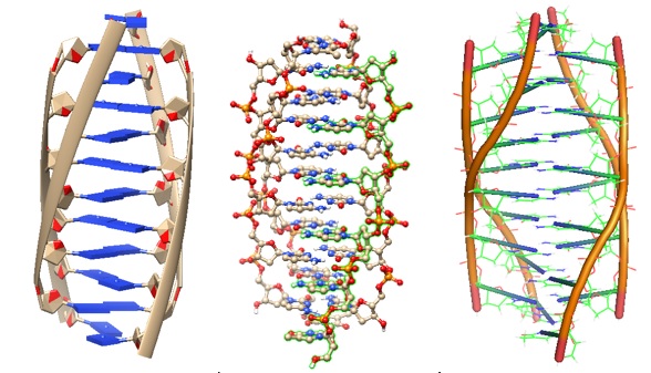
The DNA i-motif is a DNA structure in which two parallel-stranded duplexes with C.C(+) pairs are intercalated head-to-tail. Multimeric associations or intra-molecular folding can form the i-motif. However, the formation of the i-motif depends on the number of cytidine tracts, the nucleotide sequences between them, and the biochemical conditions.

The second layer of DNA structures provides regulatory control. B-from DNA stores the genetic information in mammalian cells and humans. A DNA structure called the i-motif formed in cytosine-rich regions of the genome also appears to have regulatory functions.

To probe for the presence of these structures in vivo, Zeraati et al., in 2018, generated and characterized an antibody fragment (iMAB) that recognized the i-motif form with high selectivity and affinity. Utilizing immunofluorescence staining, the research group showed that i-motif structures are present in the nuclei of human cells. The study’s results indicated that i-motif DNA structures form under physiological conditions and are highly cell-cycle specific. The iMab method revealed that the highest level of i-motif formation occurs in the late G1 phase. A high level of transcription and cellular growth characterizes the late G1 stage. Also, the researchers suggested that i-motifs play a regulatory role in the promotors of several proto-oncogenes. Compared to G4 DNA structures, i-motif structures are less stable, transient, and pH-dependent. The reported results indicate that C-rich sequences within the genome can form i-motif structures in the nuclei of human cells and control regulatory functions.

These findings suggest that mutations altering i-motif stability and conformation can affect their regulatory role. However, to confirm this notion, more research is needed.

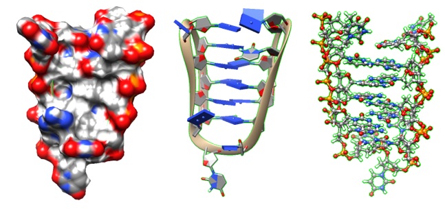
The i-motif was first observed in 1993 when Gehring et al. solved the DNA oligomer 5'-d(TCCCCC) structure at acid pH using NMR. The structure revealed a four-stranded complex in which two base-paired parallel-stranded duplexes are orientated in an antiparallel fashion so that each base pair is face-to-face with its neighbors (Figure 1). A solution structure of a modified centromeric fragment was solved in 2001 (Figure 2).

Figure 1: A tetrameric DNA structure with protonated cytosine.cytosine base pairs. PDB ID 225D (Gehring et al. 1993).

Figure 2: Solution structure of a modified human centromeric fragment. PDB ID 1G22 Nonin-Lecomte, S.,Leroy, J.-L.  (2001) J Mol Biol 309: 491-506


Reference

Gehring K., Leroy J.L., Gueron M.; A tetrameric DNA-structure with protonated cytosine.cytosine Base-Pairs. Nature. 1993; 363:561–565. [PubMed

i-Motif Wiki: 
i-motif DNA - Wikipedia

Nonin-Lecomte S, Leroy JL. Structure of a C-rich strand fragment of the human centromeric satellite III: a pH-dependent intercalation topology. J Mol Biol. 2001 Jun 1;309(2):491-506. doi: 10.1006/jmbi.2001.4679. PMID: 11371167. [Pubmed]

Zeraati, Mahdi; Langley, David B.; Schofield, Peter; Moye, Aaron L.; Rouet, Romain; Hughes, William E.; Bryan, Tracy M.; Dinger, Marcel E.; Christ, Daniel (June 2018). 
 I-motif DNA structures are formed in the nuclei of human cells. Nature Chemistry. 10 (6): 631-637.

---...---

Bio-Synthesis provides a full spectrum of oligonucleotide and peptide synthesis including bio-conjugation services as well as high quality custom oligonucleotide modification services, back-bone modifications, conjugation to fatty acids and lipids, cholesterol, tocopherol, peptides as well as biotinylation by direct solid-phase chemical synthesis or enzyme-assisted approaches to obtain artificially modified oligonucleotides, such as BNA antisense oligonucleotides, mRNAs or siRNAs, containing a natural or modified backbone, as well as base, sugar and internucleotide linkages.

Bio-Synthesis also provides biotinylated mRNA and long circular oligonucleotides.
---...---

Cytosine Protonation studied in i-Motif oligonucleotides using a Raman Tag

$
0
0

Raman Spectroscopy of cytosines modified with a vibrational tag enables the study of protonated cytosine base pairs in oligonucleotides.

A Raman tag is a vibrational tag added to a molecule that allows tracking or probing of protonation states in target molecules. During Raman scattering, an inelastic light scattering process, frequencies of the scattered photons shift from those of the incident photon frequencies. The shift in frequencies occurs according to the vibrational modes of the molecule or atomic group.


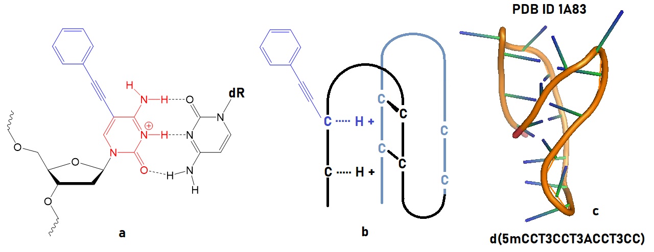
DNA i-motifs contain cytosine-hemiprotonated cytosine base pairs [C-H-C]+. These base pairing types can mediate complicated biological functions, including the transfer of genetic information, gene manipulation, and regulation. In i-motifs, an intercalating [C-H-C]+ base pair can form a quadruplex structure even at neutral pH.

Knowing detailed structural information of nucleobases in intercellular nucleic acids and their interaction with the surrounding environment is vital for designing functional molecules useful for oligonucleotide therapeutics. Unfortunately, the protonation state of cytosines in DNA is complicated and remains elusive. Since conventional spectroscopic approaches have some drawbacks, molecular vibration monitored using Raman spectroscopy allows obtaining a structural fingerprint of target molecules.

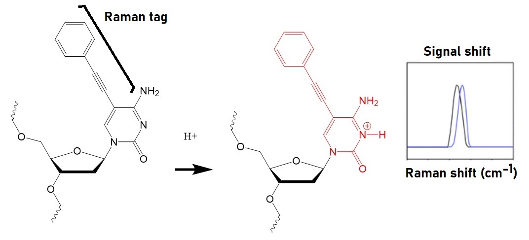
Itaya expected that the protonation of nucleic acids in oligonucleotides affects their vibrational modes. The research group, therefore, prepared a Raman tag as a vibrational tag by adding a phenyl-acetylene group to a cytosine. The protonation of this tag on the N3 position of cytosine shortens the acetylene bond.

Figure 1: Identification of protonation in a Raman-tagged cytosine oligonucleotide (Itaya et al. 2021). Protonation of the tagged cytosine results in a frequency shift of the Raman-tag.


Figure 2: Illustration of an oligonucleotide with a cytosine containing a Raman tag. a) The chemical structure of a C-C+ pair with a tagged C is shown. b) Conceptual drawing of an i-motif oligonucleotide containing the Raman tag. c) Intramolecular i-motif structure solved by NMR (Han et al. 1998; 1a83). Protonation of C-C base pairs leads to formation of an i-motif in oligonucleotides.

When protonated, cytosine can adopt specific structures allowing the formation of higher order structures. 

Examples are the formation of: 

[1] C+–G–C triplets with one protonated cytosine that interacts with a G–C base pair to form a triplex, and 

[2] cytosine·hemiprotonated cytosine base pairs ([C–H–C]+) found in DNA i-motifs.

However, determining the correct protonation status of C in complicated environments is quite tricky. Raman or vibrational tags enable specific detection of chemical species by circumventing the endogenous cellular background. Raman tags can be designed and synthesized by adding chemical bonds to chemical compounds that vibrate in the cell-silent Raman window between 1800 to 2600 cm−1, in which no other endogenous molecules vibrate.

Reference

Han X, Leroy JL, Guéron M. An intramolecular i-motif: the solution structure and base-pair opening kinetics of d(5mCCT3CCT3ACCT3CC). J Mol Biol. 1998 May 22;278(5):949-65. doi: 10.1006/jmbi.1998.1740. PMID: 9600855 [Pubmed].

Ryota Itaya, Wakana Idei, Takashi Nakamura, Tatsuya Nishihara, Ryohsuke Kurihara, Akimitsu Okamoto, and Kazuhito Tanabe; Changes of C≡C Triple Bond Vibration that Disclosed Non-Canonical Cytosine Protonation in i-Motif-Forming Oligodeoxynucleotides. ACS Omega 2021, 6, 47, 31595–31604 [ACS]

Jones RR, Hooper DC, Zhang L, Wolverson D, Valev VK. Raman Techniques: Fundamentals and Frontiers. Nanoscale Res Lett. 2019 Jul 12;14(1):231. doi: 10.1186/s11671-019-3039-2. PMID: 31300945; PMCID: PMC6626094 [PMC].


---...---
Bio-Synthesis provides a full spectrum of oligonucleotide and peptide synthesis including bio-conjugation services as well as high quality custom oligonucleotide modification services, back-bone modifications, conjugation to fatty acids and lipids, cholesterol, tocopherol, peptides as well as biotinylation by direct solid-phase chemical synthesis or enzyme-assisted approaches to obtain artificially modified oligonucleotides, such as BNA antisense oligonucleotides, mRNAs or siRNAs, containing a natural or modified backbone, as well as base, sugar and internucleotide linkages.
Bio-Synthesis also provides biotinylated mRNA and long circular oligonucleotides.

---...---

Short- and Long-term storage of purified oligonucleotides

$
0
0

How to store DNA and RNA oligonucleotides to enhance your success in all your experiments?


Isolated and purified RNA is intrinsically labile. The ubiquity of ribonuclease (RNase) activity further complicates RNA oligonucleotides' handling. The proper storage of oligonucleotides is crucial for their stability. Starting with precise amounts of DNA and RNA in the solution ensures reliable results during all downstream procedures. The correct storage conditions for RNA samples are often hotly discussed. Improper storage, for a few hours or longer, can profoundly affect experimental results.


For RNA oligonucleotides, to prevent repeated freezing and thawing, the most prudent approach is to determine the concentration first, followed by storing the remaining RNA in suitable aliquots at -80 ºC.


Storage of DNA oligonucleotides:

The purified oligonucleotides' stability depends on the nature of the storage medium and temperature. When correctly stored a -20 ºC, frozen oligonucleotides can remain stable for up to two years or longer, as a dry powder and in both TE buffer and nuclease-free water. 
Most oligonucleotides are stable for more than 60 weeks when stored dry at 4 ºC or in DNase and RNase-free medium, including TE buffer and water.

However, the best practice is the storage of oligonucleotides in a freezer in the dark at -20 ºC either in nuclease-free TE buffer or water for up to 2 years.

Storage in increased temperatures or room temperature:

Storing oligonucleotides at 37°C/98°F is possible for several weeks. However, the selected medium will influence their stability. Oligonucleotides are generally most stable when stored in TE buffer at pH 8.0. Any moisture present in dry oligonucleotides, even small traces, can cause damage to the oligonucleotides.

Reconstitution and resuspension of oligonucleotides:

Bio-Synthesis Inc. provides oligonucleotides as a lyophilized white powder. However, per special request, a normalized RNA solution is provided for an additional fee.

To reconstitute oligonucleotides, follow the next steps:

1. Centrifuge at 10,000g for 15 seconds at room temperature (RT).

2. Add the desired volume of filtered, sterile TE buffer (10 mM Tris-HCL pH 8.0, 1 mM EDTA) or DNase-free and RNAase-free H2O.

3. Allow rehydrating for 10-15 minutes at room temperature.

4. Vortex for 5 seconds, then centrifuge at 10,000g for 15 seconds at room temperature.

5. Proceed with your downstream application.

Shipping of oligonucleotides:

Bio-Synthesis Inc. ships oligonucleotides as a lyophilized dry powder, usually overnight, to allow flexibility to our customers. The shipping time will not be detrimental to the stability of dry oligonucleotides since dry oligonucleotides remain stable for up to 25 weeks when stored at 37°C (98°F). Most oligonucleotides will remain functional for multiple weeks at this higher temperature when stored in water and storage media.

Freezing and thawing of oligonucleotides:

During some experiments, oligonucleotides may need to be repeatedly frozen and thawed. If correctly dissolved in nuclease-free media, up to 30 freeze-thawing cycles may have no significant impact on the stability of an oligonucleotide.

Modified oligonucleotides:

Most modified oligonucleotides will have similar stability characteristics as unmodified oligonucleotides. However, heavily modified oligonucleotides will be significantly more stable, especially to nuclease-depended degradation.

However, it is most prudent to store modified oligonucleotides like unmodified oligonucleotides.

RNA oligonucleotides are less stable:

RNA oligonucleotides are inherently less stable than DNA. Also, the ubiquity of ribonuclease (RNase) activity complicates the handling of RNA oligonucleotides further. RNases are present in human saliva, mucus, sweat, dead hair, and skin cells and are also more prevalent in laboratories than DNases. Therefore, avoiding RNase contamination is critical to maintaining RNA stability. Even minor exposure to RNases will cause RNA degradation. We recommend, for long-term storage storing RNA as an ethanol precipitate at -80°C.

Reference

Farrell, R.E.; RNA Methodologies 5th Edition. Academic Press 2017.

Roskams, J. and Rogers, L.; Lab Ref. A handbook of recipes, reagents, and other reference tools fo use at the bench. Cold Spring Harbor Laboratory Press. 2002.


---...---
Bio-Synthesis provides a full spectrum of oligonucleotide and peptide synthesis including bio-conjugation services as well as high quality custom oligonucleotide modification services, back-bone modifications, conjugation to fatty acids and lipids, cholesterol, tocopherol, peptides as well as biotinylation by direct solid-phase chemical synthesis or enzyme-assisted approaches to obtain artificially modified oligonucleotides, such as BNA antisense oligonucleotides, mRNAs or siRNAs, containing a natural or modified backbone, as well as base, sugar and internucleotide linkages.
Bio-Synthesis also provides biotinylated mRNA and long circular oligonucleotides.

---...---


The RNA TrimethylGuanosine or TMG cap

$
0
0

The 2,2,7-trimethylguanosine (TMG or m3G) cap is one of the earliest identified RNA modifications. The TMG-cap ensures nuclear localization of small nuclear RNAs (snRNAs).

Research during recent decades has provided a better understanding of the biological roles of base and ribose modifications, as well as cellular degradation pathways. Advances in enzymatic synthesis, RNA modifications, and cellular delivery vehicles enabled the administration of exogenous mRNA into cells to express proteins in situ. A few examples illustrating this progress are stem cell reprogramming, vaccination, and the expression of therapeutic proteins.


The capping machinery of RNAs, particularly mRNAs, is quite complicated. Cap structures of the type m7GpppNmpNm are at the 5’-ends of nearly all eukaryotic cellular and viral mRNAs. The m7GpppNmpNm recruits cellular proteins and mediates cap-related biological functions such as pre-mRNA processing, nuclear export, and cap-dependent protein synthesis. Also, the 2’-O-methylated cap is an identifier of self RNA in the innate immune system against foreign RNA. The discovery of the cytoplasmic capping machinery suggested an additional control network. For example, Singh et al., in 1989, characterized the cap structure of human U6 snRNA and showed that the gamma phosphate of the 5' guanosine triphosphate is methylated.

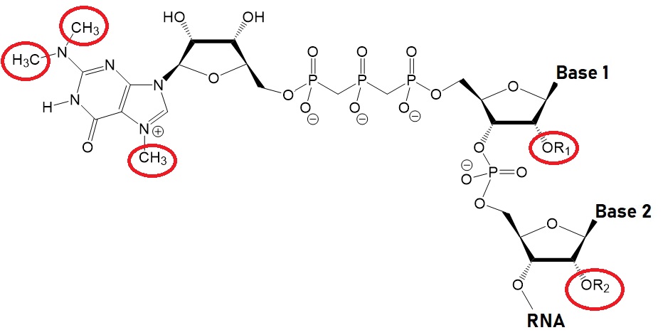
Structures of Uncapped and Capped RNA including the TMG Cap

 Uncapped RNA


 γ-methyl snRNA cap


 TMG snRNA CAP, m3GpppRNA


 m7G mRNA caps, m7GpppRNAs


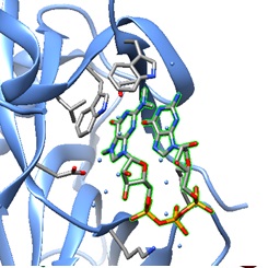
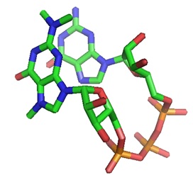
The structural models of a TMG-capped dinucleotide bound to the nuclear import receptor snurportin 1 domain are illustrated below. Snurportin 1 interacts with the m3G-cap enhancing the m3G-cap-dependent nuclear import of U snRNPs in Xenapus laevis oocytes and digitonin-permeable HeLa (human) cells.  


A: Ribbon plot of the crystal structure of the m3G-Cap-binding domain of snurportin1 in complex with a m3GpppG-Cap dinucleotide (PDB ID 1X5K).

The m3GpppG-Cap dinucleotide is shown in surface mode ( Strasser et al. [ PMC ]).

B:  The m3GpppG-Cap dinucleotide is shown in its binding pocket.

C: The m3GpppG dinucleotide is depicted in stick mode with nitrogen atoms in blue, carbons in grey, oxygens in red and phosphorus atoms in yellow.


Do TMG-capped oligonucleotides accumulate in the nucleus?

Yes, according to a recent paper by Moreno and others.

RNA oligonucleotides capped with the 2,2,7-trimethylguanosine (TMG or m3G) cap can direct nuclear accumulation of a cargo protein in mammalian cells after cytosolic delivery using a transfection reagent. Moreno et al., in 2009, reported the use of the m3G- or TMG-cap as an adaptor molecule that, when attached to oligonucleotides, enables targeted transport of large-size cargo molecules into the nucleus. Furthermore, TMG-capped 2′-O-methyl RNA antisense oligonucleotides (ASOs) showed increased efficiency in a splice correction assay. Moreno et al. suggested that synthetically capped oligonucleotides may selectively interfere with splicing events, potentially resulting in therapeutic clinical applications (Moreno et al.).

A TMG-cap structure enables nuclear import!

The spliceosomal small nuclear ribonucleoproteins snRNPs U1, U2, U4, and U5 depend on a complex nuclear localization signal (NLS) for their nuclear import. The NLS comprises the 5'-2,2,7-terminal trimethylguanosine (m3G or TMG) cap structure of the U snRNA and the Sm core domain. When injected into Xenopus laevis oocyte nuclei, U snRNA precursor molecules temporarily concentrate in Cajal bodies before their export (Suzuki et al. )

The TMG-cap structure is essential for telomere maintenance!

The TMG-cap plays a central role in recruiting telomerase to telomeres and engaging Cajal bodies in telomere maintenance. Many RNA molecules contain the TMG-cap at the 5’-end.

Buemi et al. recently showed that the formation of the TMG cap structure at the human telomerase RNA 5′-end by the Trimethylguanosine Synthase 1 (TGS1) is needed for recruiting telomerase to telomeres and engaging Cajal bodies in telomere maintenance. Depletion of the enzyme TGS1 with sinefungin blocked telomerase recruitment to telomeres. The result was exonuclease 1 (EXO1)-mediated formation of single-stranded 3′-telomere overhangs that engage in RAD51-dependent homologous recombination and APB formation and the activation of key features of the ALT pathway in telomerase-positive H1299 cells and lung tumor organoids.

The alternative lengthening telomere pathway (ALT pathway) refers to the telomerase-independent, recombination-dependent process that extends telomeres. APB refers to the ALT-associated promyelocytic leukemia nuclear body. Recent findings indicate a critical role for 2,2,7-TMG capping in telomerase-dependent telomere maintenance to restrict the formation of telomeric substrates conducive to ALT. This pathway is relevant in telomere-related diseases such as cancer and aging.

The Nuclear Import Machinery

Transporting biomolecules from the cytoplasm to the nucleus is critical for cell physiology and pathology. The nuclear envelope is a significant barrier to delivering therapeutic molecules into the nucleus. The large size of nucleic acids makes delivering therapeutic molecules into the nucleus difficult for medical applications. The deregulation of the import and export machinery is a cause of many diseases.


The nuclear envelope comprises control gates known as nuclear pore complexes (NPCs) that selectively regulate nuclear transport. Small molecules like metabolites and ions can diffuse through the NPC. However, molecules bigger than 40 kDa, like most proteins, RNA, ribosomal subunits, and viral particles, require an active transport mechanism. Soluble carrier molecules belonging to the karyopherin-β family, including importins, exportins, and transportins, are part of the transporting machinery.


The NPC is a large protein complex fusing the internal and external nuclear membrane forming an aqueous channel. Approximately 2000 to 5000 NPCs are present in a vertebrate cell. 
Extensive studies of NPC by electron microscopy and proteomic approaches are available now (Cautain et al., 2014).

The Ran system provides the energy source for efficient cargo transport. In particular, the Ran-GTP/GDP gradient across NPC regulates the transportation of cargo molecules between the nucleoplasm and the cytoplasm. RanGTP binds to a carrier importin and helps to release the cargo in the nucleus. After transporting cargo to the cytoplasm, the exportin-RanGTP-cargo complex is hydrolyzed, releasing the cargo molecule.


Large cargo molecules need a nuclear localization signal (NLS) to access the nucleus. Classical import signals (cNLS) contain a short stretch of basic residues. The adaptor protein importin-α recognizes a cargo molecule containing the cNLS. For interaction with NPS, the complex binds to importin-β. 
RanGTP mediates the translocation of the cargo into the nucleus. However, non-classical NLSs do not need an adaptor protein by binding directly to a transporter protein. Table 1 lists examples of different nuclear transport signals.

Table 1: Examples of nuclear transport signals.

Amino acid sequence

Protein

Signal

PKKKRKV

SV40 T antigen

Classical NLS

KRx{10}KKKL

Nucleoplasmin

Bipartite NLS

VRILESWFAKNIENPYLDT

Matα2

Polar/nonpolar residues NLS

PAAKRVKLD

c‐Myc

cMyc‐NLS

YNDFGNYNNSSNFGPMKGGNFGGRSSGPYGGGGQY

hnRNP A1 (M9 sequence hydrophobic subclasses)

hPY‐NLS

KVSRRG‐GHQNSYKPY

hnRNP D (basic enriched)

bPY‐NLS

RQARRNRRRRWR

VIH Rev protein

Arginine‐rich NLS

DNSQRFTQRGGGAVGKNRRGGRGGNRGGRNNNSTRFNPLAK

Nab2p

Arginine/glycine‐rich NLS

KTPGKKKKGK

Parathyroid hormone‐related protein (PTHrP)

Lysine‐rich‐NLS

QDLNSTAAPHPRLSQYKSKYSSLEQSERRRRL

Snurportin1

UsnRNPs‐NLS

LPPLERLTL

HIV Rev

Hydrophobic‐NES

LALKLAGLKI

PKI

NES

LCQAFSDVIL

Cyclin B1

NES

LQKKLEELEL

MAPKK

NES

LAEMLEDLHI

NMD3

NES


( Source: Cautain et al., made freely available through PubMed Central as part of the COVID-19 public health emergency response. It can be used for unrestricted research re-use and analysis in any form or by any means with acknowledgement of the original source, for the duration of the public health emergency. )


The non-covalent or covalent complexation of an NLS peptide with a plasmid DNA or an oligonucleotide promotes nuclear delivery. Also, transfection is possible by combining plasmid DNA with polyethylimines (PEIs) conjugated to an NLS peptide. 

Small nuclear RNAs (snRNAs) as part of small nuclear RNA binding proteins (snRNPs) are mainly found in U1, U2, U5, and U4/U6. snRNAs are transcribed by RNA polymerase II, and co-transcriptionally capped with an m7G-Cap. Uridine-rich, small nuclear ribonucleoproteins (U snRNPs) involved in pre-mRNA splicing contain the TMG-cap as an NLS signal.

The heterodimeric cap-binding complex (CBC) recognizes the m7G-Cap. It recruits the phosphorylated adaptor of export protein (PHAX), further recognized by an export receptor CRM1 for cytoplasmic localization and maturation. Next, a complex of proteins assembles around the SM core domain of snRNAs, creating a platform for trimethylguanosine synthase 1 (TGS 1). TGS 1 catalyzes the methylation of the m7G-cap to the m3G-cap (TMG-cap). The TMG-cap is recognized by the adaptor protein surportin 1. During the final maturation of snRNPs, this complex binds to importin-β for nuclear relocalization.

Sm-proteins received their name from human autoimmune serum initially used in their identification. In human cells, the assembly of the snRNAs with the Sm-proteins is highly specific and mediated by a large protein complex that includes the SMN protein (survival of motor neurons).


Reference

Buemi, V., Schillaci, O., Santorsola, M. et al. TGS1 mediates 2,2,7-trimethyl guanosine capping of the human telomerase RNA to direct telomerase dependent telomere maintenance. Nat Commun 13, 2302 (2022). https://doi.org/10.1038/s41467-022-29907-z. https://www.nature.com/articles/s41467-022-29907-z

Furuichi Y. Discovery of m(7)G-cap in eukaryotic mRNAs. Proc Jpn Acad Ser B Phys Biol Sci. 2015;91(8):394-409. doi:10.2183/pjab.91.394. https://www.ncbi.nlm.nih.gov/pmc/articles/PMC4729855/pdf/pjab-91-394.pdf


Honcharenko, M., Bestas, B., Jezowska, M., Wojtczak, B. A., Moreno, P. M. D., Romanowska, J., Baechle, S. M., Darzynkiewicz, E., Jemielity, J., Smith, C. I. E., and Stroemberg, R. (2016) Synthetic m3G-CAP attachment necessitates a minimum trinucleotide constituent to be recognised as a nuclear import signal. RSC Adv. 6, 51367-51373.


Pedro M. D. Moreno, Malgorzata Wenska, Karin E. Lundin, Örjan Wrange, Roger Strömberg, C. I. Edvard Smith, Nucleic Acids Research, Volume 37, Issue 6, 1 April 2009, Pages 1925–1935, https://doi.org/10.1093/nar/gkp048


Moteki S, Price D. Functional coupling of capping and transcription of mRNA. Mol Cell. 2002 Sep;10(3):599-609. doi: 10.1016/s1097-2765(02)00660-3. PMID: 12408827. https://www.cell.com/molecular-cell/fulltext/S1097-2765(02)00660-3?_returnURL=https%3A%2F%2Flinkinghub.elsevier.com%2Fretrieve%2Fpii%2FS1097276502006603%3Fshowall%3Dtrue


Ramanathan A, Robb GB, Chan SH. mRNA capping: biological functions and applications. Nucleic Acids Res. 2016;44(16):7511-7526. doi:10.1093/nar/gkw551.  https://www.ncbi.nlm.nih.gov/pmc/articles/PMC5027499/#!po=5.84416.


Shatkin AJ. Capping of eucaryotic mRNAs. Cell. 1976 Dec;9(4 PT 2):645-53. doi: 10.1016/0092-8674(76)90128-8. PMID: 1017010. https://pubmed.ncbi.nlm.nih.gov/1017010/


Shatkin A.J., Manley J.L., The ends of the affair: capping and polyadenylation. Nat. Struct. Biol. 2000, 7, 838-842. https://pubmed.ncbi.nlm.nih.gov/11017188/


Shih DS, Dasgupta R, Kaesberg P. 7-Methyl-guanosine and efficiency of RNA translation. J Virol. 1976 Aug;19(2):637-42. doi: 10.1128/JVI.19.2.637-642.1976. PMID: 957483; PMCID: PMC354898. https://www.ncbi.nlm.nih.gov/pmc/articles/PMC354898/


Strasser A, Dickmanns A, Lührmann R, Ficner R. Structural basis for m3G-cap-mediated nuclear import of spliceosomal UsnRNPs by snurportin1. EMBO J. 2005;24(13):2235-2243. [
PMC ].

Suzuki T, Izumi H, Ohno M. Cajal body surveillance of U snRNA export complex assembly. J Cell Biol. 2010;190(4):603-612. doi:10.1083/jcb.201004109.


Zhang JM, Yadav T, Ouyang J, Lan L, Zou L. Alternative Lengthening of Telomeres through Two Distinct Break-Induced Replication Pathways. Cell Rep. 2019 Jan 22;26(4):955-968.e3. doi: 10.1016/j.celrep.2018.12.102. PMID: 30673617; PMCID: PMC6366628.


---...---
Bio-Synthesis provides a full spectrum of oligonucleotide and peptide synthesis including bio-conjugation services as well as high quality custom oligonucleotide modification services, back-bone modifications, conjugation to fatty acids and lipids, cholesterol, tocopherol, peptides as well as biotinylation by direct solid-phase chemical synthesis or enzyme-assisted approaches to obtain artificially modified oligonucleotides, such as BNA antisense oligonucleotides, mRNAs or siRNAs, containing a natural or modified backbone, as well as base, sugar and internucleotide linkages.
Bio-Synthesis also provides biotinylated mRNA and long circular oligonucleotides.
---...---

Writers, Readers, and Erasers of RNA Modifications

$
0
0

Because of many recent improvements made in the last decades in automated synthesis of biomolecules such as oligonucleotides (DNA/RNA), polypeptides, carbohydrates, and modified derivatives of these, and in high-resolution and sensitive analytical instruments, our understanding of cells, cell structures and their dynamics at the molecular level has dramatically increased. A collection of human genomes is now available, scientists studied the 3D organization of the genome, and a data set of genomes is available at the ATCC genome portal: Human Genome Collection.Initial sequencing and analysis of the human genome | NatureGenomes | ATCC Genome Portal.

Molecular medicine aims to link “omic” approaches to cancer research. Understanding and characterizing metabolism in cancer and healthy cells is the key to developing new specific and efficient therapeutics targeting cancer cells. The knowledge of possible marks and their functions on biomolecules involved in the metabolism of human cells is expected to create a comprehensive picture of human and tumor biology at the systems and molecular level.

Multi-omics approaches enable the characterization and quantification of regulatory marks in the genome, the epigenome, the transcriptome, the epitranscriptome, the proteome, and the epiproteome. This knowledge is expected to increase our understanding of eukaryotic metabolism and physiology, helping to create improved personalized therapeutics.

Research conducted during the last decades revealed that cancer is a disease driven by genetic mutations, epigenetic modifications, a misregulated transcriptome, now known as the epitranscriptome, and a misregulated proteome, known as the epiproteome. The so-called epitranscriptome is now understood as the chemical modifications of RNA that regulates and alters the activity of RNA molecules. An example is the hypermethylation of DNA associated with silencing tumor suppressor genes and aberrant histone modifications. A signaling pathway project is underway to integrate an “omics” knowledgebase for mammalian cellular signaling pathways. This database contains curated data sets validated using alignment with the canonical literature knowledge and gene target-level integration of transcriptomic and cistromic data points. [
Signaling pathways Project]

Research in recent decades has made it clear that the addition or removal of RNA modifications in various RNA species regulates a broad spectrum of RNA regulatory processes. These RNA-regulating processes regulate specific sets of genes. Therefore, the context of the RNA molecule and the RNA effector enzymes involved determine the molecular destiny of any given RNA transcript. Interestingly, subcellar localization of both RNAs and RNA-modifying proteins, the number of transcripts of specific cellular RNAs, the various RNA types, the folding of RNAs, RNA-protein interactions, and responses to stimuli such as DNA or RNA damage determine the metabolisms of RNA modifications. Defects in any of these processes may lead to cancer progression. Table 1 illustrates the interplay of “omics” systems.


Table 1: "Omics" Systems 

Genome

DNA

Epigenome

DNA modified: 5hmC, 5mC, 5fC, 3mC, 4mC, 6mA.

Transcriptome

RNA:

rRNA, tRNA, snRNA, mRNA, lncRNA, miRNA, etc.

Epitranscriptome

RNA modified:

Ψ, m5C, m1A, m6A, m5A, etc.

Proteome

Proteins

Epiproteome

Proteins modified:

PTMs.

SNP

CNV

LOH

Rearrangement

DNA modifications:

methylation

Alternative splicing RNA editing

RNA modifications:

methylation, pseudouridylation, acetylation, A-t-I, ribose methylation, others.

Protein isoforms

Peptides and micropeptides.

Protein post-translational modifications (PTMs):

methylation, phosphorylatin, glycosylation, ubiquitination, nitrosylation, SUMOylation.

NGS, WES

WGS, FISH, CGH

ChiP-seq

DNA microarray

Targeted DNA seq

DNA methylation array

Pyrosequencing

Bisulfite sequencing (BS)

RNA seq, SLAM-seq,

RNA microarray

Targeted RNA seq,

RNA Exome Capture Seq

Ribosome profiling

qRT-PCR

Methylated RNA IP-seq, miCLIP, RNA BS-seq, m1A/m6A-seq,

DART-seq

Quantification of RNA mods by LC-MS

Mass Spectrometry

Protein Array

Immuno-precipitation

Immuno-fluorescence

Western Blot Analysis

Mapping PTMs by mass spectrometry (LC-MS/MS)

SILAC, HPLC

Phospho-Kinase array

Western Blot Analysis


Legend
: CGH = comparative genomic hybridization, ChiP-seq = chromatin immunoprecipitation DNA sequencing, CNV = copy number variation, DART-seq = Diversity Arrays Technology, FISH = fluorescence in situ hybridization, LOH = loss of heterozygosity, NGS = next generation sequencing, SILAC = Stable isotope labeling by amino acids in cell culture, SNP = single-nucleotide polymorphism, WES = whole exome sequencing.

 Table 2: RNA Modifications and Enzymes

  Important RNA modifications

  Enzymes that modify RNA

 m1A: 1-methyladenosine,

 ms2i6A: 2-methylthio-N6-isopentenyl-adenosine,

 i6A: N6-isopentenyladenosine,

 m6A: N6-methyladenosine,

 m3C: 3-methylcytosine,

 m5C: 5-methylcytosine,

 ac4C: N4-acetylcytosine,

 m7Gpp(pN): 7-methylguanosine cap,

 m7G: 7-methylguanosine internal,

 m2,2G: N2,N2,-dimethylguanosine,

 m2G: N2-methylguanosine,

 Q: queuosine, yW et al.: Wybutosine and derivatives,

 m5U: 5-methyluridine,

 ncm5U: 5-carbamoyl-methyluridine,

 mcm5U: 5-methoxycarbonyl-methyluridine,

 mcm5s2U: 5-methoxycarbonylmethyl-2-thiouridine,

 D: dihydrouridine,

 Ψ: pseudouridine,

 Nm: 2-O-Methylnucleotide,

 m(pN): 5phosphate monomethylation,

 A-to-I: Deamination of Adenosine,

 C-to-U: Deamination of Cytosine.


 ADAR1-3: Adenosine Deaminase RNA Specific 13,

 ALKBH1/3/5/8: AlkB Homolog 1/3/5/8,

 APOBEC1/3G: Apolipoprotein B mRNA Editing
 Enzyme Catalytic Subunits 1/3G,

 BCDIN3D: BCDIN3 Domain Containing
 Methyltransferase,

 BUD23: RRNA Methyltransferase And Ribosome
 Maturation Factor,

 CDK5RAP1: CDK5 Regulatory Subunit Associated Protein 1,

 CMTR1/2: Cap Methyltransferase 1/2,

 CTU1/2: Cytosolic Thiouridylase Subunit 1/2,

 DKC1: Dyskerin Pseudouridine Synthase 1,

 DNMT2: tRNA Aspartic Acid Methyltransferase 1,

 DUS2: Dihydrouridine Synthases 2,

 ELP3: Elongator Acetyltransferase Complex Subunit 3,

 FTO: FTO Alpha-Ketoglutarate Dependent Dioxygenase,

 HENMT1: HEN Methyltransferase 1,

 METTL1/2/3/6/8/14/16: Methyltransferase Like-1/2/3/6/8/16,

 NAT10: N-Acetyltransferase 10,

 NSUN1-5: NOP2/Sun RNA Methyltransferase 15,

 NUDT16: Nudix Hydrolase 16,

 RNMT: RNA Guanine-7 Methyltransferase,

 TGT: Queuine TRNA-Ribosyltransferase Catalytic Subunit 1,

 TRIT1: tRNA Isopentenyltransferase 1,

 TRMT1/2A/2B1/5/6/10C/11/61A/61B/112:
  tRNA Methyltransferase Subunits,

 TYW2: tRNA-YW Synthesizing Protein 2 Homolog.


RNA modifications known as epitranscriptomic marks


Table 3: Epitrancriptome of small non-coding RNAs


SncRNAs Species

Described Chemical Modifiction

Writers

Readers

miRNA

m6A

METTL3/ METTL14

HNRNPA2B1/ HNRNPC

m7G

METTL1

/

2′-O-Me

HEN1

/

5′Pme2

BCDIN3D

/

Uridylation

TUT7/4/2

/

A to I

ADARs

/

o8G

/

/

8-OHG

/

/

piRNA

2′-O-Me

HEN1


snRNA

Ψ

Box H/ACA RNP/ Pus1 and Pus7

/

2′-O-Me

Box C/D RNP

/

m6A

METTL16

/

m6Am

METTL4

/

TMG

TGS1

/

m5C

/

YPS

snoRNA

Ψ

Box H/ACA RNP

/

m6A

/

/

tsRNA

m5C

DNMT2/ NSUN2

/

m2G

DNMT2

/

Q

QTRT1/QTRT2

/

2′-O-Me

TRM7/FTSJ1

/

m1A

TRMT6/61A

/

m3C

METTL2/ METTL6

/

m1G

TRMT10A

/

hm5C

TET2

/

Ψ

PUS7

/

mcm5S2

/

/

 (Source: Li et al. 2021; Wang et al. 2022)


Reference

Esteve-Puig R, Bueno-Costa A, Esteller M. Writers, readers and erasers of RNA modifications in cancer. Cancer Lett. 2020 Apr 1;474:127-137. [PubMed]

Li X, Peng J, Yi C. The epitranscriptome of small non-coding RNAs. Noncoding RNA Res. 2021;6(4):167-173. [PMC]

Wang S, Li H, Lian Z, Deng S. The Role of RNA Modification in HIV-1 Infection. Int J Mol Sci. 2022;23(14):7571. [PMC]


---...---

Bio-Synthesis provides a full spectrum of oligonucleotide and peptide synthesis including bio-conjugation services as well as high quality custom oligonucleotide modification services, back-bone modifications, conjugation to fatty acids and lipids, cholesterol, tocopherol, peptides as well as biotinylation by direct solid-phase chemical synthesis or enzyme-assisted approaches to obtain artificially modified oligonucleotides, such as BNA antisense oligonucleotides, mRNAs or siRNAs, containing a natural or modified backbone, as well as base, sugar and internucleotide linkages.

Bio-Synthesis also provides biotinylated mRNA and long circular oligonucleotides.
---...---

Metastasis Inhibiting Peptides

$
0
0

Peptides inhibit metastasis-associated cancer cell adhesion.

Metastasis is a deadly aspect of cancer resulting from interconnected steps, including cell proliferation, developing new blood vessels (angiogenesis), cell adhesion, migration, and invasion of surrounding tissues. During metastasis, tumor cells spread from the primary focus to invade other cells and tissue leading to mortality. Cancerous, latent cells can lodge in the bone marrow and organs distant from the primary tumor seeding new metastatic growth later.

Cancer starts after a single tissue cell is genetically damaged. Progressive damage caused by mutations produces cells that proliferate in an uncontrolled manner. The spread of cancer from one organ or cell tissue to another is known as "metastasis." Some cancer cells can penetrate the walls of lymphatic or blood vessels allowing these cells to circulate through the bloodstream to other sites and tissues in the body. These cells are known as circulating tumor cells.

Intravascular cancer is cancer that occurs within a blood vessel or blood vascular system. Intravascular cancer cell adhesion plays a significant role in the metastatic process. The lectin galectin-3 is involved in carbohydrate-mediated metastatic cell adhesion via interaction with the tumor-specific Thomsen-Friedrich glycoantigen (TF or TFAg). The Thomsen–Friedenreich antigen (Gal-GalNAc) represents a tumor-associated molecule.

Mereiter et al., in 2018, observed a highly significant association between the expression status of the Thomsen-Friedenreich (TF) antigen and microsatellite instability in gastric cancer, identifying the first single marker for MSI in gastric cancer. In colorectal cancer, microsatellite instability (MSI) caused by deficient DNA mismatch repair (dMMR) is associated with several clinical and histopathological features.

In 2021, Leão et al. reported the results of a study that correlated the expression of the TF antigen in colorectal cancer with microsatellite instability. The study results indicated that the TF antigen is not a predictor of MSI in colorectal cancer, contrary to what has been described in gastric cancer with MSI. However, the study’s results revealed that patients harboring MSI-high tumors that express the TF antigen have significantly better survival than TF-negative cases.

The Thomsen-Friedenreich antigen, Galβ1-3GalNAcα1-O-Ser/Thr (Figure 1 ans 2), is the core one (1) structure of O-linked mucin-type glycans found in tumor-associated glycosylation. This antigen occurs in about 90% of human cancer cells and is a potential ligand for the human endogenous galectins.

In 2005, Zou et al. reported that galectin-3 plays a significant role in metastasis. Galectin-3 is a member of the galectin family of soluble animal lectins. This lectin mediates carbohydrate-mediated metastatic cell adhesion between carcinoma cells and the endothelium and between carcinoma cells. The tumor-specific Thomsen–Friedenreich glycoantigen is involved in cell-specific interactions.


Isolation of galectin-3-binding peptides

Zou et al. selected Galectin-3-specific peptides by screening a random 15 amino acid and a cysteine-constrained library displayed on coat protein VIII. After the fourth round of selection, the output phage yield from both libraries was ∼100-fold higher than the first round, indicating enrichment of galectin-3-binding phage clones in the eluted phage population. The study identified short synthetic peptides bound to the carbohydrate recognition domain of galectin-3 using a combinatorial phage display technology. The peptides inhibited the interaction between galectin-3 and its sugar ligand TFAg, interfering with metastasis-associated carcinoma cell adhesionG3-A9 and G3-C12 were the predominant output clones ( Table I ).

Table I.  Results of combinatorial peptide selections against galectin 3.Deduced peptide sequences, percent occurrence of phage clones and affinity of peptides (Zou et al.). 


Name

Sequence

Library

% clones

Affinity (Kd)(nM)

G3-A3 

SMEPALPDWWWKMFK 

f88-15 

8.8 

17.7 ± 9.4 ( Kd1 )  


4.2 ± 2.3 ( Kd2 )  

G3-A4 

DKPTAFVSVYLKTAL 

f88-15 

1.3 

NA 

G3-A9 

PQNSKIPGPTFLDPH 

f88-15 

10.0 

72.2 ± 32.8 

G3-A18 

APRPGPWLWSNADSV 

f88-15 

1.3 

NA 

G3-A19 

GVTDSSTSNLDMPHW 

f88-15 

1.3 

NA 

G3-A28 

PKMTLQRSNIRPSMP 

f88-15 

1.3 

NA 

G3-A29 

PQNSKIPGPTFLDPH 

f88-15 

1.3 

NA 

G3-A40 

LYPLHTYTPLSLPLF 

f88-15 

1.3 

NA 

G3-C4 

LTGTCLQYQSRCGNTR 

f88-Cys6 

1.3 

NA 

G3-C9 

AYTKCSRQWRTCMTTH 

f88-Cys6 

1.3 

5.7 ± 2.2 

G3-C12 

ANTPCGPYTHDCPVKR 

f88-Cys6 

80.0 

72.2 ± 32.8 

G3-C44 

NISRCTHPFMACGKQS 

f88-Cys6 

1.3 

NA 

G3-C60 

PRNICSRRDPTCWTTY 

f88-Cys6 

1.3 

NA 


Note: 
The percentage of individual clones is reported that were represented in the populations of 80 f88-15 and 60 f88-Cys6 libraries selected. 


Figure 1:  Galectin-3 in complex with Thomsen-Friedenreich antigen, Galβ1-3GalNacα1-O-Ser/Thr, and a sulfate ion.  PDB ID 3AYA 

(Bian C-F, Zhang Y, Sun H, Li D-F, Wang D-C (2011) Structural Basis for Distinct Binding Properties of the Human Galectins to Thomsen-Friedenreich Antigen. PLoS ONE 6(9): e25007. [plosone])


Figure 2:  Complex between an antitumor galectin AAL and the Thomsen-Friedenreich antigen. PDB ID 3AFK. The structural model illustrates the recognition mode between AAL and TF antigen as a unique conservative (Glu-water-Arg-water) structural motif-based hydrogen bond network.

This galectin is a target for the development of antitumor drug designs based on the AAL-TF recognition mode.

(Feng L, Sun H, Zhang Y, Li DF, Wang DC. Structural insights into the recognition mechanism between an antitumor galectin AAL and the Thomsen-Friedenreich antigen. FASEB J. 2010 Oct;24(10):3861-8. [PMC])


Reference

Khan N, Mukhtar H. Cancer and metastasis: prevention and treatment by green tea. Cancer Metastasis Rev. 2010;29(3):435-445. [PMC]

Leão B, Wen X, Duarte HO, et al. Expression of Thomsen-Friedenreich Antigen in Colorectal Cancer and Association with Microsatellite Instability. Int J Mol Sci. 2021;22(3):1340. [PMC]


Mereiter S, Polom K, Williams C, et al. The Thomsen-Friedenreich Antigen: A Highly Sensitive and Specific Predictor of Microsatellite Instability in Gastric Cancer. J Clin Med. 2018;7(9):256. [PMC]


Pinho, S., Reis, C. Glycosylation in cancer: mechanisms and clinical implications. Nat Rev Cancer 15, 540–555 (2015). [NRC]


Jun Zou, Vladislav V. Glinsky, Linda A. Landon, Leslie Matthews, Susan L. Deutscher, Peptides specific to the galectin-3 carbohydrate recognition domain inhibit metastasis-associated cancer cell adhesion, Carcinogenesis, Volume 26, Issue 2, February 2005, Pages 309–318. [Article]


---...---

Thiophosphoramidate Morpholinos, A New Class of Antisense Oligonucleotides

$
0
0

The search is on for better options in antisense technology because significant obstacles still prevent the specific delivery of antisense oligonucleotides (ASOs) into many selected tissues or cells. For the development of cost-efficient oligonucleotide-based therapeutics, the following points still need to be addressed:

  • Large-scale production,
  • the toxicity of ASOs,
  • the localization of oligonucleotides in specific cellular compartments or tissues, and
  • the high cost of treatment.

However, a new type of ASOs called Thiophosphoramidate Morpholino Oligonucleotides could solve many of these problems. The chemical structures for a PMO, a PM, and a TMO are illustrated in figure 1.

Figure 1: Chemical structures of phosphorodiamidate morpholino (left, PMO), phosphoramidate morpholino (middle, PM), and thiophosphoramidate morpholino (right, TMO).

Antisense morpholino oligomers (MOs) are molecular tools that allow selective gene knockdown. Morpholinos are synthetic antisense oligonucleotides, approximately 25 nucleotides in length. MOs have allowed antisense knockdown studies and screens in zebrafish (Danio rerio, Bill et al. 2009). The major antisense compounds utilized in zebrafish studies are morpholino phosphorodiamidate oligonucleotides. These molecules comprise a phosphorodiamidate backbone with a morpholine ring DNA bases. MOs function through the steric hindrance of the normal endogenous splicing or translation mechanisms. MOs target the splice donor site by inhibiting the binding of the U1 complex by inhibiting lariat formation and incorporation of the intron. Correctly designed MOs bind and block the translation initiation complex of messenger RNA (mRNA) sequences. ATG MOs block the initiation of protein translation. The result is a knockdown of a targeted protein. Splice-blocking MOs interfere with RNA splicing, resulting in truncated proteins, and target protector MOs block specific endogenous microRNAs (miRNA), stabilizing particular mRNA transcripts.

It appears that MOs do have a few side effects. For example, Bile et al. reported that not all MOs cause nonspecific events; however, some MOs correlate with aberrant morphologies with increased cell death, indicating off-targeting effects. Also, there is evidence that the impact of MOs only lasts up to five days, making them unsuitable for studying gene function at later stages of development. Further, MO effects do not always correspond to the observed phenotype of true mutants. Gentsch et al., in 2018, reported another example of a side effect. Embryos injected with control or target MOs to knockdown mesoderm-specific Brachyury paralogs showed a systemic GC content-dependent immune response and many off-target splicing defects. Homologs of the T-box gene Brachyury play essential roles in mesoderm differentiation and other aspects of early development in all bilaterians. Bilateral organisms contain a single plane of symmetry, body shapes that are mirror images along a midline called the sagittal plane. Examples are flatworms, clams, snails, octopuses, and others. Also, Gerety & Wilkinson, in 2011, reported that many MOs could have off-target effects mediated by the activation of tumor protein p53 (Tp53) and inducing apoptosis.

To address these issues, Langner et al. set out to develop a new type of ASOs called "Thiophosphoramidate Morpholino Oligonucleotides." Previously, in 2016, Paul and Caruthers showed that synthesizing phosphorodiamidate morpholinos (PMOs) and PMO-DNA chimeras is possible on DNA synthesizers using phosphoramidite chemistry (P.S. The paper was retracted). In this approach boronephosphoromidate morpholino internucleotide linkages are prepared first followed by oxidative substitution with different amines.

Langner et al. assumed phosphoramidates with P=S moieties will have improved hydrolytic stability under acidic conditions and increased cell nuclease resistance. 
T
he research group reported the optimized chemical synthesis of a new oligonucleotide analog called thiophosphoramidate morpholinos (TMOs). The combination of two well-studied pharmacophores, phosphorothioates (pS) and morpholinos, allowed the creation of morpholino–pS hybrid oligonucleotides. The newly simplified synthesis strategy enabled the incorporation of morpholino–pS moieties and sugar modifications in tandem to create this novel oligonucleotide (ON) analog. The research group used these new nucleic acid building blocks to synthesize TMO chimeras.

Figure 2: Building blocks for PMOs and TMOs.

Hybridization studies showed that TMO chimeras consisting of alternating TMO and DNA–pS subunits exhibited a higher binding affinity toward complementary RNA when compared to the canonical DNA/RNA duplex (∼10 °C).

Oligonucleotides that consist entirely of TMOs also showed a higher RNA binding affinity but did not recruit ribonuclease H1 (RNase H1).  Also, chimeric TMO analogs showed a high gene silencing efficacy during in vitro bioassay screens used to evaluate their potential as microRNA inhibitors of hsa-miR-15b-5p in HeLa cells.

Next, Le et al., in 2022, introduced TMOs as splice-switching oligonucleotides. The research group designed and tested a synthetic antisense TMO called TMO1 ASO targeting exon 23 in the mouse dystrophin transcript. TMO1 induced exon skipping when transfected without lipofectin or nucleofection, demonstrating the ability of TMO1 to induce exon skipping in an in vitro model.


Reference

Bill, B. R., Petzold, A. M., Clark, K. J., Schimmenti, L. A., & Ekker, S. C. (2009). A primer for morpholino use in zebrafish. Zebrafish, 6(1), 69–77. [PMC]

Gentsch GE, Spruce T, Monteiro RS, Owens NDL, Martin SR, Smith JC. Innate Immune Response and Off-Target Mis-splicing Are Common Morpholino-Induced Side Effects in Xenopus. Dev Cell. 2018;44(5):597-610. [PMC]

Gerety SS, Wilkinson DG. Morpholino artifacts provide pitfalls and reveal a novel role for pro-apoptotic genes in hindbrain boundary development. Dev Biol. 2011 Feb 15;350(2):279-89. [
PMC]

Heera K. Langner, Katarzyna Jastrzebska, and Marvin H. Caruthers; Synthesis and Characterization of Thiophosphoramidate Morpholino Oligonucleotides and Chimeras. J. Am. Chem. Soc. 2020, 142, 38, 16240–16253. [
JACS]

Le BT, Paul S, Jastrzebska K, Langer H, Caruthers MH, Veedu RN. Thiomorpholino oligonucleotides as a robust class of next generation platforms for alternate mRNA splicing. Proc Natl Acad Sci U S A. 2022 Sep 6;119(36). [
PMC]

Sibasish Paul and Marvin H. Caruthers; Synthesis of Phosphorodiamidate Morpholino Oligonucleotides and Their Chimeras Using Phosphoramidite Chemistry. J. Am. Chem. Soc. 2016, 138, 48, 15663–15672. [
JACS]

Tumor protein p53 (
Tp53)


---...---

Invader Probes Allow Selective Genomic Target Detection

$
0
0

Invader probes are oligonucleotides or modified oligonucleotides used in invader assays for genotyping of single point mutations (SNPs) and gene expression monitoring. Some invader probes also utilize intercalation, the insertion of a molecule between the planar bases of DNA, to recognize specific regions in DNA, such as chromosomal DNA.

One Invader assay uses a structure-specific flap endonuclease (FEN) to cleave a three-dimensional complex formed by hybridizing allele-specific overlapping oligonucleotides to target DNA containing a single nucleotide polymorphism (SNP) site. The basic assay utilizes two synthetic oligonucleotides, one invasive and a signal oligonucleotide probe. Both probes anneal in tandem to the target strand to form the overlapping complex. The signal probe contains two sequence regions, (i) a target-specific regioan complementary to the target sequence, and (ii) a 5'-arm or flap that is noncomplementary to both the target and the invasiv probe sequence (Figure 1).The annealing of the oligonucleotide complementary to the SNP allele in the target molecule triggers the cleavage of the oligonucleotide by cleavase, a thermostable FEN (Flap endonuclease) that recognizes the overlapping complex (Figure 2).

In 2003, Lyamichev & Neri published a protocol in Method in Molecluar Biology descibing the basic Invader assay.The assay utilizes two oligonucleotide probes that hybridize to target DNA containing a polymorphic site. The two oligonucleotides hybridize to the single-stranded target and form an overlapping invader structure at the site of the SNP. The cleavage of the oligonucleotide flap is detected by (i) a fluorescence resonance energy transfer (FRET) cassette to generate a fluorescent signal or (ii) with the help of fluorescence polarization (FP) probes or by (iii) mass spectrometry. Figure 1 illustrates the allele-specific cleavage in an invader reaction by flap endonucleases.


Figure 1: Allele-specific cleavage in an Invader reaction by flap endonucleases (FENs) (Adapted from Oliver 2005).

The structure of human flap endonuclease Fen 1 in complex with substrate Flap DNA is shown in figure 2.


Figure 2: Human flap endonuclease Fen 1 (D181a) in complex with substrate 5’-Flap DNA and K+.

Flap endonuclease (FEN1) is essential for DNA replication and repair by removing RNA and DNA 5' flaps. FEN1 5' nuclease superfamily members acting in nucleotide excision repair (XPG), mismatch repair (EXO1), and homologous recombination (GEN1) incise structurally distinct bubbles, ends, or Holliday junctions.

(Tsutakawa et al. 2011. PDB ID 3Q8M)


Reference

Germer JJ, Majewski DW, Yung B, Mitchell PS, Yao JD. Evaluation of the invader assay for genotyping hepatitis C virus. J Clin Microbiol. 2006 Feb;44(2):318-23. [PMC


Hsu TM, Law SM, Duan S, Neri BP, Kwok PY. Genotyping single-nucleotide polymorphisms by the invader assay with dual-color fluorescence polarization detection. Clin Chem. 2001 Aug;47(8):1373-7. [PubMed]

Lyamichev, V., Neri, B. (2003). Invader Assay for SNP Genotyping. In: Kwok, PY. (eds) Single Nucleotide Polymorphisms. Methods in Molecular Biology™, vol 212. Springer, Totowa, NJ. [MMB 212].

Michael Olivier; The Invader® assay for SNP genotyping.Mutation Research/Fundamental and Molecular Mechanisms of Mutagenesis. Volume 573, Issues 1–2, 3 June 2005, Pages 103-110. [PMC]

Tsutakawa SE, Classen S, Chapados BR, Arvai AS, Finger LD, Guenther G, Tomlinson CG, Thompson P, Sarker AH, Shen B, Cooper PK, Grasby JA, Tainer JA. Human flap endonuclease structures, DNA double-base flipping, and a unified understanding of the FEN1 superfamily. Cell. 2011 Apr 15;145(2):198-211. [PubMed]


---...---

Bio-Synthesis provides a full spectrum of oligonucleotide and peptide synthesis including bio-conjugation services as well as high quality custom oligonucleotide modification services, back-bone modifications, conjugation to fatty acids and lipids, cholesterol, tocopherol, peptides as well as biotinylation by direct solid-phase chemical synthesis or enzyme-assisted approaches to obtain artificially modified oligonucleotides, such as BNA antisense oligonucleotides, mRNAs or siRNAs, containing a natural or modified backbone, as well as base, sugar and internucleotide linkages.

The synthesis of FRET-oligonucleotides or peptides is also possible.

Bio-Synthesis also provides biotinylated mRNA and long circular oligonucleotides.
---...---


Circular RNA And Their Biological Functions

$
0
0

What are circular RNAs?

Circular RNAs (circRNAs) are classified as non-coding RNAs (ncRNAs). CircRNAs are covalently closed RNA molecules. Initially considered a splicing error, circRNAs appear to have essential roles in gene regulation. Thousands of circRNAs are expressed from human genomes.


CircRNAs are found in the cytoplasm, are evolutionary conserved, and are relatively stable compared to their linear versions; for example, circRNAs are stable against exonucleolytic decay. The average half-life of endogenously produced 3′-5′-linked circRNA varies from 19 to 24 hours but can be up to 48 hours. However, linear mRNAs have an average lifetime of only 4 to 9 hours. Holdt et al. suggested their use as therapeutic agents and targets.


Generally, circRNAs are generated via "back-splicing" or exon skipping of precursor mRNAs (pre-mRNAs). The 'back-splicing' structure is primarily formed via the junction of a downstream 3′-splice site with an upstream 5′-splice site (head-to-tail splicing). The spliceosome fuses a splice donor site in a downstream exon to a splice acceptor site in an upstream exon.

Figure 1:  Back-splicing and canonical splicing of a single pre-mRNA.

A single pre-mRNA can be back-spliced with the 5'-terminus upstream of exon 2 ligated to the 3'-terminus of downstream exon 3 generating a circular RNA. During canonical splicing, the exons of the pre-mRNA can be joined colinearly to form mRNAs or lncRNAs (Adapted from Yu & Kuo 2019). 

CircRNAs can resist exonucleolytic degradation by RNase R. Also, exon skipping results in a restricted lariat structure that can promote cyclization.

Based on their composition, currently, circRNAs are divided into four categories:

1. Exonic circular RNAs (EcircRNAs; ~80% or more); 

2. Circular intronic RNAs (ciRNAs); 

3. Exon–intron circRNAs (EIciRNAs); and 

4. tRNA intronic circular RNAs (tricRNAs) formed by tRNA introns.

CiRNAs and EIciRNAs are predominantly localized in the nucleus, where they may regulate gene transcription.

Exons from the flanking regions of a gene form so called "read-through" circRNAs via back-splicing. CircRNAs can participate in several physiological and pathological processes. 

Biological roles of circular RNA:

(A) RNA binding protein (RBP)-mediated circularization. 

(B) Intron pairing-driven circularization. 

(C) Lariat-driven circularization. 

(D) tRNA intronic circular RNAs (tricRNAs) are formed during the process of pre-tRNA splicing. 

(E) Exon-intron circular intronic RNAs (EIciRNAs) can interact with U1 small nuclear ribonucleoproteins. This interaction increases the transcription of their host genes by binding with RNA pol II. ciRNAs and the RNA pol II complex can directly interact and regulate parental gene transcription. 

(F) Circularization can compete with canonical splicing. 

(G) EcircRNAs can trap mRNAs. 

(H) CircRNAs can sponge miRNAs. 

(I) CircRNAs can act as protein sponges. 

(J) CircRNAs can be translated into peptides or proteins.

Holdt et al. describe two fundamentally different modes of circRNA biogenesis:

(1) Cotranscriptional back-splicing within the linear pre-mRNA and 

(2) Posttranscriptional back-splicing from within already excised exon(s)- and intron(s)-containing lariats and intra-lariat back-splicing occurs physically separated from the maturing linear mRNA molecule.

Research studies of circRNA biogenesis are ongoing and bioinformatic algorithms for mapping circRNAs will surely constantly improve. For the over-expression of circRNAs in cells, DNA constructs utilize parts of native introns with inverted repeats or artificially cloned sequences in reverse complementary orientation adjacent to circulation exons. 

Reference

Chen, R., Wang, S.K., Belk, J.A. , Amaya, L., Li, Z., Cardenas, A., Abe, B.T., Chen, C.K., Wender, P.A., Cahng, H.Y.; Engineering circular RNA for enhanced protein production. Nat Biotechnol (2022). [PubMed]


Enuka, Y., Lauriola, M., Feldman, M. E., Sas-Chen, A., Ulitsky, I., and Yarden, Y. (2016). Circular RNAs are long-lived and display only minimal early alterations in response to a growth factor. Nucleic Acids Res. 44, 1370–1383. [PMC]


Hansen, T. B. (2018). Improved circRNA identification by combining prediction algorithms. Front. Cell Dev. Biol. 6:20. [PMC]


Holdt LM, Kohlmaier A, Teupser D. Circular RNAs as Therapeutic Agents and Targets. Front Physiol. 2018 Oct 9;9:1262. [PMC]


Jeck, W. R., Sorrentino, J. A., Wang, K., Slevin, M. K., Burd, C. E., Liu, J., et al. (2013). Circular RNAs are abundant, conserved, and associated with ALU repeats. RNA 19, 141–157. [PMC]

Schwanhausser, B., Busse, D., Li, N., Dittmar, G., Schuchhardt, J., Wolf, J., et al. (2011). Global quantification of mammalian gene expression control. Nature 473, 337–342. [PubMed]

Szabo, L., and Salzman, J. (2016). Detecting circular RNAs: bioinformatic and experimental challenges. Nat. Rev. Genet. 17, 679–692. [PMC]

Yu CY, Kuo HC. The emerging roles and functions of circular RNAs and their generation. J Biomed Sci. 2019 Apr 25;26(1):29. [PMC]

Peng Zhang, Xiao-Ou Zhang, Tingting Jiang, Lingling Cai, Xiao Huang, Qi Liu, Dan Li, Aiping Lu, Yan Liu, Wen Xue, Peng Zhang, Zhiping Weng, Comprehensive identification of alternative back-splicing in human tissue transcriptomes, Nucleic Acids Research, Volume 48, Issue 4, 28 February 2020, Pages 1779–1789. [NAR]

Applications for circular oligonucleotides

Enzymatic synthesis of circular oligonucleotides

Extrachromosomal circular DNA is found in eukaryotic cells

Synthesis of circular oligonucleotides

Synthetic long single-stranded and circular DNA

What are circular oligonucleotides?


---...---

Bio-Synthesis provides circular oligonucletides of various lengths.

Bio-Synthesis provides a full spectrum of oligonucleotide and peptide synthesis including bio-conjugation services and high quality custom oligonucleotide modification services, back-bone modifications, conjugation to fatty acids and lipids, cholesterol, tocopherol, peptides as well as biotinylation by direct solid-phase chemical synthesis.

Enzyme-assisted approaches are also available to obtain artificially modified oligonucleotides, such as BNA antisense oligonucleotides, mRNAs or siRNAs.

Backbone modifications of base, sugar and internucleotide linkages are also possible.


---...---

Delivery Of Active Silencing RNA (siRNA) Into Mitochondria Is Possible

$
0
0

Gao et al., in 2020, reported that transfected siRNAs could enter the mitochondrial matrix and allow the silencing of targeted mitochondrial transcripts. The study investigated whether siRNAs and small hairpin RNAs (shRNAs) can target mitochondria DNA (mtDNA) encoded transcripts.

Small interfering RNAs (siRNAs) are powerful tools for studying gene functions and manipulating gene expression in cells. siRNAs designed following mapped argonaute 2 (Ago2)-binding peaks allowed targeting the selected mtDNA-encoded transcripts. However, since the mitoRNAi effect can be detected at the mRNA level but not on relatively unstable proteins, the research group also targeted the individual respiratory chain complex.To follow the silencing process, the research group used nuclease protection, in vitro RNA import, and click chemistry assays to detect small RNAs in the mitochondria.

Figure 1: Mitochondrial electon transport chain (Adapted from Wiki commens. METC)

Mitochondria are organelles in eukaryotic cells that function as the powerhouse of cells. Mitochondria generate adenosine triphosphate (ATP) through oxidative phosphorylation (OXPHOS). Mitochondria contain a genome with a modified genetic code. The human mitochondrial DNA (mtDNA) is a double-stranded, circular molecule of 16 569 bp and includes 37 genes coding for two rRNAs, 22 tRNAs, and 13 polypeptides. Mammalian mitochondrial genomes are transmitted exclusively through the female germ line.

Reference

Gao K, Cheng M, Zuo X, Lin J, Hoogewijs K, Murphy MP, Fu XD, Zhang X. Active RNA interference in mitochondria. Cell Res. 2021 Feb;31(2):219-228. [PMC]

Jan-Willem Taanman; The mitochondrial genome: structure, transcription, translation and replication. Biochimica et Biophysica Acta (BBA) - Bioenergetics, Volume 1410, Issue 2, 1999, Pages 103-123.
 (sciencedirect)

---...---


Targeting the amino acid tyrosine of peptides or antibodies for selective bioconjugation of therapeutics

$
0
0
 Despite the advances that have been made in molecular medicine, many of the centuries-old diseases continue to persist.  These include genetic disorders such as cancer, diabetes, and immunological diseases (ex. autoimmune disorders such as arthritis) as well as infectious diseases like malaria suffered by King Tut (Egyptian pharaoh Tutankhamen during 1332–1323 BC).  Despite the temporary containment of microbial diseases, the continued emergence of drug-resistant strains and novel pathogenic strains via molecular evolution at the genomic level [ex. HIV (human T-lymphotropic virus-III), Covid-19 (SARS-CoV-2) coronavirus] undermines the efficacy of therapeutics time after time.

 To meet these challenges, there has been a steadfast demand for pharmaceutical industries to develop innovative therapeutics.  However, the exorbitant cost associated with both the development and clinical testing of novel drugs presents an unprecedented dilemma.  To resolve this, some industries have resorted to repurposing previously FDA-approved drugs—that is, if they could meet mechanistic requirements.

 Nevertheless, many of these drugs (ex. anticancer chemotherapeutics) display acute or delayed side effects.   Hence, for their re-utilization, a molecular means to deliver them selectively to diseased cells (whether from normal organs or cancerous tissue) is increasingly sought.  To fulfill this role, various biological entities have been examined, which include antibodies, peptides, lipids as well as sugar molecules, ex. N-acetylgalactosamine (GalNAc) (Ranasinghe et al., 2022).  Even the possibility of using synthetic organic polymers (ex. polyester) to fabricate nanoparticles has been suggested (Yan et al. 2016).  As the composition of the guiding agent may differ significantly from that of the therapeutic payload, the technique of conjugating chemically without disrupting their functions has become an area of great priority.

 For conjugating to biological molecules, several commonly occurring reactive groups have been exploited.  For instance, the -NH2 group, which occurs N-terminally as well as in the side chains (ex. lysine) could be used to link to various biomolecules bearing functional groups.  The sulfhydryl group of cysteine represents another reactive moiety.  Carboxylic groups (ex. glutamic acid) could react with various functional groups to enable conjugation.  Nonetheless, the frequent occurrence of these residues in proteins has made it difficult to control the number of drugs being conjugated per biomolecule (ex. antibody), resulting in a heterogeneous mixture of conjugates with diverse therapeutic and pharmacokinetic properties.  


Further, conjugating to sulfhydryl groups may require reducing the interchain disulfide bridge a priori, which may alter the overall 3-dimensional structure of a protein.  Conjugates incorporating thiosuccinimide may not be as stable (Szijj et al., 2018) though it’s present in several FDA-approved antibody-drug conjugates (ADCs), ex. Trastuzumab emtansine (T-DM1; Genentech of Roche Pharmaceuticals, Switzerland), Trastuzumab deruxtecan (T-Dxd; AstraZeneca Pharmaceuticals, United Kingdom).  Whereas the linker used to generate T-DM1 is uncleavable, the linker for T-Dxd is proteolytically cleavable.  The latter ADC T-Dxd consisting of the antibody Herceptin (recognizes Her2 receptor) conjugated to topoisomerase I inhibitor has shown efficacy for treating metastatic breast cancer despite side effects.   Her2 is also expressed in normal cardiac tissues.

 Other amino acids being considered for bioconjugation include arginine, tryptophan, methionine, and tyrosine.  The side group of tyrosine consists of a phenyl ring and a hydroxyl group (may form H-bond).   Unlike lysine, tyrosine (which occurs at a moderate frequency) is often buried from the surface of the protein and could be used for site-specific conjugation (Dorta et al., 2020).   The concern over protein dimerization caused by the highly reactive -SH group of cysteine may not apply to tyrosine, which is nevertheless sufficiently reactive for bioconjugation.  A widely used conjugation method employs PTAD [4-phenyl-3H-1,2,4-triazoline-3,5(4H)-dione], which reacts selectivity with tyrosine even in the presence of histidine, tryptophan, or lysine.     PTAD must be oxidized from its precursor immediately before reaction as it is unstable under physiological condition (Szijj et al., 2020).  One caveat is its tendency to decompose upon exposure to water and form isocyanate product (reacts with an amino group to generate side product), which may entail the use of an isocyanate scavenger.

 Alternatively, tyrosine could be selectively modified via a ‘Mannisch-type reaction’ (imine formed from aniline and aldehyde may react with tyrosine).  Further, tyrosyl radicals generated via catalysis by transition metal complexes could be used to achieve tyrosine O-alkylation.  Sulfur fluoride exchange represents another method for conjugating to tyrosine, wherein fluoride (of sulfur fluoride) is displaced by an O-nucleophile (of tyrosine) to form a conjugate.  Oxidation of tyrosine may be achieved enzymatically if buried tyrosine could be accessed.   To externally engraft tyrosine at the surface of an antibody, a method involving recombinant protein expression plus tubulin tyrosine ligase was described (Creative Biolabs, Inc.).


The key to preventing an epidemic is the ability to diagnose the infected early to preempt further propagation.  For this, Bio-Synthesis, Inc. provides primers and probes (as well as synthetic RNA control) for COVID-19 diagnosis via RT-PCR assay.  It specializes in oligonucleotide modification and provides an extensive array of chemically modified nucleoside analogs (over ~200) including bridged nucleic acid (BNA) in addition to mRNA synthesis.  A number of options are available to label oligonucleotides (DNA or RNA) with fluorophoreseither terminally or internally as well as to conjugate to peptidesor antibodies. It provides custom conjugation of small molecules such as chemical drugs, metabolites and labeled compounds with synthetic or natural polymers (enzymes, peptide, protein, oligonucleotide, antibody, dendrimer, nanoparticle, etc).  It recently acquired a license from BNA Inc. of Osaka, Japan, for the manufacturing and distribution of BNANC, the third generation of BNA oligonucleotides.  To meet the demands of therapeutic application, its oligonucleotide products are approaching GMP grade.  It has recently entered into collaborative agreement with Bind Therapeutics, Inc. to synthesize miR-21 blocker using BNA for triple-negative breast cancer.  The BNA technology provides superior, unequaled advantages in base stacking, binding affinity, aqueous solubility and nuclease resistance.  It also improves the formation of duplexes and triplexes by reducing the repulsion between the negatively charged phosphates of the oligonucleotide backbone.  Its single-mismatch discriminating power is especially useful for diagnosis (ex. FISH using DNA probe).  For clinical application, BNA oligonucleotide exhibits lesser toxicity than other modified nucleotides.   For therapeutic consideration, peptide modifications may include labeling, conjugation, cyclization, incorporation of unusual amino acids, and modification of side chain and backbone.


https://www.biosyn.com/oligo-flourescent-labeling.aspx

https://www.biosyn.com/tew/Speed-up-Identification-of-COVID19.aspx

https://www.biosyn.com/covid-19.aspx

https://www.biosyn.com/mrna.aspx

https://www.biosyn.com/tew/Design-Guidelines-for-BNA-based-Oligonucleotide-Probes.aspx#!

https://www.biosyn.com/bioconjugation.aspx

https://www.biosyn.com/tew/Basic-Bioconjugation-Chemistry-of-Reactive-Groups-in-Biomolecules.aspx

https://www.biosyn.com/tew/Bioconjugate-Chemistry-for-Molecular-Engineering.aspx#!


References

Dorta DA, Gouin SG, et al. Tyrosine Conjugation Methods for Protein Labelling.  Chemistry. 26:14257-14269 (2020).  PMID: 32538529

Ranasinghe P, Webb DJ, et al.  Small interfering RNA (siRNA): discovery, pharmacology and clinical development - an introductory review.  Br J Pharmacol.  Oct 17, 2022.   PMID: 36250252

Szijj PA, Chudasama V, et al. Minireview: Addressing the retro-Michael instability of maleimide bioconjugates.  Drug Discov Today Technol. 30: 27-34 (2018).  PMID: 30553517

Yan Y, Siegwart DJ, et al.  Functional polyesters enable selective siRNA delivery to lung cancer over matched normal cells.  Proc Natl Acad Sci USA. 113: E5702-10 (2016).  PMID: 27621434


Pyrene Excimer Probe Based RNA Imaging

$
0
0

Pyrene and its derivatives are polycyclic aromatic hydrocarbons acting as fluorophores. Pyrene-based molecular probes display monomeric fluorescence emission peaks between 375 to 405 nm. The absorption peak for pyrene is 360 nm, and the typical emission peak is 387 nm.

Pyrene exhibits an additional excimer band at ~460 nm when two pyrene fluorophores are in spatial proximity. Excimer-forming pyrene pairs have a long 
Stokes shift, the difference between absorption and emission maxima of around > 100 nm, a long exciting lifetime, a temperature-independent emission, and good chemical stability. The pyrene excimer forms when two pyrene molecules are in proximity as a short-lived dimer in an electronically excited state. The pyrene excimer has a long fluorescence lifetime of >40 nanoseconds allowing the selective detection of the excimer using time-resolved emission spectra. The fluorescence from pyrene can be selectively detected after the fluorescence from the cellular background has decayed (Marti et al. 2006). The spectral features of pyrene allows biochemist the study of conformation and conformational changes in biomolecules, for example, in proteins (Bains et al. 2011).

Pyrene probes are sensitive to their microenvironment. Pyrene exhibits several monomer fluorescence emission peaks that report on the polarity of the probes microenvironment. The appearance of an additional band at longer wavelengths reflects the presence of another pyrene molecule in spatial proximity (~10 Å).

Because of their strong fluorescent emission in living cells, pyrene-based fluorescent probes are widely used as fluorescent probes. Their low cytotoxicity, high fluorescence quantum yield, easy modification, and good cell permeability make them the molecules of choice for the design of specific cellular probes. One example is the spatial imaging of biomolecules in cells, including various RNA molecules.

The modification of biomolecules with pyrene allows the design of specific molecular probes for the detection and study of metal cations, toxic metal cations, the sensing of anions, small neutral molecules, different pH, and fluorescence-based bioimaging in living cells.

Yamana et al. showed in 1999 that pyrene-modified oligonucleotides bind to their complementary DNA and RNA in aqueous solutions. Oligonucleotides with a 2’-pyrene-modified ribonucleoside have different fluorescence properties when bound to DNA or RNA targets than in the unbound stage.

Time-resolved emission spectra (TRES) using pyrene probes enabled Marti et al. the selective detection of mRNAs in the presence of cellular extracts. Pyrene-based probes allow imaging of mRNAs in cellular environments with background fluorescence, for example, by localizing neuronal mRNA. Optimized hybridization probes for detecting messenger RNA sequences enable their selective detection.

Excimer probes contain two oligonucleotide strands labeled with a pyrene molecule. The excitation of the free monomeric probes in solution at 350 nm yields a defined spectrum with a maximum of 390 and 410 nm. Excimer complexes are only weekly associated in the electronic ground state but interact strongly when electronically exited. As shown by Hoche et al., a characteristic red-shifted fluorescence identifies excimers with a lifetime of several ns. In fluorescence excitation spectra of pyrene and its dimer, the S1( 1B2u) ← S0( 1A1g) transition of the monomer is observed at 367.4 nm; however, the strongly vibronically coupled S2 state is observed at 321.5 nm, and the dimer (Py)2 exhibits a broad and unstructured electronic spectrum around 370 nm. Hoche et al. studied the mechanism of the pyrene excimer formation to achieve a detailed picture of the excimer formation dynamics. They showed that dynamical effects could be essential in designing novel organic functional materials such as pyrene-labeled oligonucleotides or peptides. 


Pyrene is a colorless solid. Both, the solid and solution have a slight blue fluorescence.


Figure 1:  Pyrene

Figure 2: Binary pyrene probes in the absence of target sequence (above) and after hybridization with the target sequence (below). [Marti et al.]


  • Pyrene phosphoramidites allow the incorporation of the pyrene fluorophore at specific positions within an oligonucleotide sequence. 
  • Pyrene maleimide enables attachment to cysteines present in biomolecule such as oligonucleotide, peptides, or proteins (N-1-Pyrenyl_maleimide).
  • Incorporation of bridged nucleic acids (BNAs) allows the design of quencher-free oligonucleotide probes useful for genotyping single nucleotide polymorphisms (SNPs) linked to cancer and other diseases.


In 2002 Smirnov et al. showed that DNA polymerases could specifically insert a hydrophobic pyrene deoxynucleotide opposite a tetrahydrofuran. The solutions structure of the duplex solved via NMR indicated that complementary and stacking interactions are sufficient to maintain the stability of DNA structures, including replicons. Figure 3 illustrates models of a 13mer duplex containing a pyrene-tetrahydrofuran (P-F) pair. 


Figure 3: DNA duplex with pyrene paired at abasic site (PDB ID 1FZS).


The model revealed that the pyrene ring stays inside the helix, close to its baseless partner in the duplex. The researchers suggested that the stability of a P-F pair is due to its ability to mimic the Watson-Crick structure. DNA polymerases preferentially incorporated pyrene opposite abasic sites but failed to extend primer DNA containing a P·F pair at the template/primer junction. Smirnov et al. suggested that this occurs because of the absence of a hydrogen bond acceptor in the minor groove of the crescent duplex known to facilitate primer extension.


Reference

1FZS  DNA WITH PYRENE PAIRED AT ABASIC SITE

Bains G, Patel AB, Narayanaswami V. Pyrene: a probe to study protein conformation and conformational changes. Molecules. 2011 Sep 14;16(9):7909-35. [
PMC]

Bains GK, Kim SH, Sorin EJ, Narayanaswami V. The extent of pyrene excimer fluorescence emission is a reflector of distance and flexibility: analysis of the segment linking the LDL receptor-binding and tetramerization domains of apolipoprotein E3. Biochemistry. 2012 Aug 7;51(31):6207-19. [
PMC]

Hoche, J., Schmitt, H.C., Humeniuk, A., Fischer, I., Mitrića, R., and Röhr ,M. I. S.; The mechanism of excimer formation: an experimental and theoretical study on the pyrene dimer. Physical Chemistry Chemical Physics 19(36) 2017, 25002-25015. [
RSC]

Martí AA, Li X, Jockusch S, Li Z, Raveendra B, Kalachikov S, Russo JJ, Morozova I, Puthanveettil SV, Ju J, Turro NJ. Pyrene binary probes for unambiguous detection of mRNA using time-resolved fluorescence spectroscopy. Nucleic Acids Res. 2006 Jun 12;34(10):3161-8. [PMC]

Smirnov S, Matray TJ, Kool ET, de los Santos C. Integrity of duplex structures without hydrogen bonding: DNA with pyrene paired at abasic sites. Nucleic Acids Res. 2002 Dec 15;30(24):5561-9. [
PMC]

Yamana K, Iwase R, Furutani S, Tsuchida H, Zako H, Yamaoka T, Murakami A. 2'-Pyrene modified oligonucleotide provides a highly sensitive fluorescent probe of RNA. Nucleic Acids Res. 1999 Jun 1;27(11):2387-92. [
PMC]

--...---
Bio-Synthesis provides a full spectrum of oligonucleotide and peptide synthesis including bio-conjugation services as well as high quality custom oligonucleotide modification services, back-bone modifications, conjugation to fatty acids and lipids, cholesterol, tocopherol, peptides as well as biotinylation by direct solid-phase chemical synthesis or enzyme-assisted approaches to obtain artificially modified oligonucleotides, such as BNA antisense oligonucleotides, mRNAs or siRNAs, containing a natural or modified backbone, as well as base, sugar and internucleotide linkages.

The synthesis of FRET-oligonucleotides or peptides is also possible.

Bio-Synthesis also provides biotinylated mRNA and long circular oligonucleotides.
---...---

siRNA-Conjugates prevent Marburg virus disease

$
0
0

The treatment with mannose and GalNAc conjugated small interfering RNA protects against lethal Marburg virus infection.

To prevent Marburg virus disease, Ye et al. recently identified a hexavalent mannose ligand with a high affinity to macrophages and dendritic cells. Both cells are key cellular targets of the Marburg virus (MARV). The research group developed a siRNA conjugate platform for successful subcutaneous delivery of siRNAs targeting the MARV nucleoprotein.

For selective delivery of siRNA therapeutic drugs to targets inside cells, siRNA drugs must overcome cellular defenses that keep invading RNAs outside cells.

In recent years, medicinal scientists developed a wide array of delivery methodologies. One crucial and successful approach was the development of N-acetylgalactosamine (GalNAc) siRNA conjugates for selective delivery to the liver. GalNAc-siRNA conjugates solved the siRNA delivery problem for liver hepatocytes. Tris-GalNAc binds to the hepatocyte's Asialoglycoprotein receptor resulting in rapid endocytosis.


The new approach utilized two ligands to deliver siRNA to multiple cell types. The ligands enabled the successful delivery of siRNA conjugates to macrophages in vitro and in vivo to achieve gene silencing. An endosome release polymer functionalized with mannose supported the Hexa-mannose-siRNA conjugate. This approach provides a subcutaneous route of administration for treating infection by these dangerous pathogens.


The siRNA conjugate treatment achieved 100% protection against the lethal MARV infection in the guinea pig model.


Marburg virus disease causes hemorrhagic fever with a fatality ratio of up to 88 percent. Two outbreaks in Marburg and Frankfurt in Germany and Belgrade, Serbia, in 1967 led to the discovery of the disease. The Marburg virus is in the same family as the Ebola virus. 
More prolonged exposure to the Marburg virus in mines or caves inhabited by Rousettus bat colonies resulted in the disease. After infecting an individual, the virus spreads through human-to-human transmission via direct contact, for example, through broken skin or mucous membranes, with the blood, secretions, organs, or other bodily fluids of infected people, and also with surfaces and materials such as bedding, and clothing contaminated with these fluids.

Marburg virus (MARV) is a negatively stranded RNA virus transmitted to humans from infected animals such as bats and nonhuman primates. The virus targets the mononuclear phagocyte system, macrophages, monocytes, Kupffer cells, and dendritic cells. Cells, such as hepatocytes and fibroblast-like cells, are also targeted by the virus. (Ye et al., 2022).

The genome of the virus is a 19-kb RNA encoding seven viral proteins, including nucleoprotein (NP), VP35 (polymerase cofactor), VP40 (matrix protein), glycoprotein (GP), VP30 (transcription activator), VP24 (secondary matrix protein), and an RNA-dependent RNA polymerase (L polymerase). All are potential targets for the development of anti-MARV therapies.

Reference

GalNAc

Ye X, Holland R, Wood M, Pasetka C, Palmer L, Samaridou E, McClintock K, Borisevich V, Geisbert TW, Cross RW, Heyes J. Combination treatment of mannose and GalNAc conjugated small interfering RNA protects against lethal Marburg virus infection. Mol Ther. 2022 Sep 15:S1525-0016(22)00563-9.  [
Molecular Therapy]

---.---


One-Step Biotinylation of Carbohydrates

$
0
0

Protein-carbohydrate interactions are essential in numerous biological processes. For the study of protein-carbohydrate interactions, Grün et al., in 2006, reported a simple and fast protocol for the biotinylation of carbohydrates (Figure 3). The labeling process utilizes reductive amination. Reductive amination is also part of the Maillard reaction. The Maillard reaction is a non-enzymatic browning reaction involving reduced sugars with compounds containing free amino groups such as amino acids.

The research group demonstrated that biotinylated glycans enable the production of glycan arrays to determine specific interactions of lectins. Furthermore, the study showed that fluorescent beads coated with selected biotinylated glycans bind to dendritic cell-specific ICAM-3-grabbing nonintegrin (DC–SIGN)-expressing dendritic cells in vitro. Finally, using biotinylated high-mannose N-glycans, Grün et al. visualized DC–SIGN-expressing cells in lymph node tissue.

Carbohydrates with the chemical formulas Cn(H2O)n or Cn(H2O)n-1 are commonly known as sugars or saccharides. Simple carbohydrates are monosaccharides. Simple monosaccharides are classified according to the number of their carbon atoms. The larger polysaccharides compromise chemically linked monosaccharides.

Monosaccharides with four or more carbons are usually cyclic molecules. Five-membered monosaccharides are known as furanoses, and six-membered monosaccharides are known as pyranoses. Furanoses and pyranoses are cyclic hemiacetals. The formation of a hemiacetal is illustrated in figure 2.

Since monosaccharides contain chiral carbon atoms, they occur in stereoisomers. Stereoisomers differ in the spatial arrangement of their atoms. The monosaccharide hexose D-glucose primarily exists in the pyranose ring form. Glucose can exist in an open-chain form and a cyclic structure. The two cyclic glucose enantiomers are known as D-glucose and L-glucose. 
D and L define the configuration of a monosaccharide's highest-numbered chiral carbon atom.

The human body uses the D-enantiomer for energy production. It has n = 4 stereocenters. Therefore there are 2n = 24 = 16 possible enantomers.

Alpha and beta anomers of monosaccharides usually have different specific optical rotations. The two anomers slowly interconvert in an aqueous solution. This interconversion is known as mutarotation (Figure 1).

Mutarotation of glucose in aqueous solution

Freshly prepared solutions of glucose in water gradually change in optical rotary power. The cause is a mutarotation reaction in which the dissolved glucose transforms from one form to another. Figure 1 shows the mutarotation between the α-anomer and the β-anomer of glucose.


Figure 1: Mutarotation of glucose. Mutarotation is characteristic of the cyclic forms of glucose. Aldehydes cannot undergo mutarotations. Mutarotation occurs by opening the pyranose ring to the free aldehyde form. This reaction is a reversal of a hemiacetal formation reaction. A rotation of 180° of the carbon-carbon bond to the carbonyl group allows reclosure of the hemiacetal ring via the reaction of the hydroxy group at the opposite site of the carbonyl carbon. In the glucose molecule, the two pyranose forms interconvert. However, other carbohydrates can undergo more complex mutarotations. For example, D-fructose can mutarotate into pyranose and furanose forms.

Figure 2: Formation of carbohydrate semiacetals containing N-acetyl-galactose moieties.

One-step Biotinylation of Carbohydrates

The hemiacetal form of monosaccharides allows the conjugation of commercially available biotin-hydrazides to the reducing ends of the carbohydrates. Examples are biotinylated glucose, galactose, and mannose.

Figure 3: Biotinylation of carbohydrate reducing ends via a biotin-hydrazide. The hemiacetal formed in aqueous solution reacts with the hydrazide group of a biotin-hydrazide to form a hydrazone linkage. The reductant NaCNBH3 reduces the hydrazone linkage to the stable product.

Reference

Bioconjugation [Bioconjugation at Biosynthesis]

Biotin Oligonucleotide Modification [
Biotin-oligonucleotide-modification]

Biotinylated Oligonucleotides [
Biotinylated-Oligonucleotide-Synthesis-Services]

Biotinylated RNA affinity probes [
RNA-network-analysis-using-biotinylated-RNA-affinity-probes]

Classification of carbohydrates [
IUPAC] [Nomenclature-of-carbohydrates-the-fundamentals]

Grün CH, van Vliet SJ, Schiphorst WE, Bank CM, Meyer S, van Die I, van Kooyk Y. One-step biotinylation procedure for carbohydrates to study carbohydrate-protein interactions. Anal Biochem. 2006 Jul 1;354(1):54-63. [
Pubmed]

Hemiacetal

Hydrocarbon

Site-specific-labeling-of-long-rna-with-biotin-for-diagnostic-or-other-purpose

Stereochemistry_at_Tetrahedral_Centers

The-Maillard-reaction-and-Amadori-rearrangement

Why use peptide biotinylation? [Peptide-biotinylation]


---...---


N-Acetylgalactosamine (GalNAc)-Conjugated Oligonucleotides for Cell Delivery

$
0
0

Biessen & Berkel recently reviewed the 30-year development of oligonucleotide-based therapeutics focusing on the evolution of the GalNAc technology. Basic research investigating substrate recognition by a liver-specific lectin, the asialoglycoprotein receptor (ASGPR), resulted in the design of synthetic N-acetyl galactosamine (GalNAc) terminated ligands. The ligands are now a key feature of many oligonucleotide therapeutics.

The GalNAc technology enhances the unfavorable 
pharmacokinetics of oligonucleotide therapeutics for hepatic drug targets. Close to a dozen GalNAc-conjugated oligonucleotides are already approved for clinical use. GalNAc-RNAi drugs' therapeutic half-life is quite long, resulting in monthly to half-yearly dosing regiments.

N-acetylgalactosamine (GalNAc) ligands improve oligonucleotides' cellular uptake and tissue-specific delivery. GalNAc-based delivery is possible because liver cells abundantly and specifically express the asialoglycoprotein receptor (ASGPR). ASGPR binds and uptakes circulating glycoproteins via receptor-mediated endocytosis. Recent years have seen significant progress in the field of GalNAc conjugates.

Figure 1: Structure of a 3’-trianennary GalNAc-based ASGPR ligand. This structure, when conjugated to an siRNA, allows specific delivery of the RNA drug to the liver since it targets the asialoglycoprotein receptor (ASGPR).

The attachment of a trimeric GalNAc moiety to biomolecules selectively directs extracellular molecules into the lysosomes of liver cells. The liver cells degrade the protein if the ligand is attached to a protein. Connecting the ligand to an oligonucleotide drug allows a small amount of the oligonucleotide drug to escape into the cell and block RNA or induce RNA degradation. The commercial availability of GalNAc building blocks, such as 5’-GalNAc C3 phosphoramidite and GalNAc C3 CPG, allows the efficient synthesis of oligonucleotides modified with this molecular compound during or after automated solid phase synthesis.

In 2014, Nair et al. reported the conjugation of small interfering RNAs (siRNAs) to an N-acetylgalactosamine (GalNAc) asialoglycoprotein receptor ligand. This oligonucleotide conjugate facilitates targeted siRNA delivery to liver cells (hepatocytes) in vitro and in vivo. The GalNAc-derived ligands are compatible with solid-phase oligonucleotide synthesis and deprotection conditions allowing their use in standard automated solid-phase synthesis. Nair et al. showed that injections of siRNA-GalNAc conjugates in the fatty tissue, just under the skin, resulted in robust RNAi-mediated gene silencing in the liver.

The discovery of RNA silencing (Nobel prize in 2006 to Mello and Fire) made it evident that small interfering RNA (siRNA) drugs may offer many advantages over small molecular drugs and other protein-based biologicals. Targeting lectins and other receptor systems promise selective and specific delivery of oligonucleotide-based drugs to other cells, not just liver cells. Oligonucleotide drug-target interactions are mainly based on Watson-Crick base pairing but not on the configuration or 3-dimensional structure of the drug and its target, allowing selective intervention of intracellular targets formerly considered undruggable. However, oligonucleotide-based drugs also have some setbacks. Mammalian cells, including human cells, are equipped with several defense mechanisms preventing the invasion and spreading of foreign external DNA and RNA in the host. In general, oligonucleotide-based medicines are not well suited for oral administration. Renal clearance, scavenger receptors of the reticuloendothelial system, and extra- and intracellular enzyme degradation readily eliminate these drugs. Also, oligonucleotides are hydrophilic and do not readily penetrate cell membranes.


Reference

Erik A.L. Biessen and Theo J.C. Van Berkel; N-Acetyl Galactosamine Targeting: Paving the Way for Clinical Application of Nucleotide Medicines in Cardiovascular Diseases. Arteriosclerosis, Thrombosis, and Vascular Biology. 2021;41:2855–2865. [Link]

Nair JK, Willoughby JLS, Chan A, Charisse K, Alam MR, Wang Q, Hoekstra M, Kandasamy P, Kel’in AV, Milstein S, et al. (2014) Multivalent N-Acetylgalactosamine-Conjugated SiRNA Localizes in Hepatocytes and Elicits Robust RNAi-Mediated Gene Silencing. J. Am. Chem. Soc 136 (49), 16958–16961. [
ACS]

Nobel prize 2006:  Mello and Fire

---...---

Viewing all 591 articles
Browse latest View live