Quantcast
Channel: Latest Articles of Bio-Synthesis Inc.
Viewing all 591 articles
Browse latest View live

Myokine musclin promotes exercise-induced cardiac conditioning

$
0
0

The protein musclin, known as osteocrin, regulates muscle metabolism and growth and is essential for exercise-induced cardiac protection. Inducing musclin signaling might serve as a novel therapeutic strategy for cardioprotection.

Exercise is the best way to promote physical and metabolic well-being. Exercise triggers cardiac conditioning, which is beneficial for healthy and diseased hearts. Muscles produce and secrete myokines. Myokines mediate local and systemic "crosstalk" during exercise to promote exercise tolerance and overall health, including cardiac conditioning. However, molecular mechanisms of exercise tolerance and its plasticity are only partially understood.

The myokine musclin acts as a ligand for natriuretic peptidereceptor NPR3/NPR-C and promotes bone growth and physical endurance in muscles.

In mammals, osteocrin regulates osteoblast differentiation and bone growth by binding to natriuretic peptide receptor NPR3/NPR-C, thereby preventing binding between NPR3/NPR-C and natriuretic peptides. In humans, osteocrin is a regulator of dendritic growth in the developing cerebral cortex in response to sensory inputs. Brain membrane depolarization induces the hormone. It inhibits dendritic branching in neurons of the developing cortex by binding to natriuretic peptide receptor NPR3/NPR-C, thereby preventing binding between NPR3/NPR-C and natriuretic peptides, leading to an increase in cyclic guanidine monophosphate (cGMP) production required to enhance physical endurance. Musclin (osteocrin) also may act as an autocrine and paracrine factor linked to glucose metabolism in skeletal muscle.

Using a mouse model, Harris et al., 2023, recently investigated the role and mechanisms by which the myokine musclin promotes exercise-induced cardiac conditioning. The infusion of the synthetic musclin peptide reproduced the cardioprotective benefits of exercise in sedentary wild-type (WT) and osteocrin knockout (Ostn-KO) mice.

The musclin peptide used for the study was the peptide with the sequence SFSGFGSPLDRLSAGSVEHRGKQRKAVDHSKKR.

Figure 1 shows the alignment of musclin protein sequences from rat, mouse, and human, with the sequence of the musclin peptide and sequences of several natriuretic peptides from atrial natriuretic peptide receptor-peptide complexes.


Figure 1: Alignment of sequences from rat, mouse, and human musclin with the sequences of natriuretic peptide A and B.

Relatively recently discovered, the musclin protein has been primarily studied in the context of skeletal muscle physiology. Musclin is a secreted protein predominantly expressed in skeletal muscle tissues. It belongs to the family of proteins known as "immunoglobulin-like domain-containing proteins." It is also called "IGFBP-rP10" (Insulin-like Growth Factor-Binding Protein-related Protein 10) due to its structural similarities to other proteins in the IGFBP family.

Thomas et al., 2003, utilized a viral-based signal-trap strategy to identify a novel gene the scientists called "osteocrin." A 1280-bp mRNA encoding osteocrin, producing a mature protein of 103 amino acids with a molecular mass of 11.4 kDa. The study identified two proteins in the medium of cells overexpressing osteocrin, a full-length 11.4 kDa protein and a processed approximately 5 kDa protein. Mutation of the 76KKKR79 dibasic cleavage site abolished the appearance of this smaller osteocrin fragment.

Nishizawa et al. 2004 identified musclin using a signal sequence trap of mouse skeletal muscle cDNAs. Musclin's copy or complementary DNA (cDNA) encoded 130 amino acids, including an NH2-terminal 30-amino acid signal sequence. The musclin protein contained a region homologous to the natriuretic peptide family and the amino acid sequence KKKR, a putative serine protease cleavage site similar to the natriuretic peptide family. Musclin cDNA-transfected mammalian cell cultures secreted the full-length musclin protein and a KKKR-dependent cleaved fragment. The musclin mRNA was expressed almost exclusively in the skeletal muscle of mice.

Recent research suggests that musclin has multiple functions in muscle metabolism. It enhances glucose uptake in muscle cells, promoting glucose utilization and improving insulin sensitivity. Musclin also appears to have an anti-inflammatory effect, as it can inhibit the production of specific pro-inflammatory cytokines. Additionally, studies have indicated that musclin may have anabolic effects on skeletal muscle. Musclin stimulates muscle protein synthesis and promotes muscle hypertrophy, potentially by activating signaling pathways involved in muscle growth.

The overexpression of musclin in muscle reduces cardiac dysfunction and myocardial fibrosis during pressure overload. Mechanistically, Musclin enhances the abundance of C-type natriuretic peptide (CNP), thereby promoting cardiomyocyte contractility through protein kinase A and inhibiting fibroblast activation through protein kinase G signaling.

However, more research is needed to fully understand its exact role in muscle physiology and its potential implications for various conditions such as metabolic disorders and muscle wasting diseases.

Atrial natriuretic peptide receptor/peptide complexes

Natriuretic peptides are a family of three structurally related hormones or paracrine factors. The cardiac atria, the two top chambers of the heart, and the ventricles, the two bottom chambers of the heart involved in pumping blood, secrete the atrial natriuretic peptide (ANP) and B-type natriuretic peptide (BNP). ANP signals in an endocrine and paracrine manner to decrease blood pressure and cardiac hypertrophy. BNP acts locally to reduce ventricular fibrosis. The C-type natriuretic peptide (CNP) primarily stimulates long bone growth but may also have other functions.


ANP and BNP activate the transmembrane guanylyl cyclase, natriuretic peptide receptor-A (NPR-A). CNP activates a related cyclase, natriuretic peptide receptor-B (NPR-B). Both receptors catalyze cGMP synthesis, which mediates the known effects of natriuretic peptides. A third natriuretic peptide receptor, natriuretic peptide receptor-C (NPR-C), clears natriuretic peptides from circulation through receptor-mediated internalization and degradation.

As of July 2023, several 3D structures of the atrial natriuretic peptide receptor are available in the PDB database, including 7BRK, 1YK1, 1YK0, 1JDP, and 1JDN.

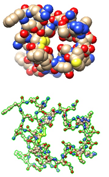
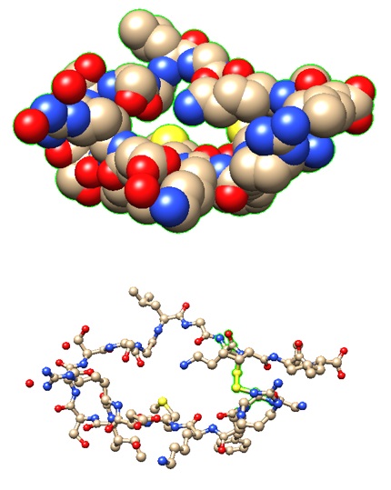
The structural models of two selected receptor-peptide complexes are shown below.

7BRK: Atrial Natriuretic Peptide Receptor complexed with deletion mutant of human Atrial Natriuretic Peptide [5-27]

Structural models of the human Atrial Natriuretic Peptide [5-27] as bound to the receptor.


>pdb|7BRK|L Chain L, Natriuretic peptides A

SSCFGGRMDRIGAQSGLGCNSFR

1YK1: Structure of Natriuretic Peptide Receptor-C complexed with Brain Natriuretic Peptide

Brain natriuretic peptide


>pdb|1YK1|E Chain E, Natriuretic peptides B

GCFGRKMDRISSSSGLGCKVL 


Reference

Harris, Matthew P., Shemin Zeng, Zhiyong Zhu, Vitor A. Lira, Liping Yu, Denice M. Hodgson-Zingman, and Leonid V. Zingman.; 2023. "Myokine Musclin Is Critical for Exercise-Induced Cardiac Conditioning." International Journal of Molecular Sciences 24, no. 7: 6525. [IJMS]

He XL, Dukkipati A, Garcia KC.; Structural determinants of natriuretic peptide receptor specificity and degeneracy. J Mol Biol. 2006 Aug 25;361(4):698-714. [ScienceDirect]

Nishizawa H, Matsuda M, Yamada Y, Kawai K, Suzuki E, Makishima M, Kitamura T, Shimomura I.; Musclin, a novel skeletal muscle-derived secretory factor. J Biol Chem. 2004 May 7;279(19):19391-5.  [
PubMed

Potter LR, Yoder AR, Flora DR, Antos LK, Dickey DM.; Natriuretic peptides: their structures, receptors, physiologic functions and therapeutic applications. Handb Exp Pharmacol. 2009;(191):341-66. [PMC]

Szaroszyk M, Kattih B, Martin-Garrido A, Trogisch FA, Dittrich GM, Grund A, Abouissa A, Derlin K, Meier M, Holler T, Korf-Klingebiel M, Völker K, Garfias Macedo T, Pablo Tortola C, Boschmann M, Huang N, Froese N, Zwadlo C, Malek Mohammadi M, Luo X, Wagner M, Cordero J, Geffers R, Batkai S, Thum T, Bork N, Nikolaev VO, Müller OJ, Katus HA, El-Armouche A, Kraft T, Springer J, Dobreva G, Wollert KC, Fielitz J, von Haehling S, Kuhn M, Bauersachs J, Heineke J.; Skeletal muscle derived Musclin protects the heart during pathological overload. Nat Commun. 2022 Jan 10;13(1):149. [PMC]

Thomas G, Moffatt P, Salois P, Gaumond MH, Gingras R, Godin E, Miao D, Goltzman D, Lanctôt C.; Osteocrin, a novel bone-specific secreted protein that modulates the osteoblast phenotype. J Biol Chem. 2003 Dec 12;278(50):50563-71. [
PubMed]

Zunner BEM, Wachsmuth NB, Eckstein ML, Scherl L, Schierbauer JR, Haupt S, Stumpf C, Reusch L, Moser O.; Myokines and Resistance Training: A Narrative Review. Int J Mol Sci. 2022 Mar 23;23(7):3501. [PMC]

---...---


2’-O-NMA phosphoramidites enable the synthesis of RNA mimics useful for antisense therapeutics

$
0
0

Therapeutic oligonucleotides affect RNA-processing reactions such as mRNA degradation, pre-mRNA splicing, or translation. Several therapeutic oligonucleotides are now approved for clinical correction of genetic mutations.

Molecular Action Platforms For Therapeutic Oligonucleotides 

Three types of molecular actions have emerged as significant platforms for therapeutic oligonucleotides:

[1] RNase H-dependent degradation: short chimeric antisense oligonucleotides called gapmers enable the correction of splicing defects.

[2] RNA-interference: Known as RNAi or siRNA, siRNA oligonucleotides mediate the degradation of the transcript to silence genes.

[3] Splicing modulation: Steric-blocking antisense oligonucleotides allow modulation of aberrant splicing events.


Therapeutic Oligonucleotides are Modified


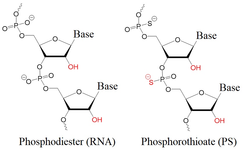
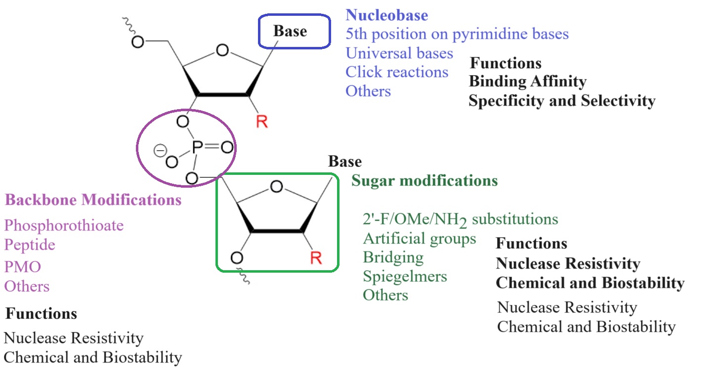
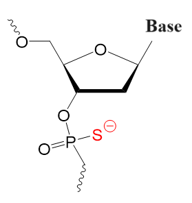
To enhance therapeutic oligonucleotides' stability and efficiency, chemical modifications in their phosphate backbone and sugar rings are usually used. Most common versions of antisense oligonucleotides (AONs) contain phosphorothioate backbones (PS). Here, a sulfur atom replaces one oxygen atom in the phosphodiester group. Also, a 2′-O-methyl group (2′-OMe) is used at the 2′-position in the ribose sugar.


However, other modifications are also investigated for their ability to enhance the function of therapeutic oligonucleotides investigated.

The 2'-O-NMA modification is a new chemical modification that enhances the stability, specificity, and therapeutic potential of RNA molecules.


Figure 1: Chemical structures of a phosphodiester (RNA) and a phosphorothioate (PS).

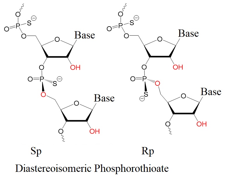
Figure 2: Rp and Sp diastereoisomers of phosphorothioates. 


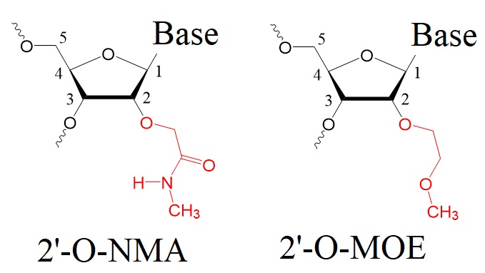
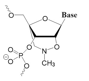
The 2'-O-NMA modification, also known as 2'-O-methyl-N-methylamino-methyl, is a chemical modification applied to nucleic acids, particularly RNA molecules. This modification involves introducing a methyl group (CH3) and an N-methylamino methyl group (-N-CH3) at the 2'-position of the ribose sugar in RNA. This modification enhances the stability, specificity, and therapeutic potential of RNA molecules.

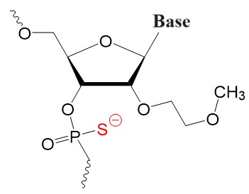
Figure 3: Structures of 2'-O-methoxyethyl-N-methylaminomethyl modified (2’-O-NMA) and 2’-O-methoxyethyl (2’-O-MOE) modified oligonucleotides.

2'-O-NMA (2'-O-methoxyethyl-N-methylaminomethyl) modified oligonucleotides enable the development of RNA mimics for antisense therapeutics. Antisense therapeutics use modified oligonucleotides to target specific RNA sequences to enable modulation of gene expression. RNA mimics imitate the function of natural RNA molecules, allowing for selective targeting and regulation of gene expression.


Incorporating 2'-O-NMA into antisense RNA oligonucleotides provides several advantages over unmodified RNA molecules. Introducing the 2'-O-NMA modification enhances the stability and resistance to degradation of RNA mimics in biological systems. This modification improves the pharmacokinetic properties of the molecule, allowing for increased cellular uptake and prolonged half-life.

Also, 2'-O-NMA enhances the affinity and specificity of RNA mimics for their target sequences. The modification strengthens the binding between the RNA mimic and its complementary RNA sequence, resulting in improved target recognition and increased potency of the therapeutic effect. The increased affinity allows lower doses of the therapeutic RNA oligonucleotide for administration while achieving the desired medicinal outcome.

Antisense oligonucleotides can designed with 2'-O-NMA modification to develop RNA-based drugs for various diseases. This modification offers several advantages over unmodified RNA molecules:


1.   Increased stability: Adding the 2'-O-NMA modification improves the stability of RNA molecules. The modification protects the RNA from degradation by cellular ribonucleases, which can rapidly break down unmodified RNA. This increased stability enables the RNA molecule to remain intact for longer, allowing it to perform its intended function more effectively.


2.   Enhanced specificity: The 2'-O-NMA modification also improves the specificity of RNA molecules. It reduces off-target effects. As a result, the modified RNA is less likely to interact with unintended cellular components or trigger unwanted immune responses. This enhanced specificity is crucial for therapeutic applications, as it increases the safety and efficacy of RNA-based drugs.


3.   Improved pharmacokinetics: Pharmacokinetics refers to how drugs are absorbed, distributed, metabolized, and eliminated by the body. The 2'-O-NMA modification can positively influence the pharmacokinetic properties of RNA molecules. It can increase their resistance to enzymatic degradation and improve cellular uptake, distribution, and retention. This results in better bioavailability and therapeutic outcomes.


4.   Reduced immunogenicity: The immune system recognizes specific RNA molecules as foreign and mounts an immune response against them, limiting the effectiveness of RNA-based therapies. The 2'-O-NMA modification helps to reduce the immunogenicity of RNA by decreasing its recognition by the immune system. By minimizing immune responses, the modified RNA has a higher chance of successfully reaching its target and exerting its therapeutic effect.


Design of 2'-O-NMA-based RNA mimics


The design and synthesis of 2'-O-NMA-based RNA mimics for antisense therapeutics require careful consideration of several factors. 

[1] Carefully identify the specific target RNA sequence.

[2] Design complementary sequences so that they can bind to the target with high affinity and specificity. Additionally.

[3] Optimize the overall secondary structure and stability of the RNA mimic to ensure efficient delivery and intracellular activity.

Overall, the 2'-O-NMA modification is a valuable tool in RNA-based therapeutics. It improves the stability, specificity, pharmacokinetics, and immunogenicity profile of RNA molecules, enhancing their potential as therapeutic agents. Ongoing research and development efforts continue to explore the full potential of this modification and its applications in treating various diseases, including genetic disorders, viral infections, and cancer. Continued research and development in this field hold promise for the advancement of RNA-based therapeutics and their application in treating various diseases.

Reference

Adachi H, Hengesbach M, Yu Y-T, Morais P. From Antisense RNA to RNA Modification: Therapeutic Potential of RNA-Based Technologies. Biomedicines. 2021; 9(5):550

Egli M, Pallan PS. Crystallographic studies of chemically modified nucleic acids: a backward glance. Chem Biodivers. 2010 Jan;7(1):60-89. [PMC]

Egli M, Manoharan M. Re-Engineering RNA Molecules into Therapeutic Agents. Acc Chem Res. 2019 Apr 16;52(4):1036-1047. [ACS]

Pattanayek R, Sethaphong L, Pan C, Prhavc M, Prakash TP, Manoharan M, Egli M. Structural rationalization of a large difference in RNA affinity despite a small difference in chemistry between two 2'-O-modified nucleic acid analogues. J Am Chem Soc. 2004 Nov 24;126(46):15006-7. [ACS]

Prakash TP, Kawasaki AM, Wancewicz EV, Shen L, Monia BP, Ross BS, Bhat B, Manoharan M. Comparing in vitro and in vivo activity of 2'-O-[2-(methylamino)-2-oxoethyl]- and 2'-O-methoxyethyl-modified antisense oligonucleotides. J Med Chem. 2008 May 8;51(9):2766-76. [PubMed]

Sheng L, Rigo F, Bennett CF, Krainer AR, Hua Y. Comparison of the efficacy of MOE and PMO modifications of systemic antisense oligonucleotides in a severe SMA mouse model. Nucleic Acids Res. 2020 Apr 6;48(6):2853-2865. [PMC]

Ziemkiewicz K, Warminski M, Wojcik R, Kowalska J, Jemielity J. Quick Access to Nucleobase-Modified Phosphoramidites for the Synthesis of Oligoribonucleotides Containing Post-Transcriptional Modifications and Epitranscriptomic Marks. J Org Chem. 2022 Aug 5;87(15):10333-10348. [PMC]

 ---...---
Bio-Synthesis provides a full spectrum of bio-conjugation services including high quality custom oligonucleotide modification services, back-bone modifications, conjugation to fatty acids and lipids, cholesterol, tocopherol, peptides as well as biotinylation by direct solid-phase chemical synthesis or enzyme-assisted approaches to obtain artificially modified oligonucleotides, such as BNA antisense oligonucleotides, mRNAs or siRNAs, containing a natural or modified backbone, as well as base, sugar and internucleotide linkages.

Bio-Synthesis also provides biotinylated mRNA and long circular oligonucleotides.
---...---

Reducing off-target effects of the siRNA therapeutics via placing a morpholino group at the 5’-end of the passenger strand

$
0
0
 Heart disease continues to cause a high rate of mortality in the U. S. as it accounts for ~20% of all deaths.  Despite investing heavily on modifying diet, ~697,000 individuals may succumb to the disease annually.  One of the major contributing factors is the accumulation of plaques within the coronary arteries (atherosclerosis), reducing the supply of oxygen to cardiac muscles.  Oxygen plays a vital role in converting energy stored in nutrients (ex. glucose) into ATPs, the main currency used by cells to enable energy-requiring tasks.  An insufficient level of oxygen may cause cardiac muscles to underperform, potentially leading to stroke, artery disease, myocardial infarction (heart attack), etc.

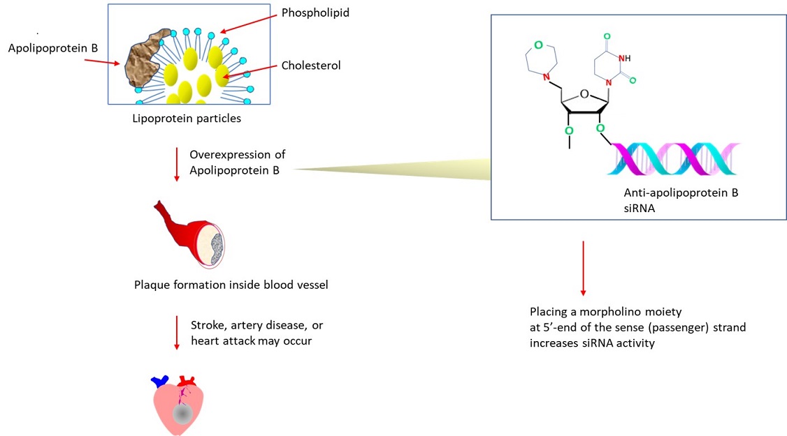
 The plaques found inside blood vessels are comprised of lipid molecules, fibrous tissues, lipid-containing cells (macrophage-like), etc.  As lipids (ex. cholesterol, triglycerides) are insoluble, they (~3000 to 6,000 molecules) are carried in spherically shaped ‘lipoprotein particles’ in plasma.  The outer shell of the transport particle consists of apolipoprotein B, phospholipid, etc.  Apolipoprotein B also serves as the ligand for the receptor that internalizes low density lipoprotein (LDL) particles via clathrin-mediated endocytosis.  Within the endosome, low pH triggers a conformational change of the receptor, releasing LDL to recycle.  Alternatively, it may get degraded in the lysosome after binding to PSCK9, which blocks its conformational change.  A key determinant for plaque formation is the overexpressed apolipoprotein B An altered expression (reduced) of apolipoprotein B may also play a role in liver cancer by upregulating metastasis-causing genes or downregulating tumor suppressor genes (Lee et al., 2018).

 The last decade has seen a marked rise in the number of siRNA-based oligonucleotide therapeutics approved by the FDA (U. S. Food & Drug Administration).  RNA interference refers to the post-transcriptional mechanism of silencing gene expression by cleaving mRNA for degradation.  Originally discovered in nematode as well as in plants, the mechanism of RNA interference has subsequently been exploited for translational medicine.  Critical to this endeavor is the ability of chemically synthesized siRNAs to elicit RNA interference in vivo (Elbashir et al., 2001).  As the siRNA therapeutics rely on Watson-Crick base pairing for target recognition, it is highly specific.

 Nonetheless, several aspects of the siRNA therapeutics may need further improvement.  One area concerns its delivery as siRNAs are highly charged and bulky, making it difficult to translocate across the cell membrane.  To facilitate, various delivery vectors are being explored, which include peptide, carbohydrate, lipid, and nanoparticle (Roberts et al. 2020).


Another potential issue concerns side effects caused by off-targeting.  To scan the mRNA (to find complementary sequence), short siRNA duplex generated by the ribonuclease Dicer are strand-separated and loaded to RNA-induced silencing complex (RISC).  In principle, as the siRNA duplex consists of two complementary strands, either the guide (antisense) strand or passenger(sense) strand could be loaded.  The loading of the passenger strand may lead to the cleavage of an unintended mRNA.

 Previous works suggest that the interaction of 5’-phosphate of the antisense (guide) strand and ‘MID domain’ of Argonaute 2 (of RISC complex) may facilitate strand selection (Elkayam et al., 2012).  It inspired the testing of various modifications at the 5’ end of sense (passenger) strand (ex. 5’-O-methylation, 5’-unlocked nucleic acid unphosphorylated monomer) to ‘suppress’ its loading (Chen et al., 2007; Snead et al., 2013).

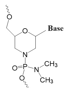
 In another strategy, the effect of blocking the 5’ phosphorylation of the sense strand with morpholino was investigated (Kumar et al., 2013).  The researchers at Alnylam Pharmaceuticals (United States) have developed siRNA targeting apolipoprotein B, which contains a morpholino group at the 5’ end of the sense or antisense strand.  Reaction steps to chemically synthesize 5’-morpholino 2′-OMe phosphoramidites are described in detail in the report (Kumar et al., 2013).  Placing morpholino on the sense strand improved the loading of anti-apolipoprotein B siRNA to the RISC complex, increasing the RNA interference activity.  


The key to preventing epidemic is the ability to diagnose the infected early to preempt further propagation.  For this, Bio-Synthesis, Inc. provides primers and probes (as well as synthetic RNA control) for COVID-19 diagnosis via RT-PCR assay.  It specializes in oligonucleotide modification and provides an extensive array of chemically modified nucleoside analogues (over ~200; ex. morpholino) including bridged nucleic acid (BNA) in addition to mRNA synthesis.  A number of options are available to label oligonucleotides (DNA or RNA) with fluorophoreseither terminally or internally as well as to conjugate to peptidesor antibodies.  It recently acquired a license from BNA Inc. of Osaka, Japan, for the manufacturing and distribution of BNANC, a third generation of BNA oligonucleotides.  To meet the demands of therapeutic application, its oligonucleotide products are approaching GMP grade.  Bio-Synthesis, Inc. has recently entered into collaborative agreement with Bind Therapeutics, Inc. to synthesize miR-21 blocker using BNA for triple negative breast cancer.  The BNA technology provides superior, unequalled advantages in base stacking, binding affinity, aqueous solubility and nuclease resistance.  It also improves the formation of duplexes and triplexes by reducing the repulsion between the negatively charged phosphates of the oligonucleotide backbone.  Its single-mismatch discriminating power is especially useful for diagnosis (ex. FISH using DNA probe).  For clinical application, BNA oligonucleotide exhibits lesser toxicity than other modified nucleotides. 


https://www.biosyn.com/tew/Thiophosphoramidate-Morpholinos,-A-New-Class-of-Antisense-Oligonucleotides.aspx

https://www.biosyn.com/tew/Peptide-conjugated-Antisense-Oligonucleotides-for-Exon-Skipping-Therapeutics.aspx

https://www.biosyn.com/tew/Phosphoryl-guanidine-oligo-2%E2%80%B2-O-methylribonucleotides-in-neutral-therapeutic-oligonucleotides-enable-enhanced-cell-penetration!.aspx


References

Elbashir SM, Tuschl T, et al.  Duplexes of 21-nucleotide RNAs mediate RNA interference in cultured mammalian cells.  Nature. 411:494-8 (2001).  PMID: 11373684

Elkayam E, Joshua-Tor L, et al. The structure of human argonaute-2 in complex with miR-20a.  Cell. 150:100-10 (2012).  PMID: 22682761

Kumar P, Manoharan M, et al. 5'-Morpholino modification of the sense strand of an siRNA makes it a more effective passenger.  Chem Commun (Camb).   55:5139-5142 (2019).  PMID: 30977478

Lee G, Yim SY, et al.  Clinical significance of APOB inactivation in hepatocellular carcinoma.  Exp Mol Med. 50:1-12 (2018).  PMID: 30429453

Roberts TC, Langer R, et al.  Advances in oligonucleotide drug delivery.  Nat Rev Drug Discov. 19:673-694 (2020).  PMID: 32782413

Snead NM, McCaffrey AP, et al. 5' Unlocked Nucleic Acid Modification Improves siRNA Targeting.  Mol Ther Nucleic Acids.   2:e103 (2013).  PMID: 23820891


The efficacy of FDA-approved drug inhibiting the ‘cap snatching’ endonuclease of RdRP polymerase is undermined by the emergence of resistant influenza virus strains

$
0
0
 With the gradual decline in the number of COVID-19 infections (total of ~103 million cases in the U. S. alone during the 3-year pandemic), the attention is returning to flu, which is projected to incur 25-51 million cases in the U. S. during 2022-2023 (https://www.cdc.gov/flu/about/burden/preliminary-in-season-estimates.htm ).  Globally, the influenza virus, which causes flu, infects 2–10% of the entire population, causing 250,000-500,000 deaths annually (El Ramahi et al., 2019).  Unlike COVID-19, flu can impact the younger and healthier populations.  The disease can deteriorate further by contracting pneumonia caused by the virus or bacterial infection, which may lead to death.  Since 1900, the influenza virus has caused pandemics 5 times, with the most notable one being  ‘The Spanish flu (1918)’, which may have led to as many as 50 million deaths globally.


For cancer patients, the death rate due to flu is significantly higher than the general public, i.e. 17.4% versus 1.3% (per 100,000 person-years), respectively.  ‘Person-year’ is defined as the number of individuals multiplied by the study duration in years (El Ramahi et al., 2019).  Over 70% of cancer patients, who contracted flu, received chemotherapy within the preceding 1 month (possibly immunocompromised) during the influenza pandemic in 2009.   Those with lung cancer suffered the worst outcome.


There are 4 types of influenza virus: A, B, C, and D.  Of these, influenza A and B are capable of infecting humans.  To infect, the virus uses its glycoprotein (hemagglutinin) to bind to the sialic acid moiety of saccharides attached to the cell surface.  To access the receptor, virally encoded neuraminidase degrades mucus; it may also assist in the release of viruses from a cell.   For influenza virus type A, 20 subtypes (H1-H18) of hemagglutinin are known.  Neuraminidases are classified into subtypes N1 through N11.   Spanish flu was caused by the H1N1 subtype.


Each year, the Center for Disease Control prepares distinct vaccines to prevent seasonal flu in the U. S.  The vaccine primarily targets the hemagglutinin antigen of the influenza virus.  The need for constantly updating flu vaccine lies in ‘antigenic drift’, which refers to amino acid changes caused by errors introduced during transcription by the virally encoded RNA polymerase (RdRP) (Taubenberger et al., 2010).

Unlike the genome of the coronavirus that caused COVID-19, the genome of the influenza virus consists of 8 segments.  The segmented genome allows shuffling of the genomic segments through a process called ‘reassortment’.  This could happen if the same cell is infected by more than 1 influenza virus.  Through the ‘antigenic shift’, drastically altered antigens can be generated, which could cause a pandemic.


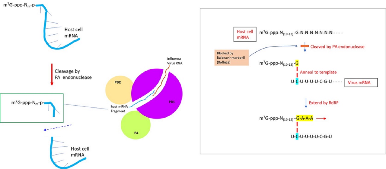
The genome of the influenza virus is composed of negative-sense single-stranded RNA, which is transcribed to generate viral mRNA (to translate into proteins) or cRNA (complementary RNA) to generate vRNA (virus RNA).  The positive-sense cRNA template is used to produce the negative-sense genomic RNA of the virus.  The synthesis of viral mRNA utilizes the ‘cap-snatching’ mechanism, in which the capped nucleotide primer is derived from the pre-mRNA of the host cell.  RNA-dependent RNA polymerase of influenza virus is comprised of 3 subunits:  PA, PB1, and PB2.  Upon binding of the PB2 subunit to m7G (N7-methyl guanosine)-capped 5’ end of cellular mRNA, the endonuclease activity of the PA subunit cleaves the host pre-mRNA at a position 10-13 nucleotides downstream of the cap structure (De Vlugt et al., 2018).  Within the PB1 subunit, the last nucleotide (usually guanidine) of capped host RNA primer is annealed to cytosine of template virus RNA and then extended by RdRP to synthesize mRNA (Li et al., 2020).  Recent works indicate that ~55% of the host RNA primers were derived from noncoding RNAs (ex. snRNA U1 and U2) (Li et al., 2020).


Previously, Tamiflu (Gilead Sciences) was developed to inhibit the neuraminidase activity of influenza viruses A and B.  More recently, Baloxavir marboxil (Xofluza), a chemical drug that inhibits the cap-dependent endonuclease activity responsible for the cap snatching process was developed.  The prodrug is hydrolyzed by arylacetamide deacetylase to release the active agent, baloxavir acid.  It was approved by U. S. FDA in 2018 as an antiviral drug to manage the influenza virus.  Nevertheless, strains resistant to the drug were detected by 2019 (Yoshino et al., 2019).


The key to preventing an epidemic is the ability to diagnose the infected early to preempt further propagation.  For this, Bio-Synthesis, Inc. provides primers and probes (as well as synthetic RNA control) for COVID-19 diagnosis via RT-PCR assay.  It specializes in oligonucleotide modification and provides an extensive array of chemically modified nucleoside analogs (over ~200) including bridged nucleic acid (BNA) in addition to mRNA synthesis.  A number of options are available to label oligonucleotides (DNA or RNA) with fluorophoreseither terminally or internally as well as to conjugate to peptidesor antibodies. It provides custom conjugation of small molecules such as chemical drugs, metabolites and labeled compounds with synthetic or natural polymers (enzymes, peptide, protein, oligonucleotide, antibody, dendrimer, nanoparticle, etc).  It recently acquired a license from BNA Inc. of Osaka, Japan, for the manufacturing and distribution of BNANC, the third generation of BNA oligonucleotides.  To meet the demands of therapeutic application, its oligonucleotide products are approaching GMP grade.  It has recently entered into collaborative agreement with Bind Therapeutics, Inc. to synthesize miR-21 blocker using BNA for triple-negative breast cancer.  The BNA technology provides superior, unequaled advantages in base stacking, binding affinity, aqueous solubility and nuclease resistance.  It also improves the formation of duplexes and triplexes by reducing the repulsion between the negatively charged phosphates of the oligonucleotide backbone.  Its single-mismatch discriminating power is especially useful for diagnosis (ex. FISH using DNA probe).  For clinical application, BNA oligonucleotide exhibits lesser toxicity than other modified nucleotides.  For therapeutic consideration, peptide synthesis or modifications may include labeling, conjugation, cyclization, incorporation of unusual amino acids, and modification of side chain and backbone.


https://www.biosyn.com/oligo-flourescent-labeling.aspx

https://www.biosyn.com/tew/Speed-up-Identification-of-COVID19.aspx

https://www.biosyn.com/covid-19.aspx

https://www.biosyn.com/mrna.aspx

https://www.biosyn.com/bioconjugation.aspx

https://www.biosyn.com/tew/Design-Guidelines-for-BNA-based-Oligonucleotide-Probes.aspx#!

Peptide Modifications, Modified Peptide Synthesis - Bio-Synthesis (biosyn.com)


References

De Vlugt C, Sikora D, Pelchat M. Insight into Influenza: A Virus Cap-Snatching.  Viruses. 10:641 (2018).  PMID: 30453478

El Ramahi R, Freifeld A.  Epidemiology, Diagnosis, Treatment, and Prevention of Influenza Infection in Oncology Patients.  J Oncol Pract. 15:177-184 (2019).   PMID: 30970229

Li L, Dai H, Nguyen AP, Hai R, Gu W. Influenza A virus utilizes noncanonical cap-snatching to diversify its mRNA/ncRNA.  RNA. 26:1170-1183 (2020).  PMID: 32444459

Taubenberger JK, Kash JC.  Influenza virus evolution, host adaptation, and pandemic formation.  Cell Host Microbe. 7:440-51 (2010).  PMID: 20542248

Yoshino R, Yasuo N, Sekijima M. Molecular Dynamics Simulation reveals the mechanism by which the Influenza Cap-dependent Endonuclease acquires resistance against Baloxavir marboxil.  Sci Rep. 9:17464 (2019).  PMID: 31767949


Aptamers-Nucleic Acid-Based Molecules with Diverse Applications

$
0
0

Aptamers are nucleic acid-based molecules that have gained significant attention in biotechnology, medicine, and diagnostics due to their unique properties and versatile applications. These molecules are often referred to as "chemical antibodies" or "synthetic receptors" because, like antibodies, they can bind specifically to target molecules, but unlike antibodies, they are entirely synthetic.

Specifically designed aptamers allow targeting a wide range of molecular targets, including proteins, small molecules, and even whole cells.

Structure of Aptamers

Aptamers are typically single-stranded DNA or RNA molecules that fold into specific three-dimensional structures. Their unique shapes allow aptamers to recognize and bind to their target molecules with high affinity and specificity. The process of selecting or synthesizing aptamers is known as the Systematic Evolution of Ligands by Exponential Enrichment (SELEX). During SELEX, a library of random sequences is subjected to multiple rounds of selection, amplification, and purification, enriching sequences that bind to the target of interest.


Applications of Aptamers

Aptamers have a wide range of applications across various fields:


Biomedical Research:
 Aptamers allow the study of protein-protein interactions, elucidating the functions of specific biomolecules, and serve as tools for developing diagnostic assays and imaging agents.


Therapeutics: Aptamers have shown promise as therapeutic agents. Some aptamers, such as Pegaptanib (Macugen), have been approved for clinical use as anti-vascular endothelial growth factor (VEGF) drugs for treating age-related macular degeneration.

Diagnostic Tools: Aptamers are valuable components of diagnostic assays, for example, to enable the detection of specific biomarkers, pathogens, or toxins in clinical samples. Aptamer-based assays are often highly sensitive and specific.

Drug Delivery: Aptamers can be conjugated to drug molecules or nanoparticles, allowing targeted drug delivery. The attachment of an aptamer that binds to a receptor on a specific cell type enables more selective delivery of drugs, reducing off-target effects.


Environmental Monitoring: Well-designed aptamers can also recognize environmental pollutants or toxins. Aptamers have been used in biosensors to detect soil, water, and air contaminants.


Food Safety: Aptamers allow the detection of pathogens or harmful substances in food products, ensuring food safety.


Nanotechnology:  Various applications, including drug delivery, imaging, and sensing, utilize aptamers incorporated into nanomaterials.


Advantages of Aptamers


Aptamers offer several advantages over traditional antibodies and other recognition molecules:

Ease of Synthesis: Aptamers can be synthesized in vitro, making them more readily available and cost-effective than antibodies, which often require animal immunization.

High Affinity and Specificity: Specifically designed aptamers bind their targets with high affinity and specificity, rivaling or surpassing the performance of antibodies.


Stability: Aptamers are stable under various environmental conditions, making them suitable for multiple applications.


Reproducibility: Aptamer synthesis is highly reproducible, ensuring consistent performance in assays and applications.


In summary, aptamers represent a versatile class of nucleic acid-based molecules with numerous applications in research, diagnostics, therapeutics, and beyond. The ability of aptamers to bind specific targets with high affinity and specificity, coupled with their ease of synthesis and adaptability, positions them as valuable tools in biological science, medicine, and technology. 

Reference

Published examples of aptamers synthesized by BSI:

(1)  Aller Pellitero M, Kundu N, Sczepanski J, Arroyo-Currás N. Os(II/III) complex supports pH-insensitive electrochemical DNA-based sensing with superior operational stability than the benchmark methylene blue reporter. Analyst. 2023 Feb 13;148(4):806-813. [PMC]

Design of electrochemical, aptamer-based (E-AB) sensors representing a specific type of E-DNA sensors that enable the continuous and real-time monitoring of molecular targets.

(2)  Zhao N, Bagaria HG, Wong MS, Zu Y. A nanocomplex that is both tumor cell-selective and cancer gene-specific for anaplastic large cell lymphoma. J Nanobiotechnology. 2011 Jan 31;9:2. doi: 10.1186/1477-3155-9-2. [PMC]

Design of a functional RNA nanocomplex as a therapeutic agent as a carrier for the selective delivery of cargo to tissues and cells.

The nanocomplexes include:

a) incorporated siRNAs into a nano-sized carrier that increase their physical size and prevent the rapid elimination of siRNA from the blood circulation in vivo;

b) incorporated CD30 aptamers enabling specific accumulation of the nanocomplexes within tumor sites and eliminate potential off target side effects of the nanocomplex components; and

c) it a potential incorporation of more than one siRNA and/or therapeutic drug into the nanocomplex to generate additive or synergistic repressive effects on tumor cells. 

(3)  Zhao N, You J, Zeng Z, Li C, Zu Y. An ultra pH-sensitive and aptamer-equipped nanoscale drug-delivery system for selective killing of tumor cells. Small. 2013 Oct 25;9(20):3477-84. [PMC]

Design of a selective chemotherapeutic using a hollow gold nanosphere (HAuNS) equipped with a biomarker-specific aptamer (Apt), and loaded with the chemotherapy drug doxorubicin (DOX).

“The 39-mer RNA aptamer specific for CD30, a lymphoma biomarker for diagnosis of Hodgkin’s lymphoma and anaplastic large cell lymphoma, was synthesized by Bio-Synthesis (Lewisville, TX), as previously described, by using the sequence:

5′-mGmAmUUCGUAUGGGUGGGAUCGGGAAGGGCUACGAACAmCmCmG-[Thiol]-3′

(mN represents 2′-O-Methyl RNA).”

(4)   Zhang P, Zhao N, Zeng Z, Feng Y, Tung CH, Chang CC, Zu Y. Using an RNA aptamer probe for flow cytometry detection of CD30-expressing lymphoma cells. Lab Invest. 2009 Dec;89(12):1423-32. [PMC]

Design and synthesis of a 39-mer nucleotide RNA aptamer with one minor modification: 5′-gauUCGUAUGGGUGGGAUCGGGAAGGGCUACGAACAccg-3′. The aptamer probe contains the most essential functional motif with the highest affinity to mouse CD30 molecules in solution. To enhance its resistance to nucleases, the first three nucleotides on each end of the aptamer were synthesized with 2′-O-methyl modification (indicated by lower case letters).

(5)   Cheng S, Jacobson O, Zhu G, Chen Z, Liang SH, Tian R, Yang Z, Niu G, Zhu X, Chen X. PET imaging of EGFR expression using an 18F-labeled RNA aptamer. Eur J Nucl Med Mol Imaging. 2019 Apr;46(4):948-956. [PMC]

Design and production of a biolayer interferometry binding assay using an alkyne modified EGFR aptamer MinE07 (ME07). The use of 18F-Fluorobenzoyl (FB) azide allowed labeling the aptamer for its use in positron emission tomography (PET) scans of tumor models. 

Others

Wang, A. Z., and O. C. Farokhzad. "Current Progress of Aptamer-Based Molecular Imaging." 2014. 
https://doi.org/10.17615/bd33-km50.

Kubiczek, C., et al. "Aptamers As Promising Agents in Diagnostic and Therapeutic Applications." 2017.
https://core.ac.uk/download/210975297.pdf.

Publications - Salk Institute for Biological Studies.
https://www.salk.edu/scientist/gerald-joyce/publications/

BSI links to aptamers

https://www.biosyn.com/aptamers.aspx Website

Custom Aptamers

https://www.biosyn.com/tew/Custom-Aptamer-Synthesis.aspx

Specifically labeled oligonucleotides

https://www.biosyn.com/tew/Specific-labeling-of-RNA.aspx#!


---...---


siRNA and the immune system

$
0
0

Double-stranded small interfering RNA (siRNA) silences the expression of specific genes. siRNAs have demonstrated their potential in treating various diseases and are a powerful tool for research and developing new therapies. However, siRNA can also interact with the immune system in several ways. Therefore, it is critical to understand the potential risks and benefits of using siRNA to modulate the immune system before using it in any therapeutic setting.

Figure 1: Molecular mechanism of RNA interference (RNAi). The process in which RNA molecules activate the cellular response to destroy specific RNA molecules such as messenger RNAs (mRNAs). (Source: Wiki Commons).

RNA interference (RNAi) therapies specifically target genes of interest. As more biological data have become available, it became apparent that in addition to mediating RNAi, siRNA molecules also have the potential to induce the innate immune system.

One significant challenge when designing siRNAs is the differentiation between therapeutic effects and non-specific innate immune system stimulation. Well-designed and considered experimental measures to establish the best design of siRNAs may avoid activating the immune system by siRNA molecules.

RNAi is an endogenous cellular mechanism by which sequence-specific siRNA induces gene silencing by targeting and cleavage of complementary messenger RNA (mRNA) within the cell's cytoplasm. The presence of double-stranded RNA (dsRNA) triggers RNAi. dsRNA is cleaved by the intracellular enzyme dicer into 21-base pair fragments of siRNA. Loading the siRNA into a protein complex called the RNA-inducing silencing complex (RISC) unwinds the siRNA and retains the antisense strand. RISC searches for any mRNA complementary to the antisense strand and cleaves it. As a result, the target gene is silenced and prevented from protein production.

Double-stranded RNA, when longer than 30 base pairs, can also be a potent activator of the innate immune interferon (IFN) response. Long dsRNA is a hallmark of viral infection. This response can limit siRNA therapies since siRNA delivery into cells introduces a foreign material into the cell's biological system. However, synthetically designed siRNA containing modified nucleic acids can circumvent Dicer mechanics and immunostimulation associated with long dsRNA.

The innate immune system is the body's first defense against infection and injury. The innate immune system cells quickly recognize and respond to various pathogens and danger signals. The mammalian immune system recognizes siRNA as a signature of viral infection; hence, siRNA can induce a potent and potentially dangerous innate immune response.


siRNA potentially activate the innate immune system in different ways.

[1] Toll-like receptors (TLRs) can recognize and bind siRNA. TLRs are proteins located on the surface of immune cells that recognize various danger signals.

[2] Other immune sensors, such as retinoic acid-inducible gene I (RIG-I), can bind siRNA.

When siRNA activates the innate immune system, it can trigger several different responses, including the production of inflammatory cytokines and the activation of immune cells. 
However, using siRNA to treat infection can induce a beneficial immune response activation. Sometimes, a harmful activation can occur when treating a chronic disease with siRNA.

[3] Another way siRNA interacts with the immune system is through the adaptive immune system. The adaptive immune system is a more sophisticated defense system that learns to recognize and respond to specific pathogens. It comprises B and T cells, which produce antibodies and other immune molecules that can neutralize or kill pathogens.

In addition, siRNA also enables modulation of the adaptive immune system. For example, siRNA allows silencing the expression of genes involved in activating or suppressing T cells. siRNA can also deliver genes to T cells that enhance their anti-tumor activity.

The mammalian immune system can recognize siRNA as a foreign molecule, likely a sign of viral infection, and respond robustly. A list of heterogeneous pattern recognition receptors (PRR) in different cell types of the mammalian system is shown in Table 1. 

Table 1: Pattern recognition receptors (PRRs) that recognize and respond to RNA.


PRR

Ligand

Cell line expression

Subcellular location

Primary response

TLR3

siRNA

dsRNA

Epithelial cells

Fibroblast

Cell surface

IFN-α

IFN-β

IFN-γ

Epithelial cells

Fibroblast

mDCs

Endosome

IFN-α

IFN-β

IFN-γ

TLR7

siRNA

ssRNA


pDCs

Endosome

IFN-α

IFN-β

Lysosome

TNF-α

IL-12

B cells


B cell differentiation

TLR8

siRNA

ssRNA

Monocytes

Macrophages

mDCs

Endosome

IFN-α

IFN-β

Lysosome

TNF-α

IL-1

IL-6

IL-12

PKR

Long dsRNA

Most mammalian cells

Cytoplasm

IFN-α

IFN-β

Inhibition of protein translation

RIG-I

Uncapped 5’-triphosphate RNA and blunt RNA

Fibroblasts

mDCs

Cytoplasm

IFN-α

IFN-β


Abbreviations: TLR, Toll-like receptor; siRNA, small interfering RNA; dsRNA, double-stranded RNA; IFN, interferon; mDCs, mature dendritic cells; ssRNA, single-stranded RNA; pDCs, plasmacytoid dendritic cells; TNF-α, tumor necrosis factor α; IL, interleukin; PKR, double-stranded RNA-dependent protein kinase; RIG-I, retinoic acid-inducible gene I protein.

Examples of immune-stimulating sequences


The nucleotide sequence of siRNAs can affect its immunostimulatory properties. Immunostimulation motifs of the TLR7-mediated interferon response, 5-UGU-3’, 5’-GUCCUUCAA-3’, induce cytokine production independent of the number of GU nucleosides. These are two examples of the many specific RNA sequences that TLR7 and 8 recognize. A ribose sugar backbone and multiple uridine residues near one another also stimulate TLR7 and 8. However, substituting guanosine with adenosine reduced TNF-α, and replacing uridine with adenosine decreased IFN-α production in plasmacytoid dendritic cells. Modifications to the 2’-position on the ribose ring of the RNA backbone can reduce innate immune activation. Also, the delivery vehicle selected to facilitate siRNA transfection potentially affects the innate immune response. As a result, researchers need to design delivery experiments for siRNA carefully.


The chemical structure of nucleic acids can influence the immune response

The chemical structure of the siRNA duplex can also influence how the innate immune system responds. For example, RIG-I can bind to ssRNA or dsRNA containing uncapped 5’-triphosphate groups, resulting in an interferon-mediated immune response. Uncapped RNA is a sign of viral infection causing a subsequently induced inflammatory action. Further, blunt-ended dsRNA can provoke an immunostimulatory activity after recognition by RIG-I. Incorporating 3’-overhangs on either or both RNA strands reduces these activities. Self microRNAs present in the cytoplasm processed by dicer include 3’-overhangs. However, viral dsRNAs typically possess blunt ends.

Chemical changes in the ribose backbone can reduce or even eliminate the innate immune response. For example, inserting bridged nucleic acids (BNAs/LNAs) into RNA strands can reduce immune recognition and response. Still, it can also compromise the potency of the siRNA depending on which point the BNA is inserted. The 2’-O-Me modification acts as a potent inhibitor of RNA-induced immune stimulation without diminishing RNAi potency.

Modulating the immune system

Examples of how siRNA can modulate the immune system:


siRNA potentially allows the development of new vaccines against cancer and infectious diseases.

siRNA may enable new treatments of autoimmune diseases, such as rheumatoid arthritis and multiple sclerosis.

siRNA may allow the development of new treatments for allergies and asthma.

siRNA potentially allows the development of new treatments for cancer by targeting genes that are involved in tumor growth and metastasis.


Reference

Kanasty RL, Whitehead KA, Vegas AJ, Anderson DG. Action and reaction: the biological response to siRNA and its delivery vehicles. Mol Ther. 2012 Mar;20(3):513-24. [PMC]

Suzuki M, Zheng X, Zhang X, Ichim TE, Sun H, Kubo N, Beduhn M, Shunnar A, Garcia B, Min WP. Inhibition of allergic responses by CD40 gene silencing. Allergy. 2009 Mar;64(3):387-97. [
Allergy]

Spurgers KB, Sharkey CM, Warfield KL, Bavari S. Oligonucleotide antiviral therapeutics: antisense and RNA interference for highly pathogenic RNA viruses. Antiviral Res. 2008 Apr;78(1):26-36. [
PMC]

Whitehead KA, Dahlman JE, Langer RS, Anderson DG. Silencing or stimulation? siRNA delivery and the immune system. Annu Rev Chem Biomol Eng. 2011;2:77-96. [
PubMed]

Zhao Y, Huang L. Lipid nanoparticles for gene delivery. Adv Genet. 2014;88:13-36. [
PMC]

Other links

https://www.biosyn.com/faq/What-are-Backbone-Modifications-in-Oligonucleotide-Synthesis.aspx#!

https://www.biosyn.com/backbone-modified-oligonucleotide-synthesis.aspx#


---...---
Bio-Synthesis provides a full spectrum of bio-conjugation services including high quality custom oligonucleotide modification services, back-bone modifications, conjugation to fatty acids and lipids, cholesterol, tocopherol, peptides as well as biotinylation by direct solid-phase chemical synthesis or enzyme-assisted approaches to obtain artificially modified oligonucleotides, such as BNA antisense oligonucleotides, mRNAs, miRNA, or siRNAs, containing a natural or modified backbone, as well as base, sugar and internucleotide linkages.

Bio-Synthesis also provides biotinylated mRNA and long circular oligonucleotides.
---...---

Signaling Aptamers with specific target binding properties

$
0
0

Designing oligonucleotides such as aptamers with specific target binding properties widens the application range of nucleic acid-based fluorescent probes for detecting many analytes, including small molecules, proteins, nucleic acids, ions, and whole cells.

Aptamer probes that exploit non-nucleic acid analytes' selective recognition allow the design of immunosensors. The design and construction of hybridization and aptamer probes are similar. Reporter groups covalently attached to oligonucleotides (DNA or RNA) with predefined sequences enable the design of fluorescence-based aptamer probes.

Fluorescent labels act as transducers, transforming biorecognition such as hybridization or ligand binding into a fluorescence signal. Fluorescent labels have several advantages when compared to radioactive labels, including high sensitivity and multiple transduction approaches such as fluorescence quenching or enhancement, fluorescence anisotropy, fluorescence lifetime, fluorescence resonance energy transfer (FRET), and excimer-monomer light switching.

Two types of fluorophores enable the labeling of oligonucleotides:

1.    Dyes that change their fluorescence properties when binding nucleic acids are used in single-labeled probes.

2.    Fluorophores with strong fluorescence that change their emission intensity when brought into contact with each other or with a quencher molecule. Examples are fluorescein and rhodamine dyes.

Standard detection modes are fluorescence resonance energy transfer, excimer–monomer switching, hybridization probes, binary probes, and molecular beacons.


Figure 1: Schematics of a fluorescent biosensor.

Oligonucleotides with defined sequences and structures can recognize specific non-nucleic acid targets - these single-stranded nucleic acid sequences obtained by the SELEX method are known as aptamers.

Aptamers with sequences characteristic of specific protein-binding regions enable protein recognition.

The anti-thrombin aptamer is an example. A 15-mer oligonucleotide, with the sequence 5′-d(GGTTGGTGTGGTTGG)-3′, forms an intramolecular quadruplex with two G-tetrads firmly binding to thrombin.

Tan and co-workers have used the pyrene excimer monomer switching (EMS) approach to design a wavelength-shifting aptamer probe.

The probe contains a platelet-derived growth factor (PDGF)-binding DNA aptamer labeled with two pyrene molecules.

In this aptamer, the two ends are far away in the absence of PDGF but close to each other in the presence of the target.

When bound to PDGF, the aptamer switches its fluorescence emission from 400 nm (pyrene monomer, with a fluorescence lifetime of ∼5 ns) to 485 nm (pyrene excimer, with a lifetime of ∼40 ns).

The wavelength-shifting and time-resolved measurement characteristics allow for sensing target molecules in complex sample matrixes.

Sensing is even possible in cell media, in which strong background fluorescence often makes intensity-based detection difficult.

Chen recently reviewed recent advances in developing excimer-based fluorescence probes useful for biological applications.


Reference


Chen Y.; Recent Advances in Excimer-Based Fluorescence Probes for Biological Applications. Molecules. 2022 Dec 6;27(23):8628. doi: 10.3390/molecules27238628. [PMC]

Gustmann H., Segler A.-L. J., Gophane D. B., Reuss A. J., Grünewald C., Braun M., et al.. (2018). Structure guided fluorescence labeling reveals a two-step binding mechanism of neomycin to its RNA aptamer. Nucl. Acids Res. 47, 15–28. 10.1093/nar/gky1110 [
PMC] [PubMed] []

Juskowiak B. (2010). Nucleic acid-based fluorescent probes and their analytical potential. Anal. Bioanal. Chem. 399, 3157–3176. 10.1007/s00216-010-4304-5 [
PMC free article] [PubMed] [CrossRef] [Google Scholar]

Li JJ, Fang X, Tan W. Molecular aptamer beacons for real-time protein recognition. Biochem Biophys Res Commun. 2002 Mar 22;292(1):31-40. [
PubMed] [Google Scholar]


---...---
Bio-Synthesis provides a full spectrum of bio-conjugation services including high quality custom oligonucleotide modification services, back-bone modifications, conjugation to fatty acids and lipids, cholesterol, tocopherol, peptides as well as biotinylation by direct solid-phase chemical synthesis or enzyme-assisted approaches to obtain artificially modified oligonucleotides, such as BNA antisense oligonucleotides, mRNAs, miRNA, or siRNAs, containing a natural or modified backbone, as well as base, sugar and internucleotide linkages.

Bio-Synthesis also provides biotinylated mRNA and long circular oligonucleotides.
---...---

Thiomorpholino oligonucleotides or TMOs – An innovative class of Antisense oligonucleotide

$
0
0

Oligodeoxynucleotide, ODN analogues, are versatile biological and therapeutic agents that have various applications such as RNA interference, splice switching, aptamer binding, and antisense technology.[1] ODN’s efficiency in delivery and gene silencing was improved tremendously by the introduction of GalNAc conjugation and lipid formulation. Antisense oligonucleotides (ASOs) have made significant progress in recent years and are beneficial for patients with both common and rare diseases due to their chemical diversity.[2]

ASOs are synthetic nucleic acid sequences that can specifically bind to certain RNA sequences using Watson-Crick base pairing. ASOs regulate the gene expression through steric blocking of RNA-protein interactions, RNase-H dependent cleavage or modulation of pre-mRNA splicing.[3] To be effective, ASOs require high nuclease resistance, high RNA targeting affinity, and better mismatch identification, leading to the development of chemical modifications on ASO that improve the pharmacokinetic profile of nucleic acid-based drugs. Since 1998, the FDA has approved nine ASO-based drugs, with many currently undergoing phase II and phase III clinical trials.[4] Some well-known chemical modifications on ASOs include 2'-OMe, 2'-O-MOE, 2'-F, PMO, LNA, PNA, tcDNA, BNA, and UNA.


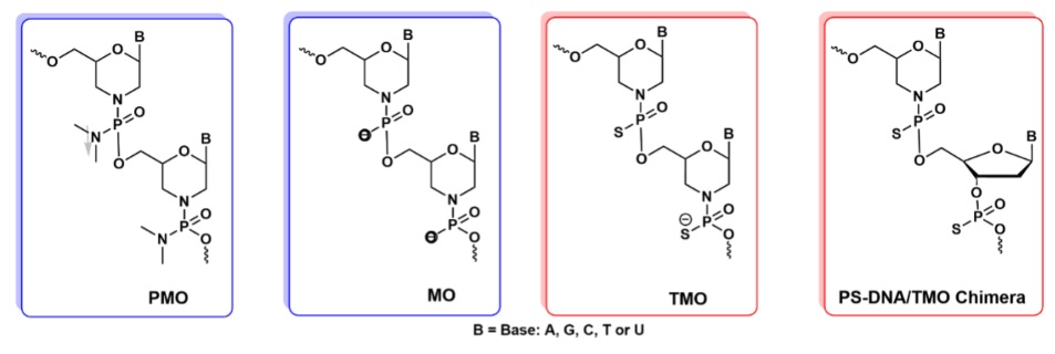
PMO, phosphorodiamidate morpholino oligomers, is an important antisense agent that is designed by the replacement of ribose sugar unit of a natural nucleotide with a morpholino ring. Neutral backbone of the PMO offers low toxicity, water solubility, and sufficient endonuclease stability.[5] Four PMO-based drugs have been approved by the FDA to treat Duchenne muscular dystrophy patients, while three PMOs have shown their effectiveness against viral and bacterial infections and cancers in preclinical models and cell-based studies.[6] However, PMOs can be challenging to synthesize on a large scale for therapeutic use, they are not suitable for synthesis of hybrid morpholinos too.[7] Also, PMOs and other regular less toxic phosphorodiester backbone oligonucleotides are rapidly excreted from the body through Urine due to their poor binding to plasma proteins. Whereas thiophosphorodiester backbone or PS-DNAs improve the resistance and in vivo circulation time of ASOs by binding to serum albumin. Stabilin class of scavenger receptors bind effectively to thiophosphorodiester backbone.[8]


When the non-bridging oxygen atom in the phosphate backbone of morpholino phosphoramidates, MOs, are replaced by sulfur atom thiomorpholino phosphoramidates, TMOs are formed. A versatile solid phase synthesis of TMO and their DNA/TMO chimeras on an automated synthesizer have been developed by Caruthers et al using morpholino phosphoramidite as precursor.[9] Morpholino phosphoramidites were prepared from 6’- DMT protected morpholinos with standard base labile protecting groups on the nucleoside bases. This chemical modification of TMO enhances their stability, resistance to nucleases, and binding affinity to RNA targets. Caruthers group reported that, compared to conventional DNA/RNA duplex, alternating PS-DNA/TMO chimeras and fully modified TMOs shows higher RNA binding affinity. High RNA affinity of fully modified TMO can block the gene expression and can be a good candidate for splicing studies. These fully modified TMOs were not recruiting RNase H1. Whereas chimeric TMO analogues like DNA/TMO chimeras can degrade the mRNA by binding with RNase H and efficient for high gene silencing compared to 2’-OMe control.[9a]

Veedu et al. reported that compared to conventional PMOs, 2’-OMe and 2’-MOE, TMOs exhibit efficient exon 23 skipping in the mouse dystrophin transcript at a lower concentration of 5-20 nM. This can improve the drug safety profile by minimizing the dosage of the drug.[10] Rinn et al. reported that TMOs effectively block splicing and affect subcellular localization and availability of the RNA. TMO could be applicable not only to specific intron transcripts, but to variety of oncogene transcripts that could be performed inert in the nucleus.[11]

References

1. a) Uhlmann, E., Peyman,A., Antisense oligonucleotides: A new therapeutic principle Chem. Rev., 1990, 90, 544-584. b) Roberts, T. C., Langer, R., Wood, M. J. A., Advances in oligonucleotide drug delivery. Nature Reviews Drug Discovery, 2020, 19, 673-694. c) Egli, M., Manoharan, M., Critical reviews and perspectives chemistry, structure and function of approved oligonucleotide therapeutics, Nucleic Acid Res., 2023, 51, 2529-2573.

2. Crooke, S. T., Baker, B. F., Crooke, R. M., Liang, X.-h., Antisense technology: an overview and prospectus, Nat. Rev. Drug Discov., 2021, 20, 427-453.

3. a) Dias, N., and Stein, C.A., Antisense oligonucleotides: basic concepts and mechanisms. Mol. Cancer Ther. 2002, 1, 347–355. b) Goyal, N., Narayanaswami, P. Making sense of antisense oligonucleotides: A narrative review, Muscle Nerve, 2018, 57, 356-370. c) Le B. T., Raguraman, P., Kosbar, T. R., Fletcher, S., Wilton, S. D., Veedu, R. N., Antisense oligonucleotides targeting angiogenic factors as potential cancer therapeutics, Molecular Therapy: Nucleic Acids, 2019, 14, 142-157. 4. Moumné, L., Marie, A., Crouvezier, A., Oligonucleotide therapeutics: From discovery and development to patentability. Pharmaceutics, 2022, 14, 260-284.

5. a) Summerton, J. E., Morpholino antisense oligomers: the case for an RNase H-independent structural type, Biochim. Biophys. Acta, 1999, 1489, 141-158. b) Summerton, J. E., Invention and Early History of Morpholinos: From Pipe Dream to Practical Products, Methods Mol. Biol. 2017, 1565, 1-15.

6. a) Nan, Y., Zhang, Y.-J., Antisense Phosphorodiamidate morpholino oligomers as novel antiviral compounds. Front. Microbiol. 2018, 9, 750-765. b) Devi, G. R., Beer, T. M., Corless, C. L., Arora, V., Weller, D. L., Iversen, P. L., In vivo Bioavailability and pharmacokinetics of a c-MYC antisense phosphorodiamidate morpholino oligomer, AVI-4126, in solid tumors. Clin. Cancer Res., 2005, 11, 3930-3938.

7. a) Summerton, J., Weller, D., Morpholino antisense oligomers: Design, preparation, and properties. Antisense Nucleic Acid Drug Dev., 1997, 7, 187-195. b) Bhadra, J., Pattanayak, S., Sinha, S., Synthesis of morpholino monomers, chlorophosphoramidate monomers, and solid-phase synthesis of short morpholino oligomers, Curr. Protoc. Nucleic Acid Chem., 2015, 62, 4.65.61-64.65.26.

8. Miller, C. M., lank, E. E., Egger, A. W., Kellar, B. M., Harris, E. N., Donner, A. J., Ostergaard, M. E., Seth, P. P., Stabilin-1 and Stabilin-2 are specific receptors for the cellular internalization of phosphorothioate – modified antisense oligonucleotides (ASOs) in the liver, Nucleic Acid. Res., 2016, 44, 2782-2794.

9. a) Langner, H. K., Jastrzebska, K., Caruthers, M. H., Synthesis and Characterization of thiophosphoramidate Morpholino Oligonucleotides and chimeras. J. Am. Chem. Soc., 2020, 142, 16240–16253. b) Paul, S., Caruthers, M. H., Synthesis of Backbone Modified Morpholino Oligonucleotides and Chimeras Using Phosphoramidite Chemistry, US Patent 11,230,565 B2, 2022.

10. Le, B. T., Paul, S., Jastrzebska, K., Langner, H., Caruthers, M. H., Veedu, R. N., Thiomorpholino oligonucleotides as a robust class of next generation platforms for alternate nRNA splicing, PNAS, 2022, 119, e2207956119.

11. Dumbovic, G., Braunschweig, U., Langner, H. K., Smallegan, M., Biayna, J., Hass, E. P., Jastrzebska, K., Blencowe, B., Cech, T. R., Caruthers, M. H., Rinn, J., Nuclear compartmentalization of TERT mRNA and TUG1 IncRNA is driven by intron retention, Nat. Comm., 2021, 12, 3308.

---...---
Bio-Synthesis provides a full spectrum of bio-conjugation services including high quality custom oligonucleotide modification services, back-bone modifications, conjugation to fatty acids and lipids, cholesterol, tocopherol, peptides as well as biotinylation by direct solid-phase chemical synthesis or enzyme-assisted approaches to obtain artificially modified oligonucleotides, such as BNA antisense oligonucleotides, mRNAs, miRNA, or siRNAs, containing a natural or modified backbone, as well as base, sugar and internucleotide linkages.

Bio-Synthesis also provides biotinylated mRNA and long circular oligonucleotides.
---...---

The antimicrobial wasp Polybia-MP1 peptide selectively kills bacterial cells!

$
0
0

The Polybia-MP1 peptide (IDWKKLLDAAKQIL-NH2) obtained from the venom of the social wasp Polybia paulista is highly selective of bacterial cells.

The peptide exhibits broad-spectrum bacterial activity and inhibits cancer cells. Specifically, it is an antimicrobial and chemotactic peptide for polymorphonucleated leukocytes, the most abundant circulating blood leukocytes, and
is also a potent antimicrobial peptide against Gram-positive bacteria B. subtilis and S. aureus and Gram-negative bacteria E. coli ATCC 25922 and P. aeruginosa ATCC 15422. The peptide has a low cytotoxicity to normal, non-cancerous cells and preferentially interacts with anionic lipid vesicles over zwitterionic ones.

This antimicrobial peptide has a decreased activity in membranes containing cholesterol. In vivo, blood biochemical parameters after mice injection suggest that the peptide induces acute renal failure, hemolysis, rhabdomyolysis, and hepatic necrosis. The death of mice appears to be due to acute renal failure resulting from rhabdomyolysis (muscle breakdown) and intravascular hemolysis. The peptide is more toxic to cancer cell lines than primary non-cancer cells (human primary lymphocytes). The peptide induces necrosis and pore-like activity on Jurkat cells with several bilayer compositions. The peptide also has a membranolytic activity on human glioblastoma multiform cells (brain tumor cells), leading to cell necrosis.

Polybia-MP1 selectively inhibits the proliferation of prostate and bladder cancer cell lines and the associated endothelial cells and is effective against multidrug-resistant leukemic cells. The peptide shows anticancer activity when tested on sarcoma xenograft tumors in vivo.

Alvares et al., in 2017, investigated the effects of Polybia MP1 on membrane lipids, specifically, the interaction with negatively charged phosphatidylserine (PS) monolayers in comparison to those formed by the zwitterionic lipid, phosphatidylcholine (PC). The study highlighted the incorporation of MP1 into PS membranes, regulating the membrane's physical properties.


The study's results indicated that the MP1 peptide affects artificial membranes containing the hopanoid diplopterol (DP) or cholesterol (CHO) differently. DP-containing membranes are less protected against leakage and peptide entry
. Diplopterol, also known as hopan-22-ol, is a lipid natural product belonging to the family of triterpenoids known as the hopanoids. Diplopterol performs similar functions involving membrane fluidity tuning.

In addition to electrostatic interactions, neutral lipids such as sterols and hopanoids appear important factors in MP1 selectivity. The observed results suggest that membranes containing DP do not rupture after peptide delivery but become permeable, leading to cell death.

In 2021, Alvares et al. reported how peptide-membrane affinity, peptide-lytic activity, and peptide effects on membrane properties are affected by DP compared to CHO. The research group used a simple membrane composition to study binary mixtures with CHO or DP, a phospholipid, and pure phospholipid membranes. The phospholipid POPC (2-oleoyl-1-palmitoyl-sn-glycero-3- phosphocholine) was chosen as a model for fluid biological membranes because it is a widely studied lipid that forms liquid-disordered membranes.

The study results showed that MP1 distinguishes DP-containing membranes from those with CHO by inducing pore formation. However, translocation of the peptide did not lead to vesicle rupture, suggesting that the presence of DP in the cell membrane is not as effective as CHO for protecting bacteria from the action of this peptide.

Reference


Alvares DS, Wilke N, Ruggiero Neto J, Fanani ML. The insertion of Polybia-MP1 peptide into phospholipid monolayers is regulated by its anionic nature and phase state. Chem Phys Lipids. 2017 Oct;207(Pt A):38-48. doi: 10.1016/j.chemphyslip.2017.08.001. Epub 2017 Aug 10. PMID: 28802697. [Pubmed], [sciencedirect]

Alvares DS, Monti MR, Ruggiero Neto J, Wilke N. The antimicrobial peptide Polybia-MP1 differentiates membranes with the hopanoid, diplopterol from those with cholesterol. BBA Adv. 2021 Jan 30;1:100002. doi: 10.1016/j.bbadva.2021.100002. PMID: 37082019; PMCID: PMC10074923. [
PMC]

Kurki M, Poso A, Bartos P, Miettinen MS. Structure of POPC Lipid Bilayers in OPLS3e Force Field. J Chem Inf Model. 2022 Dec 26;62(24):6462-6474. [PMC]

---...---
Bio-Synthesis provides a full spectrum of custom synthesis of oligonucleotides and peptides including bio-conjugation custom services for high quality custom oligonucleotide modifications, back-bone modifications, conjugation to fatty acids and lipids, cholesterol, tocopherol, peptides as well as biotinylation by direct solid-phase chemical synthesis or enzyme-assisted approaches to obtain artificially modified oligonucleotides or peptides, as well as BNA antisense oligonucleotides, mRNAs, miRNA, or siRNAs, containing a natural or modified backbone, as well as base, sugar and internucleotide linkages.

Bio-Synthesis also provides biotinylated mRNA and long circular oligonucleotides.
---...---


Lanmodulin, a rare-earth element binding protein

$
0
0

Rare-earth elements, such as permanent magnets, light-emitting diodes, and phosphors, are widely used in modern technologies. However, effective separation of lanthanides is challenging. The increased demand for rare-earth elements led to environmental exposure and water pollution from rare-earth metal mines and various commercial products. The development of safe technology for separating and adsorbing rare-earth elements promises a green chemistry approach, potentially allowing the removal of environmental pollution.

Rare-earth metals include the elements scandium, yttrium, lanthanum, cerium, praseodymium, neodymium, promethium, samarium, europium, gadolinium, terbium, dysprosium, holmium, erbium, thulium, ytterbium, lutetium. The elements lanthanum to lutetium in the periodic system are known as lanthanoids.


Analytically, rare-earth elements are virtually inseparable because of their similar chemical properties. However, their electronic and magnetic properties allow for unique technological applications.

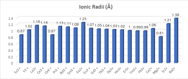
The separation of these lanthanoids is complicated because of the similar physicochemical properties of their predominating three plus (3+, or III) ions. The ionic radii decrease only 0.19 Å between LaIII and LuIII, leading to these metals co-occurring in rare-earth-bearing minerals.  

Data source for ionic radii (Shannon, R.D., 1976; Mattocks et al. 2023.).

Recently, in 2018, Cotruvo et al. reported the identification of a new highly selective lanthanum (LnIII)-binding protein in the model methylotroph, Methylobacterium extorquens, which the researchers named lanmodulin. Lanmodulin is a high-affinity lanthanide-binding protein identified in the bacterium Methylobacterioum extorquens. This lanthanide-modulated protein contains metal coordination motifs known as EF-hands. EF-hands are associated with nanomolar- to millimolar-affinity towards calcium ions. However, lanmodulin responds to lanthanum ions with a 108-fold selectivity over calcium ions. Lanthanum (La) has an atomic number of 57; its usual oxidation state is three plus (3+). A biological role for La in humans is not known but is essential in some bacteria
.

The research group purified the protein from M. extorquens AM1 cells grown in 1 μM LaCl3 and methanol. Classical ammonium sulfate precipitation followed by cation exchange chromatography (CIX) and size exclusion chromatography (SEC) resulted in a highly purified protein. After sequence analysis, the scientists expressed the protein in E. coli for biochemical characterization studies.

Mattocks et al., in 2019, created a highly selective, genetically encoded fluorescent sensor for lanthanoids called LaMP1. The researchers created LaMP1 by replacing the CaM and M13 domains of the FRET-based biosensor Cameleon D2 (ECFP and Citrine, λex = 433 nm; λem = 475 nm, λem = 529 nm) with lanmodulin (A22–R133).

LaMP1 showed FRET ratio changes (Rf/R0) from ~5.4 to 6.7 with picomolar affinity (Kd = 9.4–44 pM) for all the rare earth elements and selectivity over Ca2+ (Rf/R0 ~ 2.9; Kd = 1.2 mM) at pH 7.2. Little to no interference was observed from environmentally and biologically relevant cations, including Fe3+, Al3+, Mn2+, Cu2+, Mg2+, Na+, and K+. As a result, LaMP1 can be used as a probe for extracellular and intracellular Ln3+ ion homeostasis in live M. extorquens using a fluorescence plate reader assay.

Also in 2019, Cook et al. reported the NMR solution structure of lanmodulin complexed with yttrium III [PDB ID 6MI5]The structure revealed an unusual fusion of adjacent EF-hand motifs, resulting in a compact fold of EF-hand motifs. Also, an additional carboxylate ligand contributes to the protein's picomolar affinity for lanthanide ions, suggesting a role of unusual N i+1-H···N i hydrogen bonds, in which lanmodulin's EF-hand proline residues participate in selective lanthanide ion recognition.

The EF-hand motif is a common calcium-binding motif found in many proteins. The lanmodulin sequence contains four predicted EF-hand motifs: EF1, EF2, EF3, and EF4. The protein binds three equivalents of lanthanum 3+ ions with picomolar affinity and a fourth with approximately micromolar affinity.

The research group cloned the lanmodulin protein using the predicted sequence [residues 1−21 are a signal peptide].

Protein sequence:

>WP_253389665.1 EF-hand domain-containing protein [Methylorubrum extorquens]
MAFRLSSAVLLAALVAAPAYAAPSTTTKVDIAAFDPDKDGTIDLKEALAAGSAAFDKLDPDKDGTLDAKE
 Signal peptide                         EF1                       EF2

LKGRVSEADLKKLDPDNDGTLDKKEYLAAVEAQFKAANPDNDGTIDARELASPAGSALVNLIR
                  EF3                         EF4  
Cloned protein:

>pdb|6MI5|X Chain X, Lanmodulin

PTTTTKVDIAAFDPDKDGTIDLKEALAAGSAAFDKLDPDKDGTLDAKELKGRVSEADLKKLDPDNDGTLD KKEYLAAVEAQFKAANPDNDGTIDARELASPAGSALVNLIRHHHHHH 

Cook et al. generated a construct for the expression of the protein in Escherichia coli comprising an N-terminal methionine residue, residues 22−133, and a C-terminal hexa-histidine tag. However, according to mass spectrometric analysis, the first two residues (Met and Ala22) are cleaved during expression. The construct exhibited metal-dependent secondary structural changes and Ln3+ affinities comparable to untagged, wild-type lanmodulin.

In 2023, Mattocks et al. reported X-ray structures of lanmodulin constructs in complex with neodymium 3+ ions [PDB ID 8FNS], dysprosium 3+ ions at pH 7 [PDB ID 8FNR], and lanthanum 3+ ions at pH 7 [PDB ID 8DQ2].


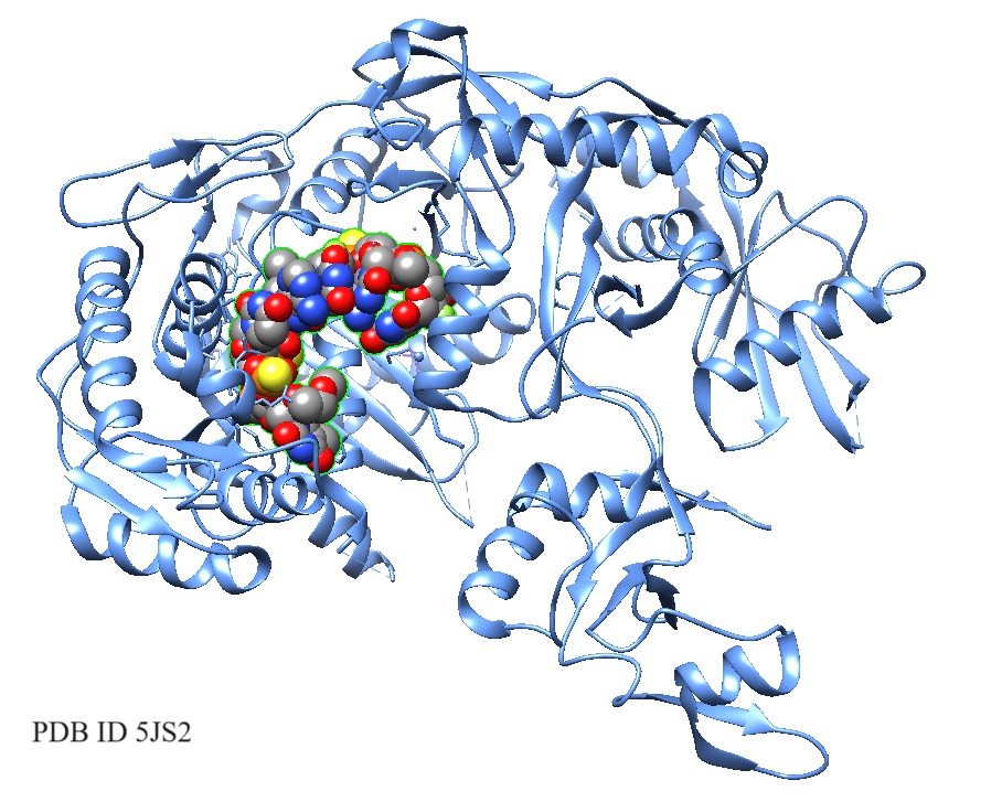
Figure 1: Lanmodulin in complex with neodymium.

PDB ID 8FNS


[X-ray crystal structure 8FNS ]


Reference

Cook EC, Featherston ER, Showalter SA, Cotruvo JA Jr. Structural Basis for Rare Earth Element Recognition by Methylobacterium extorquens Lanmodulin. Biochemistry. 2019 Jan 15;58(2):120-125. [ACS] [PDB ID: 6MI5].

Cotruvo, J. A., Jr., Featherston, E. R., Mattocks, J. A., Ho, J. V., and Laremore, T. N. (2018) Lanmodulin: A highly selective lanthanide-binding protein from a lanthanide-utilizing bacterium. J. Am. Chem. Soc., 2018, 140, 44, 15056-15061 [
ACS].

Lewit-Bentley A, Réty S. EF-hand calcium-binding proteins. Curr Opin Struct Biol. 2000 Dec;10(6):637-43. [
Sciencedirect]. 

Mattocks, J.A., Ho, J.V. and Cotruvo, J.A.Jr. (2019) J. Am. Chem. Soc., 141, 2857–2861. [PubMed]

Mattocks JA, Jung JJ, Lin CY, Dong Z, Yennawar NH, Featherston ER, Kang-Yun CS, Hamilton TA, Park DM, Boal AK, Cotruvo JA Jr. Enhanced rare-earth separation with a metal-sensitive lanmodulin dimer. Nature. 2023 Jun;618(7963):87-93. [
PMC]

Shannon, R.D., 1976. Revised effective ionic radii and systematic studies of interatomic distances in halides and chalcogenides. Acta Crystallogr. A32, 751–767.  [
AC

Snyder EE, Buoscio BW, Falke JJ. Calcium(II) site specificity: effect of size and charge on metal ion binding to an EF-hand-like site. Biochemistry. 1990 Apr 24;29(16):3937-43. [
PMC]

---...---
Rare-earth element-capture peptides!

$
0
0

Landmodulin protein- and peptide-based ion exchange chromatography may allow the capture, separation, and purification of rare-earth elements.

According to the
U.S. Secretary of Energy Rick Perry, rare-earth elements are vital for developing and manufacturing high-tech devices, including computers, cell phones, and other similar devices. Coal samples analyzed from Illinois, Northern Appalachian, Central Appalachian, Rocky Mountain Coal Basin, and the Pennsylvania Anthracite region contained high rare-earth element concentrations at greater than 300 parts per million (ppm). However, extracting and separating individual rare earth elements from rare-earth elements containing sources is challenging.

Recently, Dong et al., in 2021, reported a water-based extraction method utilizing ion exchange column chromatography with covalently conjugated lanmodulin to capture and separate rare-earth elements. The bioconjugation of biological molecules such as proteins or peptides, as well as oligonucleotides, for example, aptamers, is now a standard method in biochemistry and biotechnology.

The research group reported that immobilized lanmodulin-based column chromatography achieved a high-purity separation of the clean-energy-critical rare-earth element (REE) pair neodymium and dysprosium (Nd/Dy) from low-grade leachate (0.043 mol % REEs) into separate fractions of heavy and light REEs (88 mol % purity of total REEs) during a single column run at high capacity.

Dong et al. performed a proof-of-concept high-purity separation between REE pairs (neodymium/dysprosium (Nd/Dy), and yttrium/neodymium (Y/Nd)) and grouped separation between heavy REEs (HREE, terbium-lutetium and yttrium (Tb–Lu + Y)) and light REEs (LREE, lanthanum-gadolinium (La–Gd)). The reported approach combines primary REE extraction from non-REEs with a secondary separation between heavy and light REEs within a single, water-based adsorption/desorption cycle.


This method simplifies further processing and provides the basis for a  protein- or peptide-based method for efficient REE extraction, concentration, and separation from both high- and low-grade rare-earth sources.

Gutenthaler et al., in 2022, showed that the four short EF-hand motif peptides behaved similarly with affinities in the micromolar range for europium (EuIII) and terbium (TbIII). However, calcium ions did not bind to the peptides, as verified with circular dichroism spectroscopy.

The researchers investigated the binding characteristics of the four peptides using isothermal titration calorimetry (ITC), time-resolved laser-induced fluorescence spectroscopy (TRLFS), and molecular dynamics (MD) simulations. The following table lists observed Kd values.

Sequences comparison of the four EF hands of Mex- and Hans-LanMs

Residues canonically involved in metal binding in EF hands are in red; Pro residues are in purple.


EF-hand motifs LanM M. extorquens


EF-hand motifs LanM H. quercus

 DPDKDGTIDLKE

 DPDKDGTIDAKE

 DPDNDGTIDKKE

 NPDNDGTIDARE

 NKDNDDSLEIAE

 NPDGDTTLESGE

 NKDGDQTLEMDE

 DANKDGKLTAAE

 
Residues canonically involved in metal binding in EF hands are in blue; Proline residues are in purple.

The study showed that the lanthanoid affinity is also preserved in these short peptide sequences. However, the very high affinity of lanmodulin, reported to be in the picomolar range, was not observed for its EF-Hand loop peptides. The short peptides had affinities in the micromolar range, similar to the average affinity of the four calmodulin binding sites, known to also have a higher affinity for Eu(III) and Cm(III) over Ca(II).

More recently, in 2023, Mattocks et al. reported the characterization of a new lanmodulin protein from Hansschlegelia quercus the researchers called Hans-LanM. The researchers found that the oligomeric state of this protein is sensitive to the radii of rare-earth ions and that the lanthanum (III)-induced dimer is >100-fold tighter than the dysprosium (III)-induced dimer. X-ray crystal structures revealed how picometre-scale differences in the radius between lanthanum (III) and dysprosium (III) modulate the Hans-LanM’s quaternary structure through a carboxylate shift that rearranges a second-sphere hydrogen-bonding network.

The comparison of the lanmodulin sequence from Methylorubrum extorquens with the lamodulin sequence from Hansschlegelia quercus revealed a distinct metal coordination explaining Hans-LanM’s selectivity within the rare-earth elements. Structure-guided mutagenesis of a critical residue at the Hans-LanM dimer interface allowed modulation of the dimerization in solution, resulting in single-stage, column-based separation of neodymium (III)/dysprosium (III) mixtures to >98% individual element purity. Hans-LanM exists in a monomer/dimer equilibrium, modulated by the presence of the specific rare-earth ion bound.

However, for practical applications in medicine, for recycling and separation of rare-earth elements, further optimization may be needed.


Reference

EF-hand motif 

Dong Z, Mattocks JA, Deblonde GJ, Hu D, Jiao Y, Cotruvo JA Jr, Park DM. Bridging Hydrometallurgy and Biochemistry: A Protein-Based Process for Recovery and Separation of Rare Earth Elements. ACS Cent Sci. 2021 Nov 24;7(11):1798-1808. [
PMC]

Gutenthaler SM, Tsushima S, Steudtner R, Gailer M, Hoffmann-Röder A, Drobot B, Daumann LJ. Lanmodulin peptides - unravelling the binding of the EF-Hand loop sequences stripped from the structural corset. Inorg Chem Front. 2022 Jun 30;9(16):4009-4021. [
PMC]

Mattocks JA, Jung JJ, Lin CY, Dong Z, Yennawar NH, Featherston ER, Kang-Yun CS, Hamilton TA, Park DM, Boal AK, Cotruvo JA Jr. Enhanced rare-earth separation with a metal-sensitive lanmodulin dimer. Nature. 2023 Jun;618(7963):87-93. [
PMC]

U.S. Secretary of Energy

---...---

How does the cytoplasmic microtubule-based motor dynein move?

$
0
0

The microtubule-based motor protein cytoplasmic dynein is involved in transporting a diverse array of cargo, allowing cells to organize their contents, move, divide, and respond to stimuli. Dyneins are a family of "ATPases Associated with diverse cellular Activities" (AAA+) motors that move towards the minus end of microtubules.

Dynein holoenzymes comprise two ~500 kDa motor (or "head") proteins containing heavy chain subunits and at least six other polypeptides.


Cytoplasmic dynein transports intracellular cargos in interphase cells and mediates spindle assembly and chromosome positioning during cell division. Other dyneins transport cargo molecules in cilia and power cilia beating. Historically, dyneins are the least studied cytoskeletal motor proteins due to the difficulties in studying active dynein complexes.

In 2012 Qui et al. reported a new model for studying the dynein stepping mechanism. The researchers combined two-color, single-molecule microscopy with high-precision, two-dimensional tracking for the study. The study found that dynein has a variable stepping pattern distinct from other cytoskeletal motor proteins. Processive cytoskeletal motors usually use "hand-over-hand" mechanisms. Whereas dynein has a stochastic or randomly determined hard-to-predict stepping pattern when its two motor domains are close together. According to the study, coordination emerges as the distance between motor domains increases. A tension-based mechanism governs these steps, allowing the tuning of dynein for all its cellular functions.

To enable the study, the researchers performed a two-dimensional analysis of dynein stepping by tracking GST-dynein homodimers labeled with a single Qdot 655 placed on either tail domain via an N-terminal HaloTag or a single motor domain containing a C-terminal HaloTag. The study found that dynein's step size was larger than the previously reported 1-D step size, and many steps had an off-axis component. Also, steps were equally likely to be to the left or the right. Dynein's stepping behavior showed significant variability and flexibility. In the off-axis component, some steps are backward. Dynein's two motor domains can step independently. Qui et al. proposed that this flexibility allows dynein to navigate a crowded cytoplasm and obstacles on microtubules, providing a molecular explanation for the observation that dynein can better navigate obstacles than kinesin motors. According to Qui et al., this apparent plasticity of the dynein stepping mechanism suggests that additional layers of regulation may be used to accomplish different cell biological functions. In eukaryotic cells, dynein transports dozens, if not hundreds, of varying cargo, but there is only a single gene encoding cytoplasmic dynein 1 in all sequenced eukaryotic genomes.

Bio-Synthesis Inc. prepared the Cy3B-Halotag.

Reference

Qiu W, Derr ND, Goodman BS, Villa E, Wu D, Shih W, Reck-Peterson SL. Dynein achieves processive motion using both stochastic and coordinated stepping. Nat Struct Mol Biol. 2012 Jan 8;19(2):193-200. [PMC]


Recent methods for the analysis of miRNA pathways

$
0
0

Micro-RNAs (miRNAs) are small, single-stranded, non-coding ribonucleic acids (RNAs). First discovered in 1993 in C. elegans, they are now known to take part in regulating gene expression.

In recent years, scientists uncovered fundamental information on miRNA machinery's structural and molecular dynamics. For example, how the transcriptome selects miRNA substrates and targets and the regulation of miRNA biogenesis and turnover. Recent technological advances include massive parallel assays, cryogenic electron microscopy, single-molecule imaging, and CRISPR-Cas9 screening. These technological advances contributed heavily to the latest discoveries. Scientists now know that miRNAs regulate gene expression and control cell development and metabolism.


Scientists estimate that humans express more than 2,000 mature miRNAs; some appear to be associated with cancer, cardiovascular, inflammatory diseases, and a broad range of neurodevelopmental and autoimmune disorders. Furthermore, miRNAs are considered important biomarkers for disease assessment.


miRNAs are secreted to extracellular fluids, bound to specific proteins, or are part of extracellular vesicles. Through RNA sequencing, next-generation RNA sequencing enables their detection in liquid biopsies and biological fluids, including plasma and serum, saliva, cerebrospinal fluid, and breast milk.


Technologies recently applied for the analysis of miRNA pathways

High-throughput substrate screening

This screening method is similar to classical in vitro selection assays. The method often involves parallel processing of a large pool of endogenous substrates or a library of designed or randomized variants. Deep sequencing is used as a read-out, to infer functionally relevant features for processing. This approach allows characterization of pri-miRNA and pre-miRNA features (Auyeung et al. 2013; Fang et al. 2015; Li et al. 2020; Lee et al. 2023; Nguyen et al. 2022).

Reference

Auyeung, V. C., Ulitsky, I., McGeary, S. E. & Bartel, D. P. Beyond secondary structure: primary-sequence determinants license pri-miRNA hairpins for processing. Cell 152, 844–858 (2013). This study is the first to utilize massively parallel substrate assays to reveal motifs involved in pri-miRNA processing.

Fang, W. & Bartel, D. P. The menu of features that define primary microRNAs and enable de novo design of microRNA genes. Mol. Cell 60, 131–145 (2015).

Lee, Y. Y., Kim, H. & Kim, V. N. Sequence determinant of small RNA production by DICER. Nature 615, 323–330 (2023).

Li, S., Nguyen, T. D., Nguyen, T. L. & Nguyen, T. A. Mismatched and wobble base pairs govern primary microRNA processing by human microprocessor. Nat. Commun. 11, 1926 (2020).

Nguyen, T. D., Trinh, T. A., Bao, S. & Nguyen, T. A. Secondary structure RNA elements control the cleavage activity of DICER. Nat. Commun. 13, 2138 (2022).

Cryogenic electron microscopy (cryo-EM)

Cryo-EM uses an electron beam for imaging specimens under cryogenic conditions. Data processing of electron microscopy densities allows the assignment of atomic coordinates. Cryo-EM is suitable for studying proteins or complexes of large molecular weight molecules. Relatively small samples are needed, and the method can capture multiple conformational states in a single experiment without crystallization.

Cryo-EM allowed the study of the Mammalian Drosha/DGCR8 complex (Jin et al. 2020; Partin et al. 2020), Mammalian Dicer complexes (Liu et al 2018; Lee et al. 2023; Zapletal et al. 2022), Drosophila Dicer complexes Dicer-1 (Jouravleva et al. 2022) and Dicer-2 (Yamaguchi et al 2022; Su et al 2022), and Arabidopsis Dicer complexes: DCL1 (Wei et al 2021) and DCL3 (Wang et al. 2021).

Reference


Jin, W., Wang, J., Liu, C. P., Wang, H. W. & Xu, R. M. Structural basis for pri-miRNA recognition by Drosha. Mol. Cell 78, 423–433.e5 (2020).

Jouravleva, K. et al. Structural basis of microRNA biogenesis by Dicer-1 and its partner protein Loqs-PB. Mol. Cell 82, 4049–4063.e6 (2022).

Lee, Y. Y., Lee, H., Kim, H., Kim, V. N. & Roh, S. H. Structure of the human DICER–premiRNA complex in a dicing state. Nature 615, 331–338 (2023).

Liu, Z. et al. Cryo-EM structure of human dicer and its complexes with a pre-miRNA substrate. Cell 173, 1191–1203.e12 (2018).

Partin, A. C. et al. Cryo-EM structures of human Drosha and DGCR8 in complex with primary microRNA. Mol. Cell 78, 411–422 e414 (2020). Together with Jin et al. (2020), this work is the first cryo-EM study of Microprocessor structures.

Su, S. et al. Structural insights into dsRNA processing by Drosophila Dicer-2–Loqs-PD. Nature 607, 399–406 (2022).

Wang, Q. et al. Mechanism of siRNA production by a plant Dicer–RNA complex in dicing-competent conformation. Science 374, 1152–1157 (2021).

Wei, X. et al. Structural basis of microRNA processing by Dicer-like 1. Nat. Plants 7, 1389–1396 (2021).

Yamaguchi, S. et al. Structure of the Dicer-2–R2D2 heterodimer bound to a small RNA duplex. Nature 607, 393–398 (2022).

Zapletal, D. et al. Structural and functional basis of mammalian microRNA biogenesis by Dicer. Mol. Cell 82, 4064–4079.e13 (2022). Together with Lee et al. (Nature, 2023), this work reports new cryo-EM structures for active mammalian Dicer complex.

Single-molecule assay

Single-molecule assays allow the analysis of real-time dynamics of biological reactions. In-vitro assays usually need purified materials. Observation times depend on reaction kinetics, photostability, and the lifetime of fluorophores. However, some reaction times can be as short as microseconds. The detection of single molecules is generally limited by diffraction (~200–300 nm spatial resolution). For example, the single-molecule Förster resonance energy transfer (smFRET) operates at 1 to 10 nm. smFRET allows resolving intermolecular and intramolecular motions. Fluorescently tagged molecules utilizing multimeric tags or scaffolds for enhanced detection enable single-molecule imaging in living cells.

Single-molecule assays allowed for studying the dynamic interplay of human Dicer and TRBP (Fareh et al. 2016), in-vitro target search and interrogation of human Ago2/RISC complexes (Solomon et al. 2015; Yao et al. 2015; Chandradoss et al. 2015; Cui et al. 2019; Willkomm et al. 2022), live cell imaging of targeting and regulation by human Ago2 (Ruijtenberg et al. 2020; Cialek et al., 2022; Kobayashi & Singer, 2022), and the assembly and dynamics of Drosophila AGO2/RISC complexes (Iwasaki et al. 2015; Tsuboyama et al. 2018).

Reference

Chandradoss, S. D., Schirle, N. T., Szczepaniak, M., MacRae, I. J. & Joo, C. A dynamic search process underlies microRNA targeting. Cell 162, 96–107 (2015). This study uses the distance sensing capability of smFRET to visualize strategies of target interrogation by RISC.

Cialek, C. A. et al. Imaging translational control by Argonaute with single-molecule resolution in live cells. Nat. Commun. 13, 3345 (2022).

Cui, T. J. et al. Argonaute bypasses cellular obstacles without hindrance during target search. Nat. Commun. 10, 4390 (2019).

Fareh, M. et al. TRBP ensures e%icient Dicer processing of precursor microRNA in RNA-crowded environments. Nat. Commun. 7, 13694 (2016).

Iwasaki, S. et al. Defining fundamental steps in the assembly of the Drosophila RNAi enzyme complex. Nature 521, 533–536 (2015).

Kobayashi, H. & Singer, R. H. Single-molecule imaging of microRNA-mediated gene silencing in cells. Nat. Commun. 13, 1435 (2022).

Ruijtenberg, S. et al. mRNA structural dynamics shape Argonaute–target interactions. Nat. Struct. Mol. Biol. 27, 790–801 (2020).

Salomon, W. E., Jolly, S. M., Moore, M. J., Zamore, P. D. & Serebrov, V. Single-molecule imaging reveals that argonaute reshapes the binding properties of its nucleic acid guides. Cell 162, 84–95 (2015).

Tsuboyama, K., Tadakuma, H. & Tomari, Y. Conformational activation of argonaute by distinct yet coordinated actions of the Hsp70 and Hsp90 chaperone systems. Mol. Cell 70, 722–729.e4 (2018).

Willkomm, S. et al. Single-molecule FRET uncovers hidden conformations and dynamics of human Argonaute 2. Nat. Commun. 13, 3825 (2022). This study uses single-molecule fluorescence to dissect internal motions within human Ago2 during transitions from guide RNA binding to target capture.

Yao, C., Sasaki, H. M., Ueda, T., Tomari, Y. & Tadakuma, H. Single-molecule analysis of the target cleavage reaction by the Drosophila RNAi enzyme complex. Mol. Cell 59, 125–132 (2015).

RNA bind-n-seq (RBNS)

This method yields relative quantitative binding affinities of an RNA-binding protein (RBP) across a library of target sites. Typically, a purified RBP is incubated with a randomized pool of RNAs. Co-purified RBP targets are then analyzed by deep sequencing to identify their features.

The method was recently applied to study Ago2–miRNA complex binding affinity to target RNAs (McGeary et al. 2019 and 2022).

Reference

McGeary, S. E. et al. The biochemical basis of microRNA targeting efficacy. Science https://doi.org/10.1126/science.aav1741 (2019). This paper reveals broad features of miRNA–target interactions using RBNS.

 McGeary, S. E., Bisaria, N., Pham, T. M., Wang, P. Y. & Bartel, D. P. microRNA 3′-compensatory pairing occurs through two binding modes, with affinity shaped by nucleotide identity and position. eLife https://doi.org/10.7554/eLife.69803 (2022).

CRISPR–Cas9 screening

Genetic mutagenesis screens enable the identification of factors involved in a cellular process of interest. miRNA screening often incorporates a specific reporter as a functional read-out, allowing cell sorting before deep sequencing enriched or depleted guide RNAs.

ERH and SAFB2 in miRNA cluster assistance ZSWIM8 in target-directed miRNA degradation (TDMD) (Shie et al. 2020; Han et al. 2020).

Reference

Shi, C. Y. et al. The ZSWIM8 ubiquitin ligase mediates target-directed microRNA degradation. Science https://doi.org/10.1126/science.abc9359 (2020).

Han, J. et al. A ubiquitin ligase mediates target-directed microRNA decay independently of tailing and trimming. Science https://doi.org/10.1126/science.abc9546 (2020). Together with Shi et al. (2020), this work reveals the regulatory mechanism of TDMD, via ZSWIM8-mediated Argonaute protein degradation.

Legend: Ago, Argonaute; miRNA, microRNA; pre-miRNA, precursor miRNA; pri-miRNA, primary miRNA; RISC, RNA-induced silencing complex; TRBP, transactivation responsive RNA-binding protein.

---...---
Bio-Synthesis provides a full spectrum of bio-conjugation services including high quality custom oligonucleotide modification services, back-bone modifications, conjugation to fatty acids and lipids, cholesterol, tocopherol, peptides as well as biotinylation by direct solid-phase chemical synthesis or enzyme-assisted approaches to obtain artificially modified oligonucleotides, such as BNA antisense oligonucleotides, mRNAs, miRNA, or siRNAs, containing a natural or modified backbone, as well as base, sugar and internucleotide linkages.

Bio-Synthesis also provides biotinylated mRNA and long circular oligonucleotides.
---...---

Cell-penetrating peptides for the delivery of siRNA into cells

$
0
0

The number of efficient delivery systems for therapeutic molecules into cells still needs to be increased. SiRNA introduced into cells drives the RNA interference (RNAi) process. Because of the high molecular weight and the negative charge of the phosphate backbone, siRNA molecules cannot readily cross the cell membrane, presently limiting the clinical and therapeutic value of siRNAs.

Research projects are ongoing to find the best approaches for delivering siRNAs through the cell membrane into the cytoplasm.


Cell-penetrating peptides (CPPs) can trigger the movement of molecules across cell membranes, including mitochondrial and nuclear membranes, without destroying them. These short peptides can translocate and transport cargo molecules into the intracellular space by crossing biological membranes. Complexed or conjugated to siRNA oligonucleotides, CPPs may offer the design of efficient cell delivery systems for enhanced delivery of oligonucleotide-based therapeutics.


The plasma membrane protects cells against the entrance of external molecules. The plasma membrane separates the intracellular cytosol from the extracellular environment to maintain cellular homeostasis. This barrier makes accessing cells difficult for extracellular hydrophilic molecules such as peptides, proteins, and nucleic acids.

A variety of strategies are applied to overcome this permeability issue. Historically, viral and non-viral approaches have been the primary delivery strategies. Viral vectors utilized for gene therapy allow the delivery of genetic material across the cell membrane with high efficacy.

Besides viral delivery, electroporation, encapsulation, and association of drugs with lipids, peptides, polymers, nanotubes, liposomes, micelles, and dendrimers have been investigated for their ability to deliver therapeutic oligonucleotides into cells with high efficiency.

Still, the use of vectors encounters several challenges, such as limited cargo-carrying capacity, low delivery efficiency, the risk of mutation, high cytotoxicity, and lack of target specificity. Also, viral vectors are incompatible with all kinds of nucleic acid-based molecules, for example, the delivery of short synthetic Oligonucleotides.

Also, scientists must overcome environmental and enzymatic degradation to develop an efficient therapeutic siRNA delivery system.

siRNAs are double strand (ds) RNAs, ~19–23 base pairs with a characteristic 3′- overhang that facilitates the recognition by the RNAi enzymatic machinery.

Usually, in RNAi, Dicer cleaves long dsRNAs into siRNAs. siRNAs then bind to the RNA-induced silencing complex (RISC), which unwinds the siRNAs and removes the sense strand for degradation by the cell’s nucleases. The antisense strand targets specific mRNA sequences, directs them to the RISC, and anneals with the complementary base pair. Finally, the targeted mRNA is rapidly degraded, resulting in a decrease in protein expression.

The following figures show structural models of siRNA gleaned from crystal structures of siRNA protein complexes.
 

5JS2:  Human Argonaute-2 Bound to a Modified siRNA.

Schirle NT, Kinberger GA, Murray HF, Lima WF, Prakash TP, MacRae IJ. Structural Analysis of Human Argonaute-2 Bound to a Modified siRNA Guide. J Am Chem Soc. 2016 Jul 20;138(28):8694-7. [PMC]


Schirle et al., in 2016, solved the crystal structure of human Ago2 bound to a metabolically stable siRNA containing extensive backbone modifications. Comparison to the structure of an equivalent unmodified-siRNA complex indicated that the structure of Ago2 is relatively unaffected by chemical modifications in the bound siRNA. 

2F8S: Crystal structure of Aa-Ago with externally-bound siRNA.

Yuan YR, Pei Y, Chen HY, Tuschl T, Patel DJ. A potential protein-RNA recognition event along the RISC-loading pathway from the structure of A. aeolicus Argonaute with externally bound siRNA. Structure. 2006 Oct;14(10):1557-65. [PMC]


siRNA structural model from the crystal structures of Aa-Ago bound to 22-mer and 26-mer self-complementary siRNAs, identified as an externally bound siRNA-Ago complex.


The 2 nt 3′ overhang at one end of the siRNA inserts into a cavity positioned on the outer surface of the PAZ-containing lobe of the bilobal Aa-Ago architecture in both complexes.


Cell-penetrating peptide for cellular siRNA delivery

Cell-penetrating peptides (CPPs) are small peptides that trigger the movement of molecules across cell membranes. CPPs can penetrate the cell membrane and mitochondrial and nuclear membranes without destroying them. These short peptides can translocate and transport cargo molecules into the intracellular space by crossing biological membranes.

Known CPPs are all water soluble, relatively small, do not have more than 35 amino acid residues, and have a very low cytotoxicity. However, there exists no unique classification for these peptides. Categorization can be done according to their origin and ability to link with cargo molecules or structures.

Classification of CPPs by their structure divides CPPs into (1) cationic CPPs, (2) amphipathic CPPs, and (3) hydrophobic CPPs.

A list of CPPS is found 
here.

A ribbon model and a surface model of the cell-penetrating peptide RW16 is shown in the following figures.

 6RQS: RW16 peptide


>pdb|6RQS|A Chain A, ARG-ARG-TRP-ARG-ARG-TRP-TRP-ARG-ARG-TRP-TRP-ARG-ARG-TRP-ARG-ARG


XXRRWRRWWRRWWRRWRR


Jobin ML, Vamparys L, Deniau R, Grélard A, Mackereth CD, Fuchs PFJ, Alves ID. Biophysical Insight on the Membrane Insertion of an Arginine-Rich Cell-Penetrating Peptide. Int J Mol Sci. 2019 Sep 9;20(18):4441.  [PMC]


Mechanism of membrane insertion

Jobin et al. reported that side chain contacts, in both buried and solvent-exposed positions, contribute to the peptide's conformational stability and function. Three tryptophanes (Ws) form a hydrophobic pocket surrounding Arg15, masking the positive charges of the Arg (R) residue. The pairing of aromatic and polar residues decreased the energetic barrier for the motion of cationic side chains through a lower dielectric environment like the cell membrane bilayer. Similar π-cation interactions are also present in penetratin between R and multiple W residues.


According to Singh et al., conjugation of siRNA to most unmodified CPPs did not generate desirable effects due to either lack of serum stability, endosomal entrapment, or some other factor. However, chemical modifications of the CPPs and siRNAs enhanced the transfection efficiency.


Compared to other methods, CPP-mediated delivery methods may have advantages in terms of efficiency, siRNA-carrying capacity, and biocompatibility.

Unfortunately, our understanding of the CPP internalization pathway is complicated and needs to be fully understood.

Further research is needed to dissect the mechanism of CPP cellular uptake for designing CPPs with enhanced delivery and cell penetration capabilities. Overall, CPPs could provide new opportunities for systemic siRNA delivery.

Reference

Edinger, Daniel. "SiRNA Therapy for Cancer." 2013 Ph.D. Thesis.

Rathnayake PV, Gunathunge BG, Wimalasiri PN, Karunaratne DN, Ranatunga RJ. Trends in the Binding of Cell Penetrating Peptides to siRNA: A Molecular Docking Study. J Biophys. 2017; 2017:1059216. [PMC]

Singh T, Murthy ASN, Yang HJ, Im J. Versatility of cell-penetrating peptides for intracellular delivery of siRNA. Drug Deliv. 2018; 25(1):1996-2006. [PMC]

Takeuchi T, Futaki S. Current Understanding of Direct Translocation of Arginine-Rich Cell-Penetrating Peptides and Its Internalization Mechanisms. Chem Pharm Bull (Tokyo). 2016;64(10):1431-1437. [article].


---...---
Bio-Synthesis provides a full spectrum of bio-conjugation services including high quality custom oligonucleotide modification services, back-bone modifications, conjugation to fatty acids and lipids, cell-penetrating peptides, cholesterol, tocopherol, other peptides as well as biotinylation by direct solid-phase chemical synthesis or enzyme-assisted approaches to obtain artificially modified oligonucleotides, such as BNA antisense oligonucleotides, mRNAs, miRNA, or siRNAs, containing a natural or modified backbone, as well as base, sugar and internucleotide linkages.
Bio-Synthesis also provides biotinylated mRNA and long circular oligonucleotides.
---...---

Therapeutic Antisense Oligonucleotides (ASOs) and their Modifications

$
0
0

Antisense oligonucleotides (ASOs) are short, synthetic strands of DNA or RNA that bind to specific RNA targets and modulate gene expression. ASOs can inhibit gene expression, promote gene expression, or alter gene splicing.

Mechanism of Action

There are two main mechanisms of action for ASOs:

[1] RNase H-dependent degradation

The enzyme RNase H degrades RNA when hybridized to DNA. ASOs complementary to a target RNA can bind to it and form a hybrid duplex. RNase H then recognizes the duplex and degrades the RNA.

[2] Steric blocking

Steric blocking ASOs bind to targeted RNA, such as mRNA, and prevent it from interacting with other molecules, for example, with ribosomes or splicing factors. ASOs can block the translation of the RNA into a protein or alter RNA splicing.


Applications of ASOs

ASOs potentially allow the development of therapeutics for a wide range of diseases, including:

Genetic disorders: ASOs allow targeting and silencing mutant genes that cause genetic disorders, such as Duchenne muscular dystrophy and spinal muscular atrophy.

Infectious diseases: ASOs enable the targeting and silencing of viral RNAs, such as those of SARS-CoV-2 (COVID-19), HIV, and hepatitis C virus.

Cancer: Well-designed ASOs targeting oncogenes that promote cancer growth and progression silencing these genes.

Cardiovascular diseases: ASOs allow the targeting and silencing of disease-causing genes, including cardiovascular diseases, atherosclerosis, or heart failure.

Neurodegenerative diseases: ASOs enable the development of oligonucleotide-based therapeutics to treat neurodegenerative diseases including Alzheimer's disease and Huntington's disease.

Advantages of ASOs

ASOs appear to have several advantages over other gene therapy approaches:

ASOs are highly specific and allow targeting of any RNA sequence.

ASOs are relatively easy to synthesize and deliver to cells.

ASOs exhibit an excellent safety profile and are generally well-tolerated.

Challenges to Solve

However, ASOs are still under development. Before the use of ASOs in clinical practice, several challenges will need to be overcome. ASOs are unstable in the bloodstream and are readily degraded by enzymes. Also, to be effective, delivery to the target cells is essential. 

Researchers are in progress to develop new ASO chemistries and delivery methods to overcome these challenges. For example, researchers have developed ASOs that are resistant to degradation and that can be conjugated to carrier molecules for delivery into specific cells.

Therapeutic Antisense Oligonucleotides

Antisense oligonucleotide (ASO) therapeutics utilize short, synthetic DNA or RNA oligonucleotides to modulate gene expression for gene therapies. ASOs bind to specific RNA targets and either inhibit or promote gene expression or alter RNA splicing.


ASO therapeutics are relatively easy to synthesize, very specific, and have a good safety profile. The medical community investigates ASOs for a wide range of diseases, including genetic disorders, infectious diseases, cancer, cardiovascular diseases, and neurodegenerative diseases.


Examples of ASO therapeutics that are currently approved or in clinical trials are:

Spinal muscular atrophy (SMA): Nusinersen (Spinraza) is an ASO approved to treat SMA, a genetic disorder that causes motor neuron loss and muscle weakness. Nusinersen works by increasing the production of a protein called SMN, which is essential for motor neuron health.

Duchenne muscular dystrophy (DMD): Eteplirsen (Exondys 51) and Viltolarsen (Viltepso) are approved to treat DMD, which causes progressive muscle weakness and death. Eteplirsen and Viltolarsen promote the skipping of a specific exon in the dystrophin gene, allowing the production of a functional dystrophin protein.

Transthyretin amyloidosis (ATTR): Inotersen (Tegsedi) and Patisiran (Onpattro) are approved to treat ATTR. This rare genetic disorder causes amyloid deposits to form in various tissues throughout the body. Inotersen and Patisiran silence the transthyretin gene, which leads to a reduction in the production of transthyretin protein.


Hepatitis B virus (HBV): Givosiran (Givlaari) is approved to treat chronic HBV infection in adults with compensated liver disease. Givosiran silences the HBV gene, reducing the production of the HBV virus.


COVID-19: Other ASOs are currently in clinical trials to treat COVID-19. These ASOs target the SARS-CoV-2 virus, which causes COVID-19, and prevent it from replicating.


The rapidly developing field of ASO therapeutics may enable the treatment of many diseases.

Review Articles

Adachi H, Hengesbach M, Yu YT, Morais P. From Antisense RNA to RNA Modification: Therapeutic Potential of RNA-Based Technologies. Biomedicines. 2021 May 14;9(5):550. [
PMC]

Anderson BA, Freestone GC, Low A, De-Hoyos CL, Iii WJD, Østergaard ME, Migawa MT, Fazio M, Wan WB, Berdeja A, Scandalis E, Burel SA, Vickers TA, Crooke ST, Swayze EE, Liang X, Seth PP. Towards next generation antisense oligonucleotides: mesylphosphoramidate modification improves therapeutic index and duration of effect of gapmer antisense oligonucleotides. Nucleic Acids Res. 2021 Sep 20;49(16):9026-9041. [
PMC]

Anthony K. RNA-based therapeutics for neurological diseases. RNA Biol. 2022;19(1):176-190. doi: 10.1080/15476286.2021.2021650. [
PMC]

Bennett CF, Swayze EE. RNA targeting therapeutics: molecular mechanisms of antisense oligonucleotides as a therapeutic platform. Annu Rev Pharmacol Toxicol. 2010;50:259-93. [
Annual Reviews]

Crooke ST, Witztum JL, Bennett CF, Baker BF. RNA-Targeted Therapeutics. Cell Metab. 2018 Apr 3;27(4):714-739. doi: 10.1016/j.cmet.2018.03.004. Erratum in: Cell Metab. 2019 Feb 5;29(2):501. [
Cell-Metabolism]

Crooke ST, Liang XH, Baker BF, Crooke RM. Antisense technology: A review. J Biol Chem. 2021 Jan-Jun;296:100416. doi: 10.1016/j.jbc.2021.100416. Epub 2021 Feb 16. PMID: 33600796; PMCID: PMC8005817. [
PMC]

Damase TR, Sukhovershin R, Boada C, Taraballi F, Pettigrew RI, Cooke JP. The Limitless Future of RNA Therapeutics. Front Bioeng Biotechnol. 2021 Mar 18;9:628137. [PMC]

Deleavey GF, Damha MJ. Designing chemically modified oligonucleotides for targeted gene silencing. Chem Biol. 2012 Aug 24;19(8):937-54. [Sciencedirect]

Egli M, Manoharan M. Chemistry, structure and function of approved oligonucleotide therapeutics. Nucleic Acids Res. 2023 Apr 11;51(6):2529-2573. [
PMC]

Gagliardi M, Ashizawa AT. The Challenges and Strategies of Antisense Oligonucleotide Drug Delivery. Biomedicines. 2021 Apr 16;9(4):433. [
PMC]

Gait MJ, Agrawal S. Introduction and History of the Chemistry of Nucleic Acids Therapeutics. Methods Mol Biol. 2022;2434:3-31. [PMC]

Goodchild, J.; 2011. Therapeutic Oligonucleotides, Methods and Protocols. MIMB, volume 764. [Springer Book]

Halloy F, Iyer PS, Ćwiek P, Ghidini A, Barman-Aksözen J, Wildner-Verhey van Wijk N, Theocharides APA, Minder EI, Schneider-Yin X, Schümperli D, Hall J. Delivery of oligonucleotides to bone marrow to modulate ferrochelatase splicing in a mouse model of erythropoietic protoporphyria. Nucleic Acids Res. 2020 May 21;48(9):4658-4671. [
PMC]

Hirabayashi Y, Maki K, Kinoshita K, Nakazawa T, Obika S, Naota M, Watanabe K, Suzuki M, Arato T, Fujisaka A, Fueki O, Ito K, Onodera H. Considerations of the Japanese Research Working Group for the ICH S6 & Related Issues Regarding Nonclinical Safety Assessments of Oligonucleotide Therapeutics: Comparison with Those of Biopharmaceuticals. Nucleic Acid Ther. 2021 Apr;31(2):114-125. [PMC]

Järver P, O'Donovan L, Gait MJ. A chemical view of oligonucleotides for exon skipping and related drug applications. Nucleic Acid Ther. 2014 Feb;24(1):37-47. [
PMC]

Juliano RL. The delivery of therapeutic oligonucleotides. Nucleic Acids Res. 2016 Aug 19;44(14):6518-48. [
PMC]

Ji D, Feng H, Liew SW, Kwok CK. Modified nucleic acid aptamers: development, characterization, and biological applications. Trends Biotechnol. 2023 Nov;41(11):1360-1384. [cell]

Kilanowska A, Studzińska S. In vivo and in vitro studies of antisense oligonucleotides - a review. RSC Adv. 2020 Sep 17;10(57):34501-34516. [
PMC]

Kilikevicius A, Meister G, Corey DR. Reexamining assumptions about miRNA-guided gene silencing. Nucleic Acids Res. 2022 Jan 25;50(2):617-634. [
PMC]

Matsuo M. Antisense Oligonucleotide-Mediated Exon-skipping Therapies: Precision Medicine Spreading from Duchenne Muscular Dystrophy. JMA J. 2021 Jul 15;4(3):232-240. [
PMC]

Moumné L, Marie AC, Crouvezier N. Oligonucleotide Therapeutics: From Discovery and Development to Patentability. Pharmaceutics. 2022 Jan 22;14(2):260. [
PMC]

Ramsden D, Belair DG, Agarwal S, Andersson P, Humphreys S, Dalmas DA, Stahl SH, Maclauchlin C, Cichocki JA. Leveraging microphysiological systems to address challenges encountered during development of oligonucleotide therapeutics. ALTEX. 2022;39(2):273–296. [
Altex]

Roberts TC, Langer R, Wood MJA. Advances in oligonucleotide drug delivery. Nat Rev Drug Discov. 2020 Oct;19(10):673-694. [
PMC]

Sasaki T, Hirakawa Y, Yamairi F, Kurita T, Murahashi K, Nishimura H, Iwazaki N, Yasuhara H, Tateoka T, Ohta T, Obika S, Kotera J. Altered Biodistribution and Hepatic Safety Profile of a Gapmer Antisense Oligonucleotide Bearing Guanidine-Bridged Nucleic Acids. Nucleic Acid Ther. 2022 Jun;32(3):177-184. [
PubMed]

Shen X, Corey DR. Chemistry, mechanism and clinical status of antisense oligonucleotides and duplex RNAs. Nucleic Acids Res. 2018 Feb 28;46(4):1584-1600. [
PubMed]

Tarn WY, Cheng Y, Ko SH, Huang LM. Antisense Oligonucleotide-Based Therapy of Viral Infections. Pharmaceutics. 2021 Nov 26;13(12):2015. [
PMC]

Thakur S, Sinhari A, Jain P, Jadhav HR. A perspective on oligonucleotide therapy: Approaches to patient customization. Front Pharmacol. 2022 Oct 19;13:1006304. [
PMC]

Xiong H, Veedu RN, Diermeier SD. Recent Advances in Oligonucleotide Therapeutics in Oncology. Int J Mol Sci. 2021 Mar 24;22(7):3295. [
PMC]

Other links

Backbone modifications:  https://www.biosyn.com/faq/What-are-Backbone-Modifications-in-Oligonucleotide-Synthesis.aspx

Duplex Stability:  https://www.biosyn.com/tew/oligo-modifications-for-increased-duplex-stability-and-nuclease-resistance.aspx

Epigenetic Modifications:  https://www.biosyn.com/tew/oligonucleotides-containing-epigenetic-cytosine-modifications.aspx

https://www.biosyn.com/rna-modifications.aspx#!

Metabolic stable siRNA: https://www.biosyn.com/tew/Argonaute-2-can-bind-metabolic-stable-siRNAs.aspx#!

Modifications to avoid off-target effects:  https://www.biosyn.com/tew/Off-Target-Effects-in-small-interfering-RNA-or-siRNA.aspx#!

Nucleotide analogs: https://www.biosyn.com/tew/Therapeutic-nucleoside-and-nucleotide-analogs.aspx#!

Synthetic RNA mimics: https://www.biosyn.com/tew/2%E2%80%99-O-NMA-phosphoramidites-enable-the-synthesis-of-RNA-mimics-useful-for-antisense-therapeutics.aspx#!

Targeted delivery of siRNA: https://www.biosyn.com/tew/Targeted-delivery-of-siRNA.aspx#!
 

Common Chemical Modifications Used In Synthetic RNAs


First Generation

Modified phosphate backbone resulting in greater resistance to nucleases.

Phosphorothioate


PS


Second Generation


2’-Substituted resulting in tighter target binding. 

2’-Fluoro-phosphorothioate,

2’-O-Methyl-phosphorothioate (OMe PS),

2’-O-Methyl-O-ethyl-phosphorothioate (O-MOE PS).


2'-F PS                         2'-OMe PS                     2'-O-OME PS


Third Generation


Non-ribose with altered charge and flexibility.

Peptide Nucleic Oligomers (PNAs or PNOs),

Bridged Nucleic Acids (BNAs),

Locked Nucleic Acids (LNAs),

Phosphoramidite Morpholino Oligomers (PMOs).


  PNA/PNO                     BNA                           LNA                          PMO                        


---...---


Tyrosine- or Y-clicking of oligonucleotides and proteins useful for functional studies

$
0
0

Click reactions are high-yielding, specific, and practical chemical reactions now applied in various chemical fields, including materials science, bioconjugation, pharmaceuticals, and nanotechnology. Efficient click reactions are compatible with different functional groups, making them valuable tools in chemical synthesis.

A "tyrosine click reaction" refers to a click reaction involving the amino acid tyrosine, one of the 20 standard amino acids commonly found in proteins. Tyrosine possesses a phenolic hydroxyl group, a nucleophile in click-type reactions. Click-like tyrosine labeling reactions enable chemo-selective labeling of target molecules.

The tyrosine click reaction or Y‐Click allows the conjugation of peptides, proteins, or antibodies with glycans, oligonucleotides, fluorescent dyes, PEG‐tags, or antiviral agents.

Ban et al. (2013) utilized 4-phenyl-3H-1,2,4-triazoline-3,5(4H)-diones to create stabile conjugation linkages through tyrosine with the help of a tyrosine-click reaction (Figure 1).

Figure 1: Tyrosine selective labeling of proteins with 4-phenyl-3H-1,2,4-triazoline-3,5(4H)-diones (PTADs) (Ban et al. 2013).

According to Ban et al., the molecular linkage created by the tyrosine click reaction is stable in a wide pH range, temperature, and exposure to human blood plasma. This linkage is significantly more robust than maleimide-type linkages commonly employed in bio-conjugations.

The tyrosine click reaction allows chemo-selective modification of small molecules, oligonucleotides, peptides, and proteins under mild aqueous conditions over a broad pH range using various biological buffers.

Meyer et al. (2022) converted N,O‐Diacetyl tyrosine and 3‐(4‐hydroxy‐phenyl)‐propanoic acid into phosphoramidite derivatives, which the researchers introduced to the 5′‐end of oligonucleotides (Figure 2). The resulting oligonucleotides now contain a 4‐hydroxyphenyl alkyl group, allowing conjugation with 4‐phenyl‐1,2,4‐triazoline‐3,5‐dione using the tyrosine click reaction. The reported reaction is fast (<1 h) and efficient (>90 %).


Figure 2: Tyrosine selective labeling of oligonucleotides with 4-phenyl-3H-1,2,4-triazoline-3,5(4H)-dione (PTAD) (Meyer et al. 2022).

Meyer et al. showed that the tyrosine‐click reaction allows selective conjugation of oligonucleotides containing a tyrosine moiety using phenyl‐1,2,4‐triazoline‐3,5‐dione or its derivative. The reaction can be applied in a solution or on solid support.


Potential applications of a tyrosine click reaction include:


3'- and 5'-tyrosine oligonucleotide conjugates

Tyrosine-labeled oligonucleotides enable tyrosine click reactions. Tyrosine-modified phosphoramidites allow the automated synthesis of oligonucleotides modified with tyrosine on their terminal ends. Alternatively, tyrosine can be conjugated to the terminal ends of oligonucleotides. Oligonucleotides containing tyrosine on their 3’- or 5’-end can be used as substrates for topoisomerase I and relaxases for functional studies of nicking and repair enzymes during DNA breakage and repair.

Protein engineering and modification

Introducing specific modifications into proteins allows the study of their structure and function. A tyrosine click reaction allows attaching functional groups or reporter molecules to tyrosine residues in proteins, allowing researchers to study protein behavior, interactions, and localization.

Drug delivery systems


A tyrosine click reaction enables the development of drug delivery systems by attaching drug molecules to carrier systems, enhancing their stability and targeting capabilities.


Bioconjugation

Tyrosine click reactions can facilitate the coupling of biomolecules, such as enzymes, antibodies, or nucleic acids, to other molecules or surfaces to create diagnostic tools, biosensors, or biocompatible materials.


Surface functionalization

A tyrosine click reaction allows modifying surfaces of materials or nanoparticles with specific functional groups, enabling tailoring of their properties and interactions with biological systems.

Polymer chemistry

Incorporating tyrosine-containing monomers in polymer synthesis enables the development of functional materials with diverse applications, such as tissue engineering scaffolds or stimuli-responsive polymers.


Bio-orthogonal chemistry

If a tyrosine click reaction is bio-orthogonal, for example, if a modified DNA, RNA, or peptide molecule is delivered into a living system such as a cell, the reaction enables selective and precise labeling of biomolecules in living cells and organisms.

The success and application of any click reaction, including one involving tyrosine, depends on the development of suitable and efficient reagents, catalysts, and conditions that ensure high selectivity, yield, and biocompatibility. Therefore, further research and validation are required to determine the practical applications of a tyrosine click reaction in oligonucleotide and peptide chemistry
.

The reversible nicking reaction is catalyzed by DNA topoisomerse I

Champoux (1981) showed that DNA strand breakage by the rat liver DNA nicking-closing enzyme resulted in the covalent attachment of the 3’-end of the broken strand to the enzyme. Labeling experiments using 32P-labeled complexes revealed that a covalent linkage was associated with O4-phosphotyrosine, providing direct evidence that tyrosine was the amino acid at the end of the DNA chain it was attached to.

Shuman and Prescott (1990) showed that the Vaccinia topoisomerase formed a cleavable complex with duplex DNA in which the enzyme is covalently attached to a 3’-phosphoryl group at the site of an enzyme induced single strand nick. The study mapped the cleavage site to a pentameric consensus sequence, 5’-(T/C)CCTT.

Pluta et al. (2017) reported that DNA-nicking reactions involve a nucleophilic tyrosine residue in the active site of the enzyme. The reaction proceeds via nucleophilic attack at the scissile phosphate to result in a transient covalent intermediate. 
A relaxase-mediated nucleophilic attack at the nicking site generates a covalent linkage between a tyrosine residue and the 5′-phosphate DNA of the cleaved dinucleotide, leaving a free 3′-hydroxyl end that serves as a primer for DNA replication by conjugative rolling-circle replication.

The current model for this type of conjugation hypothesizes that the covalent phosphotyrosine DNA-relaxase complex is pumped to the recipient cell by the coupling protein and the type IV secretion system (T4SS). In the recipient cell, the transferred ssDNA is converted into double-stranded (ds) DNA molecules by replication from a lagging strand origin, followed by a second relaxase-mediated reaction to close the newly synthesized strand and supercoiling of the dsDNA by the recipient gyrase.

Reference

Ban H, Nagano M, Gavrilyuk J, Hakamata W, Inokuma T, Barbas CF 3rd. Facile and stabile linkages through tyrosine: bioconjugation strategies with the tyrosine-click reaction. Bioconjug Chem. 2013 Apr 17;24(4):520-32. [PMC]

Champoux JJ.; DNA is linked to the rat liver DNA nicking-closing enzyme by a phosphodiester bond to tyrosine. J Biol Chem. 1981 May 25;256(10):4805-9. [
pdf]

Champoux JJ.;
  (2001). DNA topoisomerases: Structure, function, and mechanism. Annual Review of Biochemistry, vol. 70, no. 1, p. 369-413.


Claeboe CD, Gao R, Hecht SM. 3'-modified oligonucleotides by reverse DNA synthesis. Nucleic Acids Res. 2003 Oct 1;31(19):5685-91. [
PMC]

DNA Repair [
book]

Li M, Liu Y. Topoisomerase I in Human Disease Pathogenesis and Treatments. Genomics Proteomics Bioinformatics. 2016 Jun;14(3):166-171. [
PMC]

Meyer, A., Baraguey, C., Vasseur, J-J., and Morvan, F., Oligonucleotide Conjugation by Tyrosine‐Click Reaction. European Journal of Organic Chemistry. Volume 2022, Issue 21, 55-64. ISSN 1434-193X, https://doi.org/10.1002/ejoc.202101361. [
sciencedirect]

Pluta R, Boer DR, Lorenzo-Díaz F, Russi S, Gómez H, Fernández-López C, Pérez-Luque R, Orozco M, Espinosa M, Coll M. Structural basis of a histidine-DNA nicking/joining mechanism for gene transfer and promiscuous spread of antibiotic resistance. Proc Natl Acad Sci U S A. 2017 Aug 8;114(32): E6526-E6535. [PMC]

Reversible Nick Reaction [
book]

Shuman S, Prescott J. Specific DNA cleavage and binding by vaccinia virus DNA topoisomerase I. J Biol Chem. 1990 Oct 15;265(29):17826-36. [
pdf]


---...---
Bio-Synthesis provides a full spectrum of bio-conjugation services including high quality custom oligonucleotide modification services, back-bone modifications, conjugation to fatty acids and lipids, cell-penetrating peptides, cholesterol, tocopherol, other peptides as well as biotinylation by direct solid-phase chemical synthesis or enzyme-assisted approaches to obtain artificially modified oligonucleotides, such as BNA antisense oligonucleotides, mRNAs, miRNA, or siRNAs, containing a natural or modified backbone, as well as base, sugar and internucleotide linkages.

Bio-Synthesis also provides biotinylated mRNA and long circular oligonucleotides.
---...---

Therapeutic tiny or short modified antisense oligonucleotides (Tiny ASOs) can block gene expression or modulate splice-switching!

$
0
0

Tiny antisense oligonucleotides and short, synthetic, antisense oligonucleotides, when forming base pairs with regulatory RNA, disrupt the normal RNA–RNA base-pairing or protein–RNA binding interactions. These interactions occur during transcription or post-transcription events.

For example, synthetic, modified nucleic acids, known as splice-switching oligonucleotides, can base pair with a pre-mRNA and disrupt the regular splicing repertoire of the transcript by blocking RNA–RNA base pairing or protein–RNA binding. Splice-switching oligonucleotides (SSOs) can block interactions between components of the splicing machinery and pre-mRNAs.

Tiny RNAs occur naturally in Caenorhabditis elegans, where they have regulatory functions in developmental timing. Lau et al. (2001) reported that the noncoding RNAs lin-4 and let-7 control developmental timing in C. elegans.

Tiny antisense oligonucleotides (Tiny ASOs) are short, modified synthetic strands of DNA or RNA designed to bind to and block the expression of specific genes. TAOs are typically 8 to 25 nucleotides in length and delivered to cells using various methods, including lipid nanoparticles, polymers, and viruses.


Once inside a cell, Tiny ASOs bind to their target mRNA molecule and prevent it from being translated into a protein.

Several approaches are possible, including: 

  • Steric blocking: Tiny ASOs can physically block the ribosome from binding to the mRNA molecule, preventing translation.

  • RNAse H recruitment: Tiny ASOs can recruit an enzyme called RNAse H, which cleaves the mRNA molecule, destroying it.

  • MicroRNA mimicry: Well-designed Tiny ASOs can mimic the structure of microRNAs, which are small RNA molecules that naturally regulate gene expression. Tiny ASO-microRNA mimics can bind to the mRNA molecule and target it for degradation. 

Tiny ASOs are promising new therapeutics for various diseases, including cancer, genetic disorders, and infectious diseases. However, therapeutic Tiny ASOs are still in the early stages of development, but clinical trials have shown encouraging results.

Medicinal scientists investigate Tiny ASOs in the treatment of several diseases:
 

  • Cancer: Tiny ASOs can target various cancer genes, including oncogenes, which promote cancer growth, and tumor suppressor genes, which inhibit cancer growth. For example, TAOs targeting the KRAS oncogene are promising oligonucleotide based therapeutics in the treatment of lung cancer and pancreatic cancer.

  • Genetic disorders: Tiny ASOs may allow the treatment of genetic disorders. Examples are Duchenne muscular dystrophy and Huntington's disease. For example, TAOs targeting the DMD gene have shown promise in treating Duchenne muscular dystrophy.

  • Infectious diseases: Tiny ASOs may also allow treatment of contagious diseases such as HIV/AIDS and hepatitis C virus infection. For example, Tiny ASOs targeting the HIV-1 genome.

Arzumanov et al. (2003) targeted the HIV-1 trans-activation responsive element (TAR) RNA stem-loop interaction with the HIV trans-activator protein Tat by inhibiting the trans-activation by steric blockage using 2'-O-methyl (OMe) oligonucleotides chimeras (mixmers) containing locked nucleic acid (LNA) units. The research group showed that OMe/LNA mixmers are steric block inhibitors of gene expression regulated by protein-RNA interactions within HeLa cell nuclei.

Kauppinen et al. (2005) reported the use of LNA-antisense, LNA-modified siRNA (siLNA), and the detection and analysis of microRNAs by LNA-modified oligonucleotide probes. LNA-antisense oligonucleotides enable gene silencing and targeting of non-coding RNAs. Also, LNA probes allow the targeting of non-coding microRNAs.

Obad et al. (2011) utilized fully LNA-modified phosphorothioate oligonucleotides, tiny LNAs, complementary to the miRNA seed regions to inhibit single miRNAs and entire miRNA families in cultured cells and in several tissues of adult mice and in a mouse breast tumor model in vivo.

Mallory and Hastings (2016) reviewed splice-switching antisense oligonucleotides that are active in vivo. The review listed several examples of advanced SSOs and their targets that have shown promise in treating disease/pathological conditions in vivo.

To enhance delivery into cells and increase the potency of ASOs, Yamamoto et al. (2016, 2021) developed a series of N-acetylgalactosamine (GalNAc)-conjugated antisense oligonucleotides. The research group conjugated the GalNAc ligand to the 5’-end of the antisense oligonucleotides and demonstrated the effect of GalNAc conjugation on anti-miRNA ASOs, specifically on tiny LNAs. The study observed an in vivo potency of ~300 to 500 fold larger than expected from previous studies of GalNAc-conjugated gamer-type ASOs. The 2021 study confirmed that GalNAc conjugation of tiny LNAs can improve poor pharmacokinetic properties of natural ASOs.

In summary, TAOs are a promising new class of therapeutics that can potentially treat many diseases. However, more research is needed to fully understand their safety and efficacy.

Clinical trials:

(1) Phase 1 Study of EZN-2968. EZN-2968, a locked nucleic acid antisense oligonucleotide against hypoxia-inducible factor 1α [NCT00466583]

(2) SPC2996 in Chronic Lymphocytic Leukemia. 
LNA Antisense Molecule Against Bcl-2, in Patients With Relapsed or Refractory Chronic Lymphocytic Leukemia. [NCT00285103]

Reference

Arzumanov A, Stetsenko DA, Malakhov AD, Reichelt S, Sørensen MD, Babu BR, Wengel J, Gait MJ. A structure-activity study of the inhibition of HIV-1 Tat-dependent trans-activation by mixmer 2'-O-methyl oligoribonucleotides containing locked nucleic acid (LNA), alpha-L-LNA, or 2'-thio-LNA residues. Oligonucleotides. 2003;13(6):435-53. [PubMed]

Katoch, Y.M. "Immunomodulators in the Treatment of HIV/AIDS and Mycobacterial Diseases." Journal of Immunology and Immunopathology, 2002, 4, 1&2, 15-19. [
pdf]

Kauppinen S, Vester B, Wengel J. Locked nucleic acid (LNA): High affinity targeting of RNA for diagnostics and therapeutics. Drug Discov Today Technol. 2005 Autumn;2(3):287-90. [
PMC]

Hayakawa, Kazushige. "[Radiation Therapy in the Treatment of Lung Cancer]." 
Nihon Igaku Hoshasen Gakkai Zasshi. 2003 Nov;63(9):533-8. [PubMed]

Lau NC, Lim LP, Weinstein EG, Bartel DP. An abundant class of tiny RNAs with probable regulatory roles in Caenorhabditis elegans. Science. 2001 Oct 26;294(5543):858-62. [
PubMed]

Mallory A. Havens, Michelle L. Hastings, Splice-switching antisense oligonucleotides as therapeutic drugs, Nucleic Acids Research, Volume 44, Issue 14, 19 August 2016, Pages 6549–6563. [
NAR]

Obad S, dos Santos CO, Petri A, Heidenblad M, Broom O, Ruse C, Fu C, Lindow M, Stenvang J, Straarup EM, Hansen HF, Koch T, Pappin D, Hannon GJ, Kauppinen S. Silencing of microRNA families by seed-targeting tiny LNAs. Nat Genet. 2011 Mar 20;43(4):371-8. [PMC]

Yamamoto T, Sawamura M, Wada F, Harada-Shiba M, Obika S. Serial incorporation of a monovalent GalNAc phosphoramidite unit into hepatocyte-targeting antisense oligonucleotides. Bioorg Med Chem. 2016 Jan 1;24(1):26-32. [
PubMed]

Yamamoto T, Mukai Y, Wada F, Terada C, Kayaba Y, Oh K, Yamayoshi A, Obika S, Harada-Shiba M. Highly Potent GalNAc-Conjugated Tiny LNA Anti-miRNA-122 Antisense Oligonucleotides. Pharmaceutics. 2021 May 31;13(6):817 [
PMC]

Web links:


Dave Bartel’s lab: http://bartellab.wi.mit.edu/

Chris Burge’s lab: http://genes.mit.edu/burgelab

splice-switching oligonucleotides


---...---
Bio-Synthesis provides a full spectrum of bio-conjugation services including high quality custom oligonucleotide modification services, back-bone modifications, conjugation to fatty acids and lipids, cell-penetrating peptides, cholesterol, tocopherol, other peptides as well as biotinylation by direct solid-phase chemical synthesis or enzyme-assisted approaches to obtain artificially modified oligonucleotides, such as BNA antisense oligonucleotides, mRNAs, miRNA, or siRNAs, containing a natural or modified backbone, as well as base, sugar and internucleotide linkages.

Bio-Synthesis also provides biotinylated mRNA and long circular oligonucleotides.
---...---

RNA Interference Studies in Cell Cultures

$
0
0

RNA interference allows the knockdown of gene expression in cells, for example, in cell cultures. During RNA interference (RNAi), double-stranded RNA (dsRNA) induces sequence-specific gene silencing by targeting mRNA for degradation. For the use of siRNA in cell cultures, cells need to be evaluated for the expression level of the gene of interest. For optimized siRNA knockdown conditions, cell lines expressing relatively high levels of the targeted gene need to be selected.

RNAi is a widespread natural gene-silencing phenomenon conserved across fungi, plants, and animals. Exogenous siRNAs, endogenous small RNAs, microRNAs (miRNAs), and piwi-interacting RNAs induce RNAi. They are involved in several cellular processes, such as cell growth, tissue differentiation, hetero-chromatin formation, and cell proliferation.


RNAi enables the knockdown of gene expression to study the effect of loss-of-function mutations. RNAi has a broad therapeutic potential for various human diseases, including infection and cancer. The use of synthetic siRNAs shows excellent promise in the development of RNA-based therapeutics. However, challenges with delivery and harmful off-target effects will need to be resolved.


Also, RNAi allows the silencing of individual genes post-transcriptionally to study the cellular function of genes. RNAi, when used in cell cultures, allows studying the function of individual genes.


Introducing short interfering RNAs (siRNAs) or short hairpin RNAs (shRNAs) into cells allows specific silencing of the expression of a target gene to study the effects of gene knockdowns on cellular processes, such as cell growth, differentiation, and death.


The introduction of double-stranded RNA (dsRNA) into cells triggers RNAi. In the cell, dsRNA is first recognized and processed into 21 to 23 base-pair small interfering RNAs (siRNA) by the RNase III family ribonuclease Dicer. Next, the resulting short interfering RNAs are incorporated into the RNA-inducing silencing complex (RISC), where they direct RISC to the target RNA. The nuclease complex RISC is responsible for destroying the target RNA, resulting in gene silencing in a sequence-specific manner.


Structure of human Dicer Platform-PAZ-Connector Helix cassette in complex with 14-mer siRNA having 5'-pUU and UU-3' ends (2.55 Angstrom resolution)

https://www.rcsb.org/3d-view/4NH5/1


Tian et al. (2014) solved crystal structures of the human Dicer "platform-PAZ-connector helix" cassette in complex with small interfering RNAs (siRNAs).


The structures have two adjacently positioned pockets: a 2 nt 3'-overhang-binding pocket within the PAZ domain (3' pocket) and a phosphate-binding pocket within the platform domain (phosphate pocket). The structures revealed that the transition from the cleavage-competent to the postulated product release/transfer state may involve the release of the 5'-phosphate from the phosphate pocket while retaining the 3' overhang in the 3' pocket.


How are siRNAs produced?

Chemical synthesis: The chemical synthesis of siRNAs allows the production of 21 to 22 base-pair siRNA oligonucleotide duplexes. Chemical synthesis is a relatively simple and quick way to generate siRNAs.

In vitro transcription (IVT): IVT uses T7 RNA polymerase to produce siRNAs. 

Endogenous expression:  The endogenous expression of siRNAs produces short hairpin RNAs (shRNAs)delivered to cells via plasmids, viral or bacterial vectors.

RNAi, as a tool, allows for studying gene function in cell cultures. Introducing siRNAs or short hairpin RNAs (shRNAs) into cells specifically silences the expression of a target gene. RNAi used in cell cultures allows studying the effects of gene knockdown on cellular processes, such as cell growth, differentiation, and death.

How are siRNAs delivered into cells?

The delivery of chemically synthesized siRNAs to mammalian cells is possible through several strategies, such as direct conjugation to cell-surface binding ligands, encapsulation into lipids, and electroporation. However, plasmid-based shRNA vectors are usually delivered with the help of lipids or electroporation, and infection allows the delivery of virus-based vectors into cells.

Three principal delivery methods are possible: 

  • Delivery of naked siRNA.
  • Delivery using siRNA packaged in lipids.
  • Delivery as siRNA-conjugates.

During transfection, siRNAs or shRNAs are introduced into cells using chemical transfection, electroporation, or lipofection. However, transduction introduces nucleic acids into cells with the help of a viral vector.

RNAi has several advantages over other gene silencing methods, such as antisense oligonucleotides and gene knockout mice. RNAi is more efficient and specific than antisense oligonucleotides, and it does not require the generation of knockout mice. RNAi is also a relatively quick and easy method to perform.


RNAi has been used to study various biological processes, including cell growth, differentiation, death, and signaling. It has also enabled the development of new therapeutic approaches for multiple diseases, such as cancer and viral infections.
 

Benefits of using RNAi in cell cultures:

  • Specificity: RNAi allows silencing the expression of a single gene without disturbing the expression of other genes.
  • Efficiency: RNAi can efficiently silence gene expression, even for genes expressed at low levels.
  • Versatility: RNAi allows silencing gene expression in various cell types, including primary cells, immortalized cell lines, and stem cells.
  • Speed: RNAi enables silencing gene expression within a few hours or days.

Challenges of using RNAi in cell cultures:

  • Off-target effects: siRNAs and shRNAs can sometimes target unintended genes, leading to off-target effects.
  • Delivery: Delivery of siRNAs and shRNAs into cells can be challenging, especially for primary and stem cells.
  • Stability: Endogenous enzymes in cells can degrade siRNAs and shRNAs, limiting their duration of action.

Despite these challenges, RNAi is a powerful tool for studying gene function in cell cultures. With careful design and optimization, RNAi can generate reliable and reproducible results.

References

Han H. RNA Interference to Knock Down Gene Expression. Methods Mol Biol. 2018;1706:293-302. [PMC]

Mocellin S, Provenzano M. RNA interference: learning gene knock-down from cell physiology. J Transl Med. 2004 Nov 22;2(1):39. [
PMC]

Paddison PJ, Caudy AA, Hannon GJ. Stable suppression of gene expression by RNAi in mammalian cells. Proc Natl Acad Sci U S A. 2002 Feb 5;99(3):1443-8. [PMC]

Paddison PJ, Hannon GJ. RNA interference: the new somatic cell genetics? Cancer Cell. 2002 Jul;2(1):17-23. [Cancer-Cell]

Perrimon N, Ni JQ, Perkins L. In vivo RNAi: today and tomorrow. Cold Spring Harb Perspect Biol. 2010 Aug;2(8):a003640. [
PMC]

Yang C, Qiu L, Xu Z. Specific gene silencing using RNAi in cell culture. Methods Mol Biol. 2011;793:457-77. [PMC]

Huppi K, Martin SE, Caplen NJ.; Defining and assaying RNAi in mammalian cells. Mol Cell. 2005 Jan 7;17(1):1-10. [Cell[

---...---
Bio-Synthesis provides a full spectrum of bio-conjugation services.

Services include high quality custom oligonucleotide modification services, back-bone modifications, conjugation to fatty acids and lipids, cell-penetrating peptides, cholesterol, tocopherol, other peptides as well as biotinylation by direct solid-phase chemical synthesis or enzyme-assisted approaches to obtain artificially modified oligonucleotides, such as BNA antisense oligonucleotides, mRNAs, miRNA, or siRNAs, containing a natural or modified backbone, as well as base, sugar and internucleotide linkages.

Bio-Synthesis also provides biotinylated mRNA and long circular oligonucleotides.
---...---

Glycol nucleic acids (GNAs) and their applications

$
0
0

Glycol Nucleic Acids (GNAs), sometimes also called glycerol nucleic acids, are unnatural nucleic acid analogs, based on a glycol monomer unit, with an acyclic three-carbon sugar-phosphate backbone that contains one stereogenic center per repeating unit. Stereogenic compounds or centers in molecules consist of a central atom and four distinguishable ligands. The interchange of any two of these ligands results in a stereoisomer. Stereoisomers only differ in the spatial arrangement of their atoms.

Glycol Nucleic Acid (GNA) is a xeno nucleic acid (XNA) with a 3'-2' linked glycol-phosphate backbone. A GNA molecule contains a 3-carbon unit fused with a nucleobase.

The polymer structure of GNAs is similar to DNA and RNA but differs in the composition of its sugar-phosphodiester backbone. Compared to DNA and RNA, the GNA backbone is shortened by one atom. The GNA unit is a simple phosphodiester-based oligomer building block.

While DNA and RNA have a deoxyribose and ribose sugar backbone, GNA comprises repeating glycol units linked by phosphodiester bonds.


Glycol Nucleic Acid Structure


Figure 1: Chemical structures of GNA, DNA, and RNA. Several crystal structures of GNA duplexes have been determined between 0.97 and 1.83 Å resolution (Schlegel et al. and Johnson et al.).

The groups of Ueda and Imoto first synthesized racemic GNA nucleosides in 1971 and 1972. The Holy group synthesized enantiomerically pure compounds in 1974 and Cook et al. and the Wengel group synthesized the first GNA phosphoramidites and GNA-containing oligonucleotides in 1995 and 1999. A few years later, in 2006 and 2009, Meggers and colleagues published improved and simplified methods.


To further reduce the total number of synthetic steps for GNA phosphoramidites and allow for the synthesis on a kilogram scale, the Alnylam group developed a procedure that allows for the ring opening of enantiopure DMT-glycidol using protected purine nucleobases.


Unlike its natural counterparts, GNA is chemically stable and not known to occur naturally.

GNA potentially allows for a wide range of applications, including:

  • Antisense therapy: GNA's ability to form stable duplexes with RNA makes it a promising candidate for designing antisense oligonucleotides to inhibit the expression of specific genes.
  • Development of aptamers: GNA aptamers can bind to specific targets with high affinity and specificity. Possible applications are molecular diagnostics, therapeutics, and biosensors.
  • Gene therapy: GNAs may allow the delivery of genes into cells for therapeutic purposes.
  • Design of artificial molecules: GNA could enable the creation of synthetic DNA or RNA molecules.
  • siRNA: Schlegel et al. (2020) observed that a siRNA duplex design with a single GNA substitution at position 6 of the guide strand, and without any further changes in sequence or chemistry, minimized off-target dysregulation without compromising on-target activity. 

GNAs are well-suited for applications in which stability is critical, such as in antisense therapy and aptamer development. 

Antisense Therapy

Antisense therapy uses nucleic acids to inhibit the expression of specific genes. GNA is a promising candidate for synthesizing antisense oligonucleotides used in antisense therapy because it can form stable duplexes with RNA, thereby preventing the targeted RNA from being translated into a protein. GNA oligonucleotides can inhibit the expression of various genes in vitro and in vivo.

Aptamers

Aptamers recognize and bind to specific targets with high affinity and specificity. GNA aptamers are particularly attractive because they are more stable than natural nucleic acid-based aptamers. Hence, GNAs are well-suited for developing diagnostics, therapeutics, and biosensors. GNA aptamers can bind to various targets, including proteins, cells, and viruses.

Gene Therapy

Gene therapy uses nucleic acids to deliver genes into cells for therapeutic purposes. GNAs may allow the delivery of genes into cells because it is stable and can be modified to incorporate targeting sequences.

siRNA

siRNA duplex designs with a single GNA substitution at position 6 of the guide strand minimize off-target effects without compromising on-target activity.

Other Applications

In addition to the applications mentioned above, GNAs could be helpful for a variety of other applications, including:

  • Nanotechnology: The use of GNAs may allow the creation of self-assembling nanostructures.
  • Biocatalysis: GNA may enable the development of new enzymes with improved catalytic activity.
  • Imaging: GNA could be used to develop new imaging probes.

GNAs are versatile molecules with a wide range of potential applications. As research into GNA continues, scientists may discover more applications for these molecules.
 

Reference

Borman, Stu. "MINING THE GENOME." Chemical & Engineering News, 2002. [CEN]

Chen JJ, Cai X, Szostak JW. N2'-->p3' phosphoramidate glycerol nucleic acid as a potential alternative genetic system. J Am Chem Soc. 2009 Feb 18;131(6):2119-21. [
PMC]

Cook PD, Acevedo OL, Davis PD, Ecker DJ, Hebert N. 1999. Phosphate linked oligomers. U.S. patent no. 5,886,177. [Google Patents]

Egli M, Schlegel MK, Manoharan M. Acyclic (S)-glycol nucleic acid (S-GNA) modification of siRNAs improves the safety of RNAi therapeutics while maintaining potency. RNA. 2023 Apr;29(4):402-414. [
PMC]

Heystek, Lauren E., et al. "Control over the Localization of Positive Charge in DNA: The Effect on Duplex DNA and RNA Stability." Journal of the American Chemical Society, 1998. [
ACS]

Holý A, Ivanova GS. 1974. Aliphatic analogs of nucleotides. Synthesis and affinity towards nucleases. Nucleic Acids Res 1: 19–34. 10.1093/nar/1.1.19 [
PMC] [PubMed]

Johnson AT, Schlegel MK, Meggers E, Essen LO, Wiest O. 2011. On the structure and dynamics of duplex GNA. J Org Chem 76: 7964–7974. 10.1021/jo201469b [
PubMed] [ACS]

Maeda Y, Niwa N, Ueno Y. Synthesis of the Phosphoramidite Units for Benzene-Glycol Nucleic Acid. Curr Protoc Nucleic Acid Chem. 2017 Sep 18;70:1.42.1-1.42.18. [
Current Protocols]

Meggers E, Zhang L. Synthesis and properties of the simplified nucleic acid glycol nucleic acid. Acc Chem Res. 2010 Aug 17;43(8):1092-102. [
ACS]

Nielsen P, Dreiøe LH, Wengel J. 1995. Synthesis and evaluation of oligodeoxynucleotides containing acyclic nucleosides: introduction of three novel analogues and a summary. Bioorg Med Chem 3: 19–28. [
PubMed] [BMC]

Schlegel MK, Meggers E. 2009. Improved phosphoramidite building blocks for the synthesis of the simplified nucleic acid GNA. J Org Chem 74: 4615–4618. [
PubMed] [ACS]

Schlegel MK, Essen LO, Meggers E. 2008. Duplex structure of a minimal nucleic acid. J Am Chem Soc 130: 8158–8159. [
PMC] [PubMed]

Schlegel MK, Essen L-O, Meggers E. 2010. Atomic resolution duplex structure of the simplified nucleic acid GNA. Chem Commun (Camb) 46: 1094–1096. [
PubMed] [Chem. Com.]

Schlegel MK, Foster DJ, Kel'in AV, Zlatev I, Bisbe A, Jayaraman M, Lackey JG, Rajeev KG, Charisse K, Harp J, et al. 2017. Chirality dependent potency enhancement and structural impact of glycol nucleic acid modification on siRNA. J Am Chem Soc 139: 8537–8546. [
PubMed] [ACS]

Schlegel MK, Matsuda S, Brown CR, Harp JM, Barry JD, Berman D, Castoreno A, Schofield S, Szeto J, Manoharan M, et al. 2021. Overcoming GNA/RNA base-pairing limitations using isonucleotides improves the pharmacodynamic activity of ESC+ GalNAc-siRNAs. Nucleic Acids Res 49: 10851–10867. [
PMC] [PubMed]

Schlegel MK, Janas MM, Jiang Y, Barry JD, Davis W, Agarwal S, Berman D, Brown CR, Castoreno A, LeBlanc S, et al. 2022. From bench to bedside: improving the clinical safety of GalNAc-siRNA conjugates using seed-pairing destabilization. Nucleic Acids Res 50: 6656–6670. 10.1093/nar/gkac539 [
PMC] [PubMed]

Schmidt M. Xenobiology: a new form of life as the ultimate biosafety tool. Bioessays. 2010 Apr;32(4):322-31. [
PMC]

Seita T, Yamauchi K, Kinoshita M, Imoto M. 1972. Condensation polymerization of nucleotide analog. Makromol Chem 154: 255–261. [
Wiley]

Ueda N, Kawabata T, Takemoto K. 1971. Synthesis of N-(2,3-dihydroxypropyl) derivatives of nucleic bases. J Heterocycl Chem 8: 827–829. [
Wiley]

Zhang L, Peritz A, Meggers E. 2005. A simple glycol nucleic acid. J Am Chem Soc 127: 4174–4175. 10.1021/ja042564z [
PubMed] [CrossRef]

Zhang L, Peritz AE, Carroll PJ, Meggers E. 2006. Synthesis of glycol nucleic acids. Synthesis (Mass) 4: 645–653. [Thieme]


---...---

 " Bio-Synthesis provides a full spectrum of high quality custom oligonucleotide modification services including back-bone modifications, conjugation to fatty acids, biotinylation by direct solid-phase chemical synthesis or enzyme-assisted approaches to obtain artificially modified oligonucleotides, such as BNA antisense oligonucleotidesmRNAs or siRNAs, containing a natural or modified backbone, as well as base, sugar and internucleotide linkages.
Bio-Synthesis also provides biotinylated mRNA and long circular oligonucleotides".

---...---

What are Threofuranosyl Nucleotides or TNAs?

$
0
0

Threofuranosyl nucleotides are the building blocks of α-l-threose nucleic acids (TNAs). The TNA moiety has one less atom in its backbone than the DNA backbone. The B-family polymerase Kod-RI, a laboratory-evolved polymerase derived from the archaeal hyperthermophilic species Thermococcus kodakarensis (Kod), allows chemical synthesis of TNAs.

Figure 1: Chemical structures and models of DNA and TNA.

Components of TNA are

Threofuranose: A threofuranose moiety is part of the sugar backbone of threofuranosyl nucleotides. Unlike the ribose sugar in RNA and deoxyribose in DNA, threofuranose has four carbon atoms arranged in a furan ring, resulting in a slightly different shape. TNA oligomers contain α-L-threofuranosyl nucleotide repeats connected by 3′-,2′-phosphodiester linkages.

Nucleobase: Like RNA and DNA, threofuranosyl nucleotides also have nucleobases attached to the sugar. These can include adenine (A), guanine (G), cytosine (C), and thymine (T), uracil (U) or modified bases.

Phosphate group: The phosphate group connects the threofuranosyl nucleotide units to form TNA strands.


Unique Features of TNA

Backbone arrangement: TNA strands have a different backbone linkage than RNA and DNA (see chemical structures in Figure 1). The phosphate group connects the 2'-carbon of one threofuranose to the 3'-carbon of the next instead of the usual 3'- to 5'-linkage in RNA and DNA.

Hybridization: Despite the different backbone, TNA can still form double helices by pairing with itself, RNA, and DNA. These characteristics make it a potential evolutionary alternative to RNA that might have played a role in the early stages of life.


Applications of TNA

As a research tool: TNA's unique properties make it a valuable tool for studying the evolution of nucleic acids and understanding how information storage and replication work.

As potential biomedicals: Scientists are exploring TNA's potential for designing new drugs and gene therapy tools, for example, TNA aptamers or XNAs. Its stability and ability to resist enzymes make it an attractive candidate for these applications, particularly for developing new aptamer types.


TNA libraries: Engineered polymerases allow the synthesis of TNA libraries, enabling in vitro selection of sequences that bind to ligands or have catalytic activity.

Reference

Chaput J.C., Ichida,J.K. and Szostak,J.W. (2003); DNA polymerase-mediated DNA synthesis on a TNA template. J. Am. Chem. Soc., 125, 856–857. [
PubMed]

Chim, N., Shi, C., Sau, S.P. et al. Structural basis for TNA synthesis by an engineered TNA polymerase. Nat Commun 8, 1810 (2017). [PMC]

Jackson LN, Chim N, Shi C, Chaput JC. Crystal structures of a natural DNA polymerase that functions as an XNA reverse transcriptase. Nucleic Acids Res. 2019 Jul 26;47(13):6973-6983. [PMCPDB ID 6MU5] Reported crystal structures of natural Bst DNA polymerase that capture the post-translocated product of DNA synthesis on templates composed entirely of 2′-deoxy-2′-fluoro-β-d-arabino nucleic acid (FANA) and α-l-threofuranosyl nucleic acid (TNA).

Kempeneers V, Vastmans K, Rozenski J, Herdewijn P. Recognition of threosyl nucleotides by DNA and RNA polymerases. Nucleic Acids Res. 2003 Nov 1;31(21):6221-6. [PMC]

Lee EM, Setterholm NA, Hajjar M, Barpuzary B, Chaput JC. Stability and mechanism of threose nucleic acid toward acid-mediated degradation. Nucleic Acids Res. 2023 Oct 13;51(18):9542-9551. [
PMC] “Lee et al. compared the stability and mechanism of acid-mediated degradation of α-l-threose nucleic acid (TNA) to that of natural DNA and RNA and found that under acidic conditions and elevated temperature (pH 3.3 at 90°C), TNA was significantly more resistant to acid-mediated degradation than DNA and RNA.“

Li Q, Maola VA, Chim N, Hussain J, Lozoya-Colinas A, Chaput JC. Synthesis and Polymerase Recognition of Threose Nucleic Acid Triphosphates Equipped with Diverse Chemical Functionalities. J Am Chem Soc. 2021 Oct 27;143(42):17761-17768. [PubMedPDB ID 7RSU]

Liao JY, Bala S, Ngor AK, Yik EJ, Chaput JC. P(V) Reagents for the Scalable Synthesis of Natural and Modified Nucleoside Triphosphates. J Am Chem Soc. 2019 Aug 28;141(34):13286-13289. [
JACS]

Liu LS, Leung HM, Tam DY, Lo TW, Wong SW, Lo PK. α-l-Threose Nucleic Acids as Biocompatible Antisense Oligonucleotides for Suppressing Gene Expression in Living Cells. ACS Appl Mater Interfaces. 2018 Mar 21;10(11):9736-9743. [PubMed]


Lynnette N Jackson, Nicholas Chim, Changhua Shi, John C Chaput, Crystal structures of a natural DNA polymerase that functions as an XNA reverse transcriptase, Nucleic Acids Research, Volume 47, Issue 13, 26 July 2019, Pages 6973–6983. [
NAR]

McCloskey CM, Li Q, Yik EJ, Chim N, Ngor AK, Medina E, Grubisic I, Co Ting Keh L, Poplin R, Chaput JC. Evolution of Functionally Enhanced α-l-Threofuranosyl Nucleic Acid Aptamers. ACS Synth Biol. 2021 Nov 19;10(11):3190-3199. [
ACS]

Mei H, Chaput J. Synthesis of a Fluorescent Cytidine TNA Triphosphate Analogue. Methods Mol Biol. 2019;1973:27-37. [
PubMed]

Schöning K, Scholz P, Guntha S, Wu X, Krishnamurthy R, Eschenmoser A. Chemical etiology of nucleic acid structure: the alpha-threofuranosyl-(3'-->2') oligonucleotide system. Science. 2000 Nov 17;290(5495):1347-51. [PubMed]

---...---

 " Bio-Synthesis provides a full spectrum of high quality custom oligonucleotide modification services including back-bone modifications, conjugation to fatty acids, biotinylation by direct solid-phase chemical synthesis or enzyme-assisted approaches to obtain artificially modified oligonucleotides, such as BNA antisense oligonucleotidesmRNAs or siRNAs, containing a natural or modified backbone, as well as base, sugar and internucleotide linkages.

Bio-Synthesis also provides biotinylated mRNA and long circular oligonucleotides".

---...---

Viewing all 591 articles
Browse latest View live