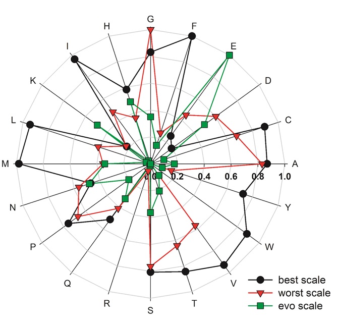
Hydrophobicity refers to the physical property of molecules that repel water. Hydrophobic molecules tend to be nonpolar and therefore prefer to interact other neutral and nonpolar solvents. In water, hydrophobic molecules often cluster together to form micelles. The hydrophobicity of molecules such as amino acids can be experimentally measured as reported by Richard Wolfenden.
Physiochemical properties routinely allow the characterization of peptide or protein sequences of known or unknown function. “Peptide Property Calculators,” such as the one found at BSIs website for the structural prediction or detection of membrane-associated or embedded β-sheets or α-helices, mostly present in larger peptides or proteins, are based on the Kyte-Doolittle Hydrophobicity scale. Several experimentally defined hydrophobicity parameters for amino acids now allow the calculation of peptide properties. In addition, many different scales for calculating peptide properties using hydrophobicity parameters have been developed in recent decades. However, the Kyte-Doolittle Hydrophobicity scale appears to be used the most in peptide calculators.
Often, to achieve a more accurate prediction for the peptide or protein structure of unknowns, five different hydrophobicity parameters are employed:
1) the overall hydrophobicity,
2) the hydrophobic moment for detection of α-helical and β-sheet membrane segments,
3) the alternating hydrophobicity,
4) the alternating hydrophobicity, and
5) the exact β-strand score.
Simm et al. in 2016 reported that most scales allow discriminating between transmembrane α-helices and transmembrane β-sheets. However, using the five different hydrophobicity parameters enables the assignment of peptides to pools of soluble peptides of different secondary structures. But using alternating hydrophobicity is not significantly beneficial. All scales appear to have limitations in separation capacity. Simm et al. observed that the scales derived from the evolutionary approach performed best in separating different peptide pools. The 98 hydrophobicity scales known today have differently defined hydrophobicity values for the 20 amino acids. Unfortunately, because different experimental approaches were used for their definition, a high variance between different scales is observed. Importantly, Simm et al. found that the scale, as defined by Naderi-Manesh, developed in 2001, performed somewhat better than the other hydrophobicity scales. Therefore Simm et al. proposed a rule of thumb for the use of a hydrophobicity scale for the identification of peptides with transmembrane segments from a pool of peptides. Place the hydrophobicity value of arginine (Arg, R) and tyrosine (Tyr, Y) most distant from the value for glutamate (Glu, E). Select the hydrophobicity values for asparagine (Asn, N), aspartate (Asp, D), histidine (His, H), and lysine (Lys, K) such that they are in the center of the scale.
{Simm S, Einloft J, Mirus O, Schleiff E. 50 years of amino acid hydrophobicity scales: revisiting the capacity for peptide classification. Biol Res. 2016 Jul 4;49(1):31. doi: 10.1186/s40659-016-0092-5. PMID: 27378087; PMCID: PMC4932767. https://www.ncbi.nlm.nih.gov/pubmed/27378087}.

Figure 1: Hydrophobicity scale as defined by Naderi-Manesh.
Selected sources for hydrophobicity scales
http://www.genome.jp/aaindex/; Kawashima S, Pokarowski P, Pokarowska M, Kolinski A, Katayama T, Kanehisa M. AAindex: amino acid index database, progress report 2008. Nucleic Acids Res. 2008;2008(36):D202–D205. [PMC free article] [PubMed] [Google Scholar].
http://split4.pmfst.hr/split/scales.html; Juretić D. Protein secondary structure conformations and associated hydrophobicity scales. J Math Chem. 1993;1993(14):35–45. doi: 10.1007/BF01164453. [CrossRef] [Google Scholar].
http://web.expasy.org/protscale/;Gasteiger E, Hoogland C, Gattiker A, Duvaud S, Wilkins MR, Appel RD, Bairoch A. Protein identification and analysis tools on the ExPASy server. In: Walker JM, editor. The proteomics protocols handbook. Totowa: Humana Press Inc.; 2005. pp. 571–607. [Google Scholar].
Table 1: Hydrophobicity Scales commonly used
Residue Type | kdHydrophobicitya | wwHydrophobicityb | hhHydrophobicityc |
Ile | 4.5 | 0.31 | -0.60 |
Val | 4.2 | -0.07 | -0.31 |
Leu | 3.8 | 0.56 | -0.55 |
Phe | 2.8 | 1.13 | -0.32 |
Cys | 2.5 | 0.24 | -0.13 |
Met | 1.9 | 0.23 | -0.10 |
Ala | 1.8 | -0.17 | 0.11 |
Gly | -0.4 | -0.01 | 0.74 |
Thr | -0.7 | -0.14 | 0.52 |
Ser | -0.8 | -0.13 | 0.84 |
Trp | -0.9 | 1.85 | 0.30 |
Tyr | -1.3 | 0.94 | 0.68 |
Pro | -1.6 | -0.45 | 2.23 |
His | -3.2 | -0.96 | 2.06 |
Glu | -3.5 | -2.02 | 2.68 |
Gln | -3.5 | -0.58 | 2.36 |
Asp | -3.5 | -1.23 | 3.49 |
Asn | -3.5 | -0.42 | 2.05 |
Lys | -3.9 | -0.99 | 2.71 |
Arg | -4.5 | -0.81 | 2.58 |
a A simple method for displaying the hydropathic character of a protein. Kyte J, Doolittle RF. J Mol Biol. 1982 May 5;157(1):105-32.
b Experimentally determined hydrophobicity scale for proteins at membrane interfaces. Wimley WC, White SH. Nat Struct Biol. 1996 Oct;3(10):842-8.
c https://www.cgl.ucsf.edu/chimera/docs/UsersGuide/midas/hydrophob.html#cnote
Amino Acid Hydrophobicity Scale used in Chimera, a modelling software from UCSF
UCSF Chimera is an example of a software package that allows visualization and analysis of molecular structures and related data. In Chimera, amino acid residues are automatically assigned an attribute named kdHydrophobicity, with values according to the hydrophobicity scale of Kyte and Doolittle.
The other scales in the following table are not assigned automatically, but input files to assign them with Define Attribute are linked below. A simple text format allows users to create custom attributes with ease.
Table 2: Chimera Amino Acid Hydrophobicity Scale
Residue Type | kdHydrophobicitya | wwHydrophobicityb | hhHydrophobicityc | mfHydrophobicityd | ttHydrophobicitye |
Ile | 4.5 | 0.31 | -0.60 | -1.56 | 1.97 |
Val | 4.2 | -0.07 | -0.31 | -0.78 | 1.46 |
Leu | 3.8 | 0.56 | -0.55 | -1.81 | 1.82 |
Phe | 2.8 | 1.13 | -0.32 | -2.20 | 1.98 |
Cys | 2.5 | 0.24 | -0.13 | 0.49 | -0.30 |
Met | 1.9 | 0.23 | -0.10 | -0.76 | 1.40 |
Ala | 1.8 | -0.17 | 0.11 | 0.0 | 0.38 |
Gly | -0.4 | -0.01 | 0.74 | 1.72 | -0.19 |
Thr | -0.7 | -0.14 | 0.52 | 1.78 | -0.32 |
Ser | -0.8 | -0.13 | 0.84 | 1.83 | -0.53 |
Trp | -0.9 | 1.85 | 0.30 | -0.38 | 1.53 |
Tyr | -1.3 | 0.94 | 0.68 | -1.09 | 0.49 |
Pro | -1.6 | -0.45 | 2.23 | -1.52 | -1.44 |
His | -3.2 | -0.96 | 2.06 | 4.76 | -1.44 |
Glu | -3.5 | -2.02 | 2.68 | 1.64 | -2.90 |
Gln | -3.5 | -0.58 | 2.36 | 3.01 | -1.84 |
Asp | -3.5 | -1.23 | 3.49 | 2.95 | -3.27 |
Asn | -3.5 | -0.42 | 2.05 | 3.47 | -1.62 |
Lys | -3.9 | -0.99 | 2.71 | 5.39 | -3.46 |
Arg | -4.5 | -0.81 | 2.58 | 3.71 | -2.57 |
a A simple method for displaying the hydropathic character of a protein. Kyte J, Doolittle RF. J Mol Biol. 1982 May 5;157(1):105-32.
b Experimentally determined hydrophobicity scale for proteins at membrane interfaces. Wimley WC, White SH. Nat Struct Biol. 1996 Oct;3(10):842-8. Attribute assignment file wwHydrophobicity.txt.
c Recognition of transmembrane helices by the endoplasmic reticulum translocon. Hessa T, Kim H, Bihlmaier K, Lundin C, Boekel J, Andersson H, Nilsson I, White SH, von Heijne G. Nature. 2005 Jan 27;433(7024):377-81, supplementary data. Attribute assignment file hhHydrophobicity.txt. In this scale, negative values indicate greater hydrophobicity.
d Side-chain hydrophobicity scale derived from transmembrane protein folding into lipid bilayers. Moon CP, Fleming KG. Proc Natl Acad Sci USA. 2011 Jun 21;108(25):10174-7, supplementary data. Attribute assignment file mfHydrophobicity.txt. In this scale, negative values indicate greater hydrophobicity.
e An amino acid “transmembrane tendency” scale that approaches the theoretical limit to accuracy for prediction of transmembrane helices: relationship to biological hydrophobicity. Zhao G, London E. Protein Sci. 2006 Aug;15(8):1987-2001. Attribute assignment file ttHydrophobicity.txt (contributed by Shyam M. Saladi).
Table 3: Distribution Coefficients of the 19 Amino Acid Side Chains
Amino acid | RH (side chain) | v>wa | c>wb | VHc | CHd | GUe | WWf | WWth g |
Least polar | ||||||||
ILE | n-butane | 2.15 | 4.92 | −0.60 | 0.24 | 2.04 | 2.16 | −1.12 |
LEU | isobutane | 2.28 | 4.92 | −0.55 | −0.02 | 1.76 | 2.29 | −1.25 |
PHE | toluene | −0.76 | 2.98 | −0.32 | 0.00 | 2.09 | 2.68 | −1.71 |
VAL | propane | 1.99 | 4.04 | −0.31 | 0.09 | 1.18 | 1.61 | −0.46 |
CYS | methanethiol | −1.24 | 1.28 | −0.13 | 0.00 | ND | 1.23 | −0.02 |
MET | methylethylsulfide | −1.48 | 2.35 | −0.10 | −0.24 | 1.32 | 1.71 | −0.67 |
ALA | methane | 1.94 | 1.81 | 0.13 | −0.29 | 0.52 | 0.87 | 0.50 |
TRP | 3-methylindole | −5.88 | 2.33 | 0.30 | −0.59 | 2.51 | 2.96 | −2.09 |
THR | ethanol | −4.88 | −2.57 | 0.52 | −0.71 | 0.27 | 0.95 | 0.25 |
TYR | 4-methylphenol | −6.11 | −0.14 | 0.68 | −1.02 | 1.63 | 1.67 | −0.71 |
GLY | hydrogen | 2.39 | 0.94 | 0.74 | −0.34 | 0.00 | 1.01 | 1.15 |
SER | methanol | −5.06 | −3.40 | 0.84 | −0.75 | 0.04 | 0.85 | 0.46 |
ASN | acetamide | −9.68 | −6.64 | 2.05 | −1.18 | −0.01 | 0.30 | 0.85 |
HIS | 4-methylimidazole | −10.27 | −4.66 | 2.06 | −0.94 | 0.95 | 0.92 | 2.33 |
GLN | propionamide | −9.38 | −5.54 | 2.36 | −1.53 | −0.07 | 0.30 | 0.77 |
ARG | N-propylguanidine | −19.92 | −14.92 | 2.58 | −2.71 | −1.32 | 2.99 | 1.81 |
GLU | propionic acid | −10.24 | −6.81 | 2.68 | −0.90 | −0.79 | −2.53 | 3.63 |
LYS | n-butylamine | −9.52 | −5.55 | 2.71 | −2.05 | 0.08 | 2.49 | 2.80 |
ASP | acetic acid | −10.95 | −8.72 | 3.49 | −1.02 | ND | −2.46 | 3.64 |
Most polar | ||||||||
r2 versus VH h | 0.73 | 0.83 | (1.0) | 0.66 | 0.59 | 0.30 | 0.77 | |
r2 versus CH h | 0.82 | 0.80 | 0.66 | (1.0) | 0.48 | 0.00 | 0.35 | |
p versus VHi | 0.000001 | <0.0000001 | 0.00003 | 0.003 |
Distribution coefficients of the 19 amino acid side chains (RH) at pH 7, expressed in kcal/mol at 25°C, sorted according to their decreasing tendencies (VH) to be found in a transmembrane helix (Hessa et al., 2005). Experimental scales are shown in bold, theoretical scale in normal type.
aSide-chain Kd values for side-chain transfer from vapor to water (Wolfenden et al., 1981) ; bSide-chain Kd values for transfer from cyclohexane to water (Radzicka and Wolfenden, 1988); cTendency of amino acid residue to be found in a transmembrane helix (Hessa et al., 2005); dTendency of amino acid residue to be buried in the interior of a globular protein (Chothia, 1976); eAmino acid side-chain Kd values for transfer from wet octanol to water (Guy, 1985); fPentapeptide Kd values for transfer from water to wet octanol (Wimley et al., 1996); gTheoretical pentapeptide Kd values for transfer from water to wet octanol, after adjustment for the estimated effects of occlusion by neighboring residues (Wimley et al., 1996, Table II, column 3); hValue of the correlation coefficient (r) obtained by linear regression of distribution coefficients against the VH or the CH scales; iProbability that this experimental scale is not related to the VH scale, i.e., that the null hypothesis is true.
{Source: Wolfenden R. Experimental measures of amino acid hydrophobicity and the topology of transmembrane and globular proteins. J Gen Physiol. 2007 May;129(5):357-62. doi: 10.1085/jgp.200709743. Epub 2007 Apr 16. PMID: 17438117; PMCID: PMC2154378.}