Quantcast
Channel: Latest Articles of Bio-Synthesis Inc.
Viewing all 591 articles
Browse latest View live

Antiviral peptides for SARS-C0V-2 (COVID-19)

$
0
0

The surface spike (S) protein is essential for coronavirus binding and entry into host cells. Molecular biology studies indicate that the heptad repeats 1 and 2 (HR1 and HR2) in the S protein induce the fusion of the viral membrane with the host's cell membrane. Therefore, inhibiting SARS-CoV-2 membrane fusion will potentially allow the prevention of COVID-19 infections or even treat it.

What do antiviral peptides do?

Antiviral blocking peptides targeting the viral fusion core can inhibit viral membrane fusion, thereby inhibiting the virus's entry into the host cell.

Presently we do not understand how SARS-CoV-2 enters a host cell in detail. However, models for the fusion core formation are available. The S protein is part of the viral capsid and contains two subunits S1 and S2. The S1 subunit contains the receptor-binding domain (RBD). Subunit S2 mediates the fusion between the virus and the host cell and cell entry. When the SARS-CoV-2 virus particle encounters the host cell, the RBD as part of S1 binds to the host's angiotensin-converting enzyme 2 (ACE2) receptor protein. Furin, an enzyme present in human cells, is thought to cleave the S protein into S1 and S2 subunits. This cleavage event exposes the fusion peptide (FP) of S2 and inserts it into the target cell membrane. The assembly of three HR1 and three HR2 domains form the fusion core by pulling on the cell membrane allowing fusion with the host cell membrane allowing entry of the virus into the cell.

An antiviral peptide designed to bind more tightly to the HR1 domain prevents the formation of the fusion core, and the virus cannot enter the host cell. Hence infection of the host cell is prevented.  

In the spring of 2020, Xia et al. reported the X-ray crystal structure of a six-helical bundle (6-HB) core of the HR1 and HR2 domains present in the S2 subunit of the SARS-CoV-2 spike protein. Several mutated amino acid residues present in the SARS-CoV-2 HR1 sequence appear to allow enhanced interactions with the HR2 domain. A SARS-CoV-2 spike (S) protein-mediated cell–cell fusion assay revealed that SARS-CoV-2 has a superior plasma membrane fusion capacity compared to that of SARS-CoV. Xia et al. designed a series of lipopeptides derived from EK1.  The study identified EK1C4 as the best inhibitor in preventing S protein-mediated membrane fusion and pseudovirus infection. EK1C4 also inhibited membrane fusion and infection of other human coronavirus and pseudoviruses.  EK1C4 inhibited SARS-CoV, MERS-CoV, SARSr-CoVs, five live human coronaviruses, and SARS-CoV-2.

Intranasal application of EK1C4 before or after challenge with HCoV-OC43 protected mice from infection. These findings suggest that EK1C4 potentially prevents infections by the currently circulating SARS-CoV-2 and other emerging SARS coronaviruses. It is also possible to use EK1C4 for the treatment of COVID-19.

A second study performed by Ling et al. using in-silico analysis predicted the HR1 and HR2 regions in the S protein via sequence alignments. A computational model allowed modeling the binding energies of HR1 and HR2 of the fusion core. The model guided the design of antiviral peptides. 

The study identified a homologous sequence region in the heptad repeats (HR) region of SARS-CoV-2 and SARS-CoV. Molecular dynamics-based modeling allowed the design of an antiviral peptide that potentially prevents virus membrane fusion with the host cell membrane. The study reported calculated binding energies for HR1 and HR2 in the SARS-CoV-2 spike protein as well.

Table 1:  Antiviral Peptides

Peptide

Sequence

Reference


EK1

SLDQINVTFLDLEYEMKKLEEAIKKLEESYIDLKEL

Xia et al.

EK1C4

SLDQINVTFLDLEYEMKKLEEAIKKLEESYIDLKELGSGSG-PEG4-cholesterol

Xia et al.

Scramble

LKVLLYEEFKLLESLIMEILEYQKDSDIKENAEDTK

Xia et al.


Linker Peptide

SGGRGG

Xia et al.


HR1

NQKLIANQFNSAIGKIQDSLSSTASALGKLQDVVNQNAQALNTLVKQ

Ling et al.

HR2

GINASVVNIQKEIDRLNEVAKNLNESLIDL

Ling et al.

Linker Peptide

LVPRGSGGSGGSGGLEVLFQGP

Ling et al.

Fusion Peptide

HR1-Linker-HR2

Ling et al.


HR2 antiviral

DISGINASVVNIQKEIDRLNEVAKNLNESLIDLQEL

Ling et al.


Other types of antiviral peptides inhibit the binding of the SARS coronavirus spike RBD to the cellular receptor, ACE2. Often these peptides are also called "
Coronavirus Inhibitory Peptides."

Interaction of EK1 with HCoV.


Figure 1: Interaction of EK1 with HCOV HR1. Xia et al. observed that peptide OC43-HR2P, derived from the HR2 domain of HCoV-OC43, exhibited broad fusion inhibitory activity against multiple human coronaviruses (HCoVs). The optimized peptide EK1 showed substantially improved pan-CoV fusion inhibitory activity and pharmaceutical properties. The crystal structures indicated that EK1 could form a stable six-helix bundle structure with short α-HCoV and long β-HCoV HR1s. This finding supported the role of the HR1 region as a viable pan-CoV target site further. The structural models illustrate that EK1 snugly fits into the hydrophobic grooves formed between two adjacent HR1 helices of the 3HR1 core from MERS-CoV, SARS-CoV, and 229E. The EK1 peptide is shown as a green ribbon.

Interaction of HR peptides illustrating the post fusion core.

Figure 2: Model of the HR1/HR2 complex. The favored model of virus entry into the host cell assumes that furin cleaves the S protein into the S1 subunit and the S2 subunit. The cleavage exposes the fusion peptide (FP) of S2 and inserts it into the target cell membrane. Three HR1s and three HR2s combine to form the fusion core, pulling the viral membrane to fuse with the host cell membrane. Ling et al. suggested that the designed and computational-optimized anti-virus peptide bind to HR1 more tightly, thereby preventing the HR1s and HR2s from forming the fusion core. Hence, the fusion peptide is a candidate for an antiviral peptide.

Reference

Coronavirus Inhibitory Peptides

Rongsong Ling, Yarong DaI, Boxuan Huang, Wenjie Huang, Jianfeng Yu, Xifeng Lu, Yizhou Jiang; In silico design of antiviral peptides targeting the spike protein of SARS-CoV-2. PeptidesVolume 130, August 2020, 170328. https://www.sciencedirect.com/science/article/pii/S0196978120300772 . Published in August 2020.

Xia, S., Liu, M., Wang, C. et al. Inhibition of SARS-CoV-2 (previously 2019-nCoV) infection by a highly potent pan-coronavirus fusion inhibitor targeting its spike protein that harbors a high capacity to mediate membrane fusion. Cell Res 30, 343–355 (2020). https://doi.org/10.1038/s41422-020-0305-x . Published in March 2020.
---...---


Peptide therapeutics target dynamic protein-to-protein interaction underlying human diseases such as hypertension, cancer, Alzheimer’s disease and potentially COVID-19

$
0
0
Increasingly, peptide-based drugs are finding utility in medicine.  This is not surprising given that many of the physiological processes are carried out by endogenously expressed peptides, ex. hormones, neurotransmitters, naturally occurring antibiotic molecules, etc.—with insulin being one of the earliest peptide drugs.  With ~60 peptide drugs having been approved in the U. S. and ~20 new peptide drugs entering clinical trial yearly (~400 peptide-based drugs are being tested globally), it has become a leading pharmaceutical industry (Lee et al., 2019).

For pharmacological application, peptides present multiple advantages.  Peptides are less toxic as they can be biologically degraded. Unlike chemical drugs, they do not accumulate in body.  Peptides are less immunogenic and can penetrate tissue (ex. tumor) more efficiently than antibodies.  Further, peptide can be engineered to translocate across the cell membrane to reach intracellular targets, which most monoclonal antibodies cannot.  As a result, pharmaceutical industries have devoted major efforts to improve its pharmacodynamic and pharmacokinetic properties by increasing its stability in vivo, reducing its clearance by the kidney, etc. (Davenport et al., 2020).

Within cells, many of the biological processes require protein-to-protein interaction.  Examples include filaments polymerized from monomers, enzymes assembled from subunits, DNA repair complexes comprised of multiple distinct proteins, receptors interacting with associating proteins for cell signaling, etc.  Of great significance is the ability of peptides to bind to large, extensive and flat interfaces, which offers unique pharmacological opportunities.  It provided a major advantage over chemical drugs that have been used primarily to target well defined pockets formed through protein folding.

There has been a paradigm shift in how the receptor-ligand interactions are portrayed.  The conventional, static view is being replaced by a fluid model as receptors were shown to adopt multiple conformations in vivo.   This has also impacted peptide drugs.  The classic view portrayed them as mere agonist or antagonist, taking into account of both the binding affinity and the effect on the function of the receptor.  An emerging view may incorporate its interaction with multiple key positions within the binding domain of the receptor occurring in a dynamic manner (Lee et al., 2019).


A significant portion (>40%) of peptide drugs target G protein-coupled receptors (GPCR).  GPCRs are seven-transmembrane domain receptors present on cell surface and respond to external stimulus such as odor, light, hormones, neurotransmitters, etc.  Upon binding to a ligand, it undergoes conformational change to activate the associating protein (G protein) to exchange GDP for GTP, resulting in the dissociation of the latter’s alpha subunit to initiate intracellular signaling.  GPCRs are involved in diverse physiological functions including inflammation, behavior, vision, etc. and its dysfunction leads to various human diseases including asthma, high blood pressure, cancer, obesity, mental illness, infectious diseases and others.  Additionally, peptide drugs have targeted transcriptional complexes regulating the genes involved in cell proliferation (i.e. oncogenesis) or cell differentiation (Inamoto et al., 2017).   Peptides can be used as therapeutic or for drug delivery.  In 2018, the largest segment (>37%) of its application was for cancer and the market is projected to increase to USD 51 billion by the year 2026.

With the recent COVID-19 pandemic, molecular targeting has picked up pace to find suitable therapeutics to counter the coronavirus.  Greater than 20 peptide drugs are in pipeline to treat COVID-19—among them, 15 synthetic peptides are being developed to treat Acute Respiratory Distress Syndrome (ARDS) and other respiratory illnesses resulting from COVID-19 infection.  Through computer simulation, the investigators at the Massachusetts Institute of Technology (USA) have synthesized peptide derivatives corresponding to an alpha helix of ACE2 that interacts with the spike protein (S) of COVID-19 to block infection (Zhang et al., 2020).  Another group developed peptide inhibitors formed by 2 sequential self-supporting alpha helices of ACE2 to bind to S protein (Han et al. 2020).  As an alternative strategy, other researchers employed supercomputers (Texas Advanced Computing Center) to simulate biomolecular environment to refine the chemical structure of candidate peptide drugs to inhibit “main protease” necessary for the propagation of COVID-19.  To treat lung injury, a number of synthetic peptide drugs are being repurposed for COVID-19 clinical trials such as Solnatide to treat alveolar edema, Plitidepsin originally developed to treat cancer (multiple myeloma), etc.  For vaccine development, peptide libraries have been developed consisting of peptides derived from COVID-19 polypeptides to facilitate B-cell or CTL (cytotoxic T cell) epitope mapping.

The key to preventing epidemic is the ability to diagnose the infected early to preempt further propagation.  For this, Bio-Synthesis, Inc. provides primers and probes (as well as synthetic RNA control) for COVID-19 diagnosis via RT-PCR assay.  For medicinal chemistry, it specializes in peptide synthesis, characterization, modification, purification to generate various peptide-based building blocks as well as pharmaceutical intermediates—in addition to peptide libraries, peptide arrays, peptidomimetics.   Antibody purification, characterization/quantification, modification and labeling are also offered.  It specializes in oligonucleotide modification and provides an extensive array of chemically modified nucleoside analogues (over ~200) including bridged nucleic acid (BNA).  A number of options are available to label oligonucleotides (DNA or RNA) with fluorophoreseither terminally or internally as well as conjugate to peptidesIt recently acquired a license from BNA Inc. of Osaka, Japan, for the manufacturing and distribution of BNANC, a third generation of BNA oligonucleotides.  To meet the demands of therapeutic application, its oligonucleotide products are approaching GMP grade.  Bio-Synthesis, Inc. has recently entered into collaborative agreement with Bind Therapeutics, Inc. to synthesize miR-21 blocker using BNA for triple negative breast cancer.  The BNA technology provides superior, unequalled advantages in base stacking, binding affinity, aqueous solubility and nuclease resistance.  It also improves the formation of duplexes and triplexes by reducing the repulsion between the negatively charged phosphates of the oligonucleotide backbone.  Its single-mismatch discriminating power is especially useful for diagnosis (ex. FISH using DNA probe).  For clinical application, BNA oligonucleotide exhibits lesser toxicity than other modified nucleotides.


https://www.biosyn.com/oligo-flourescent-labeling.aspx

https://www.biosyn.com/tew/Speed-up-Identification-of-COVID19.aspx

https://www.biosyn.com/covid-19.aspx

https://www.biosyn.com/peptide-synthesis.aspx

https://www.biosyn.com/tew/Potential-Peptide-Targets-for-a-COVID-19-Vaccine.aspx


References

Davenport AP, Scully CCG, de Graaf C, Brown AJH, Maguire JJ.  Advances in therapeutic peptides targeting G protein-coupled receptors. Nat Rev Drug Discov. 19:389-413 (2020). PMID: 32494050

Han Y, Kral P.  Computational Design of ACE2-Based Peptide Inhibitors of SARS-CoV-2.  ACS Nano 4, 5143-5147 (2020). https://pubs.acs.org/doi/10.1021/acsnano.0c02857

Inamoto I, Shih JA.  Peptide therapeutics that directly target transcription factors.  Peptide Sci.  111:1-11 (2018).  https://onlinelibrary.wiley.com/doi/epdf/10.1002/pep2.24048

Lee AC, Harris JL, Khanna KK, Hong JH.   A Comprehensive Review on Current Advances in Peptide Drug Development and Design.  Int J Mol Sci. 20:2383 (2019).  PMID: 31091705

Zhang G, Pomplun S, Loftis AR, Tan X, Loas A, Pentelute BI.  Investigation of ACE2 N-terminal fragments binding to SARS-CoV-2 Spike RBD.  bioRxiv (2020).

 https://www.biorxiv.org/content/10.1101/2020.03.19.999318v2


Innate Immunity, RNA Sensing and COVID-19

$
0
0

The recent COVID-19 pandemic has rapidly expanded to infect more than 14 million people by July 21, 2020, and has had a significant impact on our lives. The innate immune system is one of the first lines of defense against pathogens, including viruses. The new coronavirus called severe acute respiratory syndrome coronavirus 2 (SARS-CoV-2) is the cause of COVID-19 infections. SARS-CoV-2 emerged in China in late 2019 in the Wuhan region. COVID-19 has been declared a pandemic by the World Health Organization in March 2020.

Recently Qiu et al. developed a biosensor for the selective detections of SARS-CoV-2 nucleic acids. The research group designed a dual-functional localized surface plasmon resonance (LSPR) based biosensor by combining the photothermal effect with plasmonic sensing transduction for SARS-CoV-2 viral nucleic acid detection. The new plasmonic chip contains a two-dimensional distribution of nanoabsorbers (AuNIs). The chip generates a local plasmonic photothermal heat (PPT) transduced in situ hybridization, allowing a highly sensitive and accurate SARS-CoV-2 detection.

Cells, both prokaryotic and eukaryotic, also have mechanisms for sensing RNA, for example the entry of foreign RNA into cells. Human epithelial cells contain RNA sensing molecules outside and inside the cell body. Epithelial cells are at the interface where self and non-self meets. In humans, in the respiratory tract, these cells are the front-line physical barrier between the environment and the body. The respiratory system is integral to innate and adaptive immunity. The epithelium plays an important role in host defense. Epithelium cells secrete a wide spectrum of protective, antimicrobial, homeostatic, and immune regulatory factors. Secreted molecules include antioxidants, defensins, collectins, complement components, lactoferrin, lysozyme, and protease inhibitors. In humans, oral epithelial cells line the oral cavity and are an effective and dynamic barrier to invading pathogens. Epithelial cells contain an extensive repertoire of innate defenses.

In 2012, Ghosh et al. studied the proteome of healthy human oral epithelial cells. The use of two-dimensional electrophoresis followed by liquid chromatography on-line with tandem mass spectrometry (2DE-LC-MS/MS) and database searches allowed the identification of 237 most abundant proteins present in oral epithelial cells. Nucleic acid-binding proteins (20.4%), transferases (11%), cytoskeletal proteins (7.6%), and enzyme modulators (7.2%) are the three most abundant protein classes identified. Also, the study identified hydrolases, isomerases, phosphatases, proteases, transcription factors, and chaperones.
Querying the innate database (innateDB), the research group identified 64 proteins associated with innate immunity. The 64 observed proteins associated with innate immunity confirmed that oral tissue acts as a primary active barrier to infection.

RNA sensors in the cytosol contain three members of the RIG-I-like receptors (RLRs). These are the retinoic acid-inducible gene I (RIG-I), the melanoma differentiation factor 5 (MDA5), and the laboratory of genetics and physiology 2 (LGP2). The three proteins share a similar organization. These proteins contain three distinct domains:

(i) a C-terminal repressor domain (RD) embedded within the C-terminal domain (CTD);
(ii) a central ATPase containing DExD/H-box helicase domain that can bind RNA; and
(iii) a N-terminal tandem CARD domain that mediates downstream signaling.

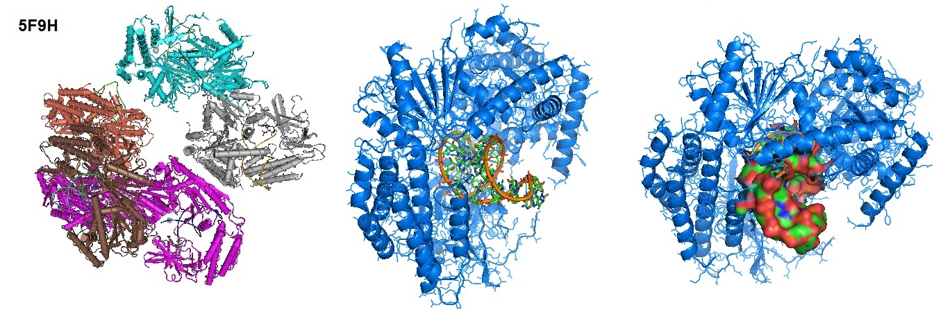
Each retinoic acid-inducible gene I like receptor senses different viral infections. Different replicating viral RNA are recognized by the receptors in the cytoplasm. The Retinoic Acid Inducible Gene-I (RIG-I) detects pathogenic RNAs carrying a 5′-triphosphate (5′ppp). RIG-I, when activated, triggers a Type I interferon (IFN) response. RIG-I does not recognize self RNAs such as mRNAs because they are capped post-transcriptionally on the 5′-end with 7-methyl guanosine (m7G) and 2′-O-methylation of 5′-end nucleotides. Viruses have evolved mechanisms to mimic these modifications for evasion of the innate immune response. Studies performed by Devakar et l. provide structural and mechanistic insights into the roles of the m7G cap and 2′-O-methylation in RIG-I evasion. They also showed that RIG-I accommodates the m7G base while maintaining 5′ppp contacts and can recognize Cap-0 RNAs but not Cap-1. Cap-0 and 5′ppp double-stranded (ds) RNAs bind to RIG-I with nearly identical Kd values and activate RIG-I’s ATPase and cellular signaling response to similar extents.

Figure 1: Structure of RIG-I Helicase-Repressor Domain in complex with 5'-ppp hairpin (HP) RNA.

The CARD domain is present in RIG-I and MDA5 but absent in LGP2. The activation by RNA ligands recruits RIG-I and MDA5 to the adaptor protein Mitochondrial Antiviral Signaling (MAVS) via a CARD–CARD, which in turn activates NF-κB and IRFs.

Toll-like receptors (TLRs) are receptors of the innate immune system that recognize conserved “pathogen-associated molecular patterns” of microbes and viruses. Activation of TLRs establishes an antiviral immune response. The antiviral immune response coordinates long-lasting adaptive immunity to control viral infection. However, after microbe-induced damage to the host tissues, “danger-associated molecular patterns” that also activate TLRs can lead to an overreacting inflammatory response, which ultimately leads to tissue damage.

The new SARS-CoV2 coronavirus is a positive-sense single-stranded RNA virus. In vitro experiments on spike proteins of the virus indicated affinity with the angiotensin-converting enzyme receptor 2 (ACE2). ACE2 appears to be the primary receptor protein allowing host cell entry of the virus. Following cell entry, the virus distributes through the circulatory stream. In some patients, viral infection triggers a systemic response leading to hyper inflammation. The hyperactivation of the immune system occurring in some infected people appears to significantly damage the lung, ultimately leading to the patients’ death. During the entry of host cells by the coronavirus SARS-CoV-2, the virus trimeric spike glycoprotein interacts with its cellular receptor angiotensin-converting enzyme 2 (ACE2). Host proteases, for example, cathepsins, furin, or members of the type II transmembrane serine proteases (TTSP) family, such as Transmembrane protease serine 2 (TMPRSS2), are thought to enable virus entry by proteolytically activating virus ligands.

The innate immune system uses Germ-line-encoded pattern recognition receptor proteins (PRRs).  A variety of cells express PRRs. These cells are responsible for sensing the presence of pathogen invasions. During infection, Toll-Like Receptor (TLR) family members upregulate anti-viral and pro-inflammatory mediators. Pro-inflammatory mediators include interleukin (IL)-6, IL-8, type I and type III Interferons, and others. Activation occurs through Nuclear Factor (NF)-kB. In some cases, when the cellular entry of the virus and innate immune responses are uncontrolled, a deleterious systemic response can sometimes occur in infected patients. These molecular events can lead to a “cytokine storm.” The downregulation of dendritic cells, macrophages, and T-cell functions initiate multiple organ failure, ultimately leading to death.

According to Li et al., the highest ACE2 expression levels are found in the small intestine, testis, kidneys, heart, thyroid, and adipose tissue. Lowest levels are observed in the blood, spleen, bone marrow, brain, blood vessels, and muscle. Medium expression levels of ACE2 are found in the lungs, colon, liver, bladder, and adrenal gland.

Humans are exposed to millions of pathogens daily. The adaptive immune system helps us to avoid infections. However, adaptive immune responses are slow to develop after exposure to a new pathogen. To respond, specific clones of B and T cells have to become activated and expand. Therefore, it can take a week or longer before the responses are effective. During the first critical hours and days after we are exposed to a new pathogen, our body relies on the innate immune system to protect us from infection. The innate immune system is one of the two immune systems found in vertebrates, including in humans.
Innate immunity is a nonspecific defense mechanism reacting to an antigen in the human body within hours of its appearance. Defense mechanisms of the innate immune system include physical barriers such as skin, chemicals in the blood, and immune system cells that attack foreign cells. However, innate immune responses are not specific to pathogens in the same way that adaptive immune responses are. Vertebrates require innate immune responses for the activation of the adaptive immune responses.

The skin and epithelial surfaces and tissue cells forming the outer layer of the human body and layers of cells that line hollow organs and glands provide a physical barrier between the body and the outside world. A mucus layer covers interior surfaces. The slimy mucus primarily containing mucin and other glycoproteins help prevent pathogens from adhering to the epithelium, the tissue forming the outer layer of the body surface. Also, the mucus layer contains substances that kill pathogens or inhibit their growth, such as defensins. Defensins are small cysteine-rich cationic peptides or proteins typically 12 to 50 amino acids in length produced by the innate immune system. Defensins kill phagocytosed pathogens. However, it is still unclear how exactly they do this. The thought is that they insert themselves into the cell membrane of their victims by disrupting membrane integrity.   

A cell of the innate immune system recognizes pathogen-associated molecules through germline-encoded pattern recognition receptors (PRRs) present on the cell surface or within distinct intracellular compartments. PRRs identify viral pathogens by engaging pathogen-associated molecular patterns (PAMPs). These include the Toll-like receptors (TLRs), the retinoic acid-inducible gene I-like receptors (RLRs), the nucleotide oligomerization domain-like receptors (NLRs, also called NACHT, LRR and PYD domain proteins) and cytosolic DNA sensors.

The TLR family is a crucial component of mammalian immunity and acts as an early surveillance mechanism of infections. TLRs sense infection via pattern recognition of specific molecules. TLRs are proteins involved in the development and activation of innate immunity. The toll-like receptor family of 11 transmembrane receptor proteins recognizes pathogen-associated molecular patterns (PAMPs). Different types of ligands originating from a variety of pathogens activate TLRs.
Coronaviruses appear to trigger a cytokine release in the human body. The release of primarily interleukin 6 (Il-6) and other proteins of the acute phase activates the immune response. 

Often homo- or heterodimers of TLR help clear infections. Some TLR types are expressed on the cell surface. Others are present on intracellular membranes of endosomes (TLR3, 7, 8, and 9). TLRs react to viral infections by detecting foreign types of nucleic acids. In healthy immune cells and epithelial cells, TLR 1, 2, 4, 5, 6, and 10 are usually expressed on the cell surface. However, TLR 3, 7, 8, and 9 are mainly expressed on endosomes, lysosomes, and endoplasmic reticulum surfaces. Coronaviruses contain single-stranded RNA (ssRNA). TLR3 and 9 interact with double-stranded viral RNA and unmethylated CpG DNA from bacteria and viruses, respectively. TLR3 is thought to respond to infections by the West Nile virus in a rodent model. The West Nile virus is a single-stranded virus.

TLR3, -7, and -8 found in the intracellular compartments recognize nucleic acid motifs. Intercellular compartments are found in the endoplasmic reticulum (ER), endosomes, lysosomes, and endolysosomes.
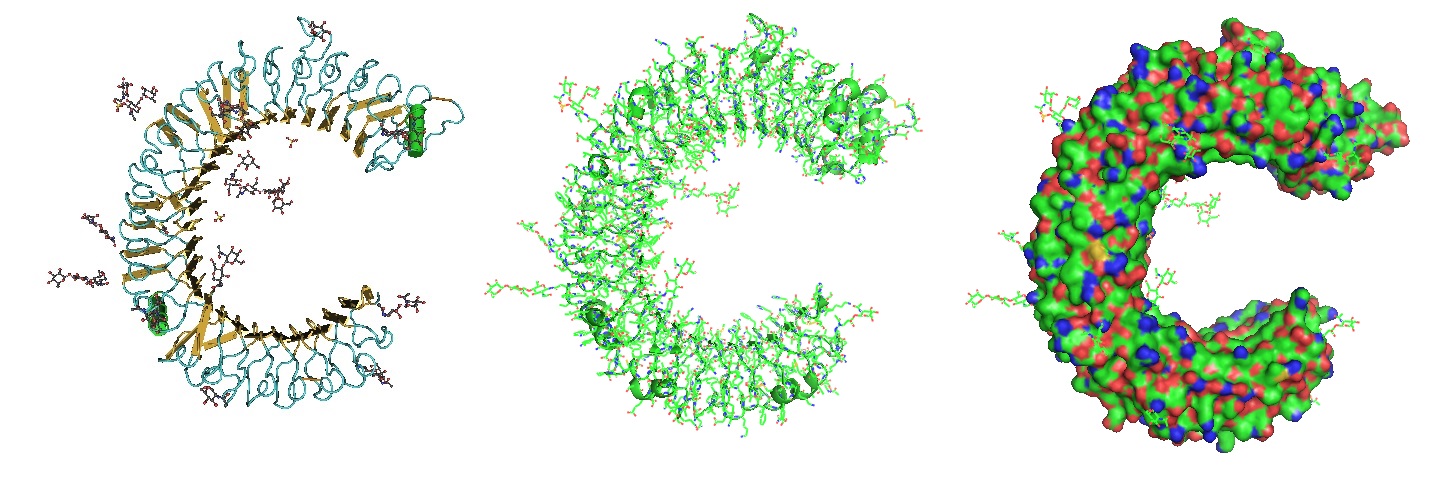
.

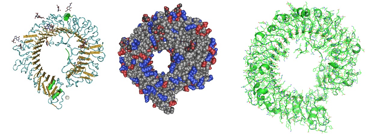
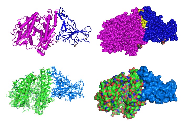
Figure 2: Structure of the Toll Like Receptor 3 Ectodomain (TLR3-ECD). Three renderings of the TlR3-ECD crystal structure at a resolution of 2.4 Å solved by Bell et al. in 2005 is shown. TLR3 senses a variety of ligands ranging from lipopolysaccharide to double stranded RNA through the ligand-binding domain composed of leucin-rich repeats. The N-linked glycans are shown as green ball-and-stick (Bell et al. 2005).


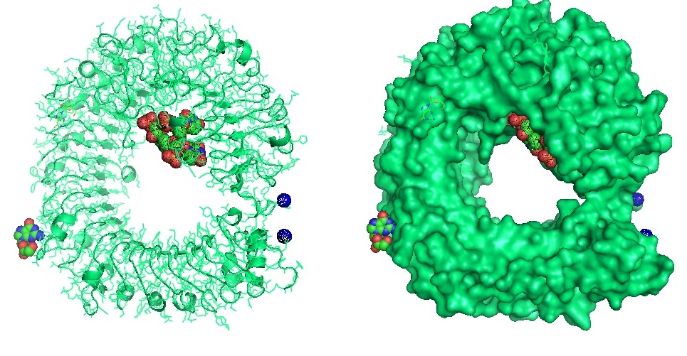
Figure 3: Structure of the Toll Like Receptor 7 (Zhang et al. 2016). Toll-like receptor 7 (TLR7) is a single-stranded RNA (ssRNA) sensor in innate immunity. TLR7 also responds to guanosine and chemical ligands, such as imidazoquinoline compounds since it contains two binding sites. One site that binds small ligands and a second site for ssRNA. TLR7 is a sensor for guanosine and uridine-containing ssRNA and is activated by both guanosine and ssRNA.


Figure 4: Structure of the Toll Like Receptor 7 with complexed with guanosine and a small uridine-containing single stranded RNA. Zhang et al. reported that the crystal structures of three TLR7 complexes form an activated m-shaped dimer with two ligand-binding sites. The first site is conserved in TLR7 and TLR8. This binding site is used for small ligand-binding and is essential for its activation. The second site differs that of TLR8 binds ssRNA. Binding to ssRNA enhances affinity of the first-site ligands. The first site preferentially recognizes guanosine and the second site specifically binds uridine moieties in ssRNA. Structural, biochemical, and mutagenesis studies revealed that TLR7 is a dual receptor for guanosine and uridine-containing ssRNA.

Table 1:  Toll Like Receptors recognizing RNA  

Receptor

Virus

Ligand

TLR7

Influenza A virus

ssRNA


Vesicular stomatitis virus

ssRNA


Human immunodeficiency virus

ssRNA


Dengue virus

ssRNA


Sendai virus

ssRNA


Lactate dehydrogenase-elevating virus

ssRNA


Mouse mammary tumor virus

ssRNA


Murine leukemia virus

ssRNA

TLR8

Human immunodeficiency virus

ssRNA

TLR3

Reoviridae

dsRNA


Respiratory syncytial virus

dsRNA


West Nile virus

dsRNA


Coxsackievirus B3

dsRNA


Poliovirus

dsRNA


Influenza A virus

dsRNA


Punta Toro virus

dsRNA

TLR2

Measles virus

HA


Lymphocytic choriomeningitis virus

?


Hepatitis C virus

Core protein/NS3

TLR4

Respiratory syncytial virus

Fusion protein


Coxsackievirus B4

?


Mouse mammary tumor virus

Envelope protein


Murine leukemia virus

Envelope protein


Reference

Akira S, Uematsu S, Takeuchi O; 2006. Pathogen recognition and innate immunity. Cell 124:783–801.

Jessica K. Bell, Istvan Botos, Pamela R. Hall, Janine Askins, Joseph Shiloach, David M. Segal, David R. Davies; The molecular structure of the Toll-like receptor 3 ligand-binding domain. Proceedings of the National Academy of Sciences Aug 2005, 102 (31) 10976-10980; DOI: 10.1073/pnas.0505077102  . https://www.pnas.org/content/102/31/10976

Birra D, Benucci M, Landolfi L, Merchionda A, Loi G, Amato P, Licata G, Quartuccio L, Triggiani M, Moscato P. COVID 19: a clue from innate immunity. Immunol Res. 2020 Jun;68(3):161-168. doi: 10.1007/s12026-020-09137-5. PMID: 32524333; PMCID: PMC7286633. https://www.ncbi.nlm.nih.gov/pmc/articles/PMC7286633/

de Groot NG, Bontrop RE. COVID-19 pandemic: is a gender-defined dosage effect responsible for the high mortality rate among males? Immunogenetics. 2020 Jul;72(5):275-277. doi: 10.1007/s00251-020-01165-7. PMID: 32342146; PMCID: PMC7186185.

Devarkar SC, Wang C, Miller MT, et al. Structural basis for m7G recognition and 2'-O-methyl discrimination in capped RNAs by the innate immune receptor RIG-I. Proc Natl Acad Sci U S A. 2016;113(3):596-601. doi:10.1073/pnas.1515152113. https://www.ncbi.nlm.nih.gov/pmc/articles/PMC4725518/

Fitzgerald KA, Kagan JC. Toll-like receptors and the control of immunity. Cell. 2020;180(6):1044–1066. doi: 10.1016/j.cell.2020.02.041.

Ghosh SK, Yohannes E, Bebek G, Weinberg A, Jiang B, Willard B, Chance MR, Kinter MT, McCormick TS. Proteomic and bioinformatic profile of primary human oral epithelial cells. J Proteome Res. 2012 Nov 2;11(11):5492-502. doi: 10.1021/pr3007254. Epub 2012 Oct 16. PMID: 23035736; PMCID: PMC3508721. https://www.ncbi.nlm.nih.gov/pmc/articles/PMC3508721/

Guangyu Qiu, Zhibo Gai, Yile Tao, Jean Schmitt, Gerd A. Kullak-Ublick, and Jing Wang; Dual-Functional Plasmonic Photothermal Biosensors for Highly Accurate Severe Acute Respiratory Syndrome Coronavirus 2 Detection. ACS Nano 2020 14 (5), 5268-5277 . DOI: 10.1021/acsnano.0c02439. https://pubs.acs.org/doi/10.1021/acsnano.0c02439

Innate Immunity in Molecular Biology of the Cell, 4th edition.    https://www.ncbi.nlm.nih.gov/books/NBK26846/

Søren Jensen, Allan Randrup Thomsen; Sensing of RNA Viruses: a Review of Innate Immune Receptors Involved in Recognizing RNA Virus Invasion. Journal of Virology Feb 2012, 86 (6) 2900-2910; DOI: 10.1128/JVI.05738-11.

Jensen S, Thomsen AR. Sensing of RNA viruses: a review of innate immune receptors involved in recognizing RNA virus invasion. J Virol. 2012 Mar;86(6):2900-10. doi: 10.1128/JVI.05738-11. Epub 2012 Jan 18. PMID: 22258243; PMCID: PMC3302314.

Jouhi L, Renkonen S, Atula T, Mäkitie A, Haglund C, Hagström J. Different Toll-Like Receptor Expression Patterns in Progression toward Cancer. Front Immunol. 2014 Dec 15;5:638. doi: 10.3389/fimmu.2014.00638. PMID: 25566251; PMCID: PMC4266018.

Hoffmann M, Kleine-Weber H, Schroeder S, et al. SARS-CoV-2 cell entry depends on ACE2 and TMPRSS2 and is blocked by a clinically proven protease inhibitor [published online ahead of print, 2020 Mar 4]. Cell. 2020;S0092–8674(20)30229–4. doi:10.1016/j.cell.2020.02.052

Khan S, Siddique R, Shereen MA, et al. The emergence of a novel coronavirus (SARS-CoV-2), their biology and therapeutic options [published online ahead of print, 2020 Mar 11]. J Clin Microbiol. 2020;JCM.00187–20. doi:10.1128/JCM.00187-20.

Kumar H, Kawai T, Akira S. Toll-like receptors and innate immunity. Biochem Biophys Res Commun (2009) 388(4):621–5.10.1016/j.bbrc.2009.08.062 [PubMed] [CrossRef] [Google Scholar]

Li, M., Li, L., Zhang, Y. et al. Expression of the SARS-CoV-2 cell receptor gene ACE2 in a wide variety of human tissues. Infect Dis Poverty 9, 45 (2020). https://doi.org/10.1186/s40249-020-00662-x

Thompson MR, Kaminski JJ, Kurt-Jones EA, Fitzgerald KA. Pattern recognition receptors and the innate immune response to viral infection. Viruses. 2011 Jun;3(6):920-40. doi: 10.3390/v3060920. Epub 2011 Jun 23. PMID: 21994762; PMCID: PMC3186011.

Parker LC, Prince LR, Sabroe I. Translational mini-review series on Toll-like receptors: networks regulated by Toll-like receptors mediate innate and adaptive immunity. Clin Exp Immunol. 2007 Feb;147(2):199-207. doi: 10.1111/j.1365-2249.2006.03203.x. PMID: 17223959; PMCID: PMC1810480. https://www.ncbi.nlm.nih.gov/pmc/articles/PMC1810480/

Sallenave JM, Guillot L. Innate Immune Signaling and Proteolytic Pathways in the Resolution or Exacerbation of SARS-CoV-2 in Covid-19: Key Therapeutic Targets? Front Immunol. 2020 May 28;11:1229. doi: 10.3389/fimmu.2020.01229. PMID: 32574272; PMCID: PMC7270404.

Wang T, Town T, Alexopoulou L, Anderson JF, Fikrig E, Flavell RA. Toll-like receptor 3 mediates West Nile virus entry into the brain causing lethal encephalitis. Nat Med. 2004;10:1366–1373. doi: 10.1038/nm1140. [PubMed] [CrossRef] [Google Scholar]

Zhang Z, Ohto U, Shibata T, et al. Structural Analysis Reveals that Toll-like Receptor 7 Is a Dual Receptor for Guanosine and Single-Stranded RNA. Immunity. 2016;45(4):737-748. doi:10.1016/j.immuni.2016.09.011 . https://pubmed.ncbi.nlm.nih.gov/27742543/


---...---

mRNA vaccines and innate immunity

$
0
0

Vaccination prevents many millions of illnesses and saves many lives every year. The smallpox virus vaccine is an excellent example of a successful vaccination approach.

Messenger RNA (mRNA) vaccines are a promising vaccine approach for high potency, rapid development, and potential low-cost manufacture and safe administration. mRNA is the intermediate between transcription and translation of a gene. A single strand of DNA is decoded by RNA polymerase during transcription, resulting in the biosynthesis of mRNA. mRNA transfers genetic information encoded in DNA to the ribosomal translational machinery in the cytoplasm to produce proteins.


Figure 1:  Mature messenger RNA.


Until recently, instability and inefficient in vivo delivery of mRNA restricted the application of mRNA vaccines. Recent technological advances help to overcome these issues. Multiple platforms against infectious diseases and several types of cancer no exist. 

Classical vaccines that protect against many dangerous diseases include living attenuated and inactivated pathogens and subunit vaccines. However, for newly emerging viral infections, such as COVID-19, more rapid development and large-scale deployment is needed. 

Nucleic acid-based therapies are promising alternatives to conventional approaches. Recently, mRNA became a promising therapeutic tool in vaccine development and protein replacement therapy.

Beneficial features for using mRNA as a vaccination platform are:

  • Safety.  mRNAs are non-infectious, non-integrating, with no potential risk of infection or insertional mutagenesis. Also, normal cellular processes degrade mRNA. Using a variety of modifications and delivery methods allow regulating the in vivo half-life of mRNA. For increased safety, inherited immunogenicity of mRNA needs to be downregulated.
  • Efficacy. A variety of modifications allow increasing mRNA stability and translatability to produce a desired and satisfactory mRNA product. To ensure improved in vivo delivery, formulating mRNA into carrier molecules allows rapid uptake and expression in the cytoplasm. The use of mRNA-based vaccines avoids anti-vector immunity and enables repeated administration.
  • Productions.  Messenger RNA vaccines potentially allow rapid, inexpensive, and scalable manufacturing due to the high yields of in vitro transcription reactions. 

Recent results suggest that mRNA vaccines potentially solve many challenges in vaccine development for infectious diseases and cancer.

Production of optimally translated in vitro transcript mRNA

Starting from a linear DNA template with the help of T7, a T3 or an Sp6 phage RNA polymerase, in vitro transcription (IVT) produces mRNA. The IVT product needs to contain an open reading frame (ORF) that encodes the target protein, flanking untranslated regions (UTRs), a 5’-cap and a poly(A) tail. Engineering of the mRNA results in a fully processed mature mRNA molecule as it occurs naturally in the cytoplasm of eukaryotic cells.

Extracellular RNases quickly degrade naked mRNA. Naked mRNA is also not efficiently internalized. To solve this problem, newly developed in vitro and in vivo transfection reagents facilitate cellular uptake of mRNA and protect it from degradation. After successful uptake and transit of mRNA into the cytosol, the cellular machinery produces protein. Post-translational modifications result in a correctly folded, functional protein. Finally, the protein is delivered to the correct cellular compartment by normal physiological processes for proper presentation or function. Degradation by normal physiological processes reduces the risk of metabolite toxicity.

Exogenous or cell foreign mRNA can stimulate an immune response. A variety of cell surfaces recognize foreign RNA through endosomal and cytosolic innate immune receptors. This effect of RNA on the cell can be beneficial or detrimental. Vaccination can drive dendritic cells (DCs) to maturity to elucidate a robust T and B cell responses. Unfortunately, innate immune sensing of mRNA can also inhibit antigen expression, thereby negatively affecting the desired immune response.

mRNA and Innate Immunity

How innate immune sensing works is not well understood. IVT mRNA show an immunostimulatory profile that can be shaped by purification, the introduction of modified oligonucleotides, and the complexing of mRNA with a variety of carrier molecules. Enzymatically synthesized mRNA contains double-stranded (dsRNA) contaminations. Pattern recognition receptors present in multiple cellular compartments sense dsRNA as a potent pathogen-associated molecular pattern (PAMP). IVT mRNA that contains dsRNA results in robust type I interferon production. Type I interferon upregulates the expression and activation of protein kinase R (PKR or EIF2AK2) and 2’-5’-oligoadenylate synthetase (OAS), resulting in inhibited translation, and degradation of cellular mRNA and ribosomal RNA. However, chromatographic purification methods allow efficient removal of contaminated dsRNA. Both reversed-phase fast protein liquid chromatography (FPLC) or high-performance liquid chromatography (HPLC) remove dsRNA to achieve a highly pure product. The result is an increased protein production in primary human dendritic cells from IVT mRNAs.      

Externally delivered single-stranded RNAs are also PAMPs. Endosomal Toll-like receptors 7 (TLR7) and TLR8 sense degradation products of RNAs resulting in type I interferon production. Incorporation of naturally occurring chemically modified nucleosides, including pseudouridine and 1-methylpseudouridine as well as others, prevents RNA sensing by TLR7, TLR8, and other innate immune sensors, resulting in a reduction of type I interferon signaling. Nucleoside modification also partially suppress the recognition of dsRNA species.

Recent studies showed increased in vitro translation of nucleoside-modified mRNA compared to unmodified mRNA, which is also the case in vivo in mice. High levels of protein production can be achieved in DCs using purified and nucleoside-modified mRNA. 

These findings advanced our understanding of innate immune sensing and how to avoid the adverse effects of innate immunity.

Lipid-encapsulated or naked forms of sequence-optimized mRNA vaccines can potentially produce potent vaccines against a variety of viruses such as SARS-CoV-2, influenza virus, Zika virus, rabies virus, and many others.

Different types of mRNA vaccines are cancer vaccines, dendritic cell vaccines, and various kinds of directly injectable mRNA vaccines.

However, for therapeutic considerations, good manufacturing practice (GMP) production must be established to guarantee the safety and increased efficacy of mRNA vaccines.

Reference


Pardi, N., Hogan, M., Porter, F. et al., 2018; mRNA vaccines— a new era in vaccinology. Nat Rev Drug Discov 17, 261–279 (2018). [nature]

World Health Organization Immunization for Diseases

Contact us for more info


---...---

Liquid biopsies containing circulating tumor DNA (ctDNA) for diagnosing biomarkers for early detection, metastatic recurrence or targeted therapy: implication for COVID-19 diagnosis

$
0
0
Biomarkers play an indispensable role for cancer management.  Biomarkers include specific molecules or genetic mutations whose presence is indicative of the disease progression.  One major area of its application has been for early detection as late-stage diagnosis portends poor survival for many cancer types.  The early detection of predisposing genotypic biomarkers provides an opportunity to take preemptive measures such as surgery to avoid tumor development.  Another key area has been to detect the onset of recurrent cancer after the initial treatment.  The changes in the expression level of molecular biomarkers could signal the presence of recurrent cancer when it is not visually detectable through imaging.  Further, biomarkers are being used to prognosticate the therapeutic outcome.  Given the adverse side effects associated with various anticancer drugs, there has been a drive to predict the response of specific drugs prior to treatment.

Typically, diagnostic analysis has been performed on solid samples, i.e. tumor specimens resected from cancer patients. Though the data obtained from primary tumor biopsies may accurately reflect the disease status, the approach is being marred by the need for invasive surgery, insufficient specimen for analysis, difficulty of monitoring repeatedly, etc.  Another key finding is that, in the event of recurrence, data obtained from a single biopsy may not reflect the mechanism of drug resistance for the overall cancer.

A solution to the above dilemma may be found in liquid biopsy.  Previous works have shown that primary tumors shed cancer cells into vasculature albeit in a low number (1-10 cells per ml of blood).  For screening, circulating tumor cells (CTCs) can be detected through microscopic imaging using tumor-specific antibodies, which could be indicative of early stage carcinogenesis (Ried et al., 2017).  The changing level of CTC counts could be used to assess therapy efficacy as well as survival prognosis.  The presence of CTCs have been associated with late recurrence (5 year after diagnosis), which occurs in ~50% of hormone receptor-positive breast cancer cases (Sparano et al., 2018). For personalized medicine, CTCs allow gene expression analysis at the single cell level or could be grown into a 3D model to test the efficacy of drugs to tailor the treatment (Potdar et al., 2015). 

To capture, antibody-functionalized carriers that are recovered through magnetic field can be used to separate CTCs from other components using microchips (Hoshino et al., 2011).  With microfluidic channels, unique physical parameters such as greater size, flow velocity, shear force/drag potential have been exploited to distinguish CTCs from normal cells (Nagrath et al., 2007).  Nevertheless, the heterogeneous nature of CTCs and the extreme rarity (1 CTC per 109 hematologic cells in blood) have made them very difficult to isolate.


The field of liquid biopsy has advanced further to allow detection of genetic biomarkers without isolating CTCs  Circulating tumor DNA (ctDNA) represent fragments of DNA released into the blood stream by dying tumor cells due to necrosis or apoptosis.  Presumably, ctDNA can be found in other bodily fluids including saliva, urine and spinal fluid.  Like CTCs, the amount of ctDNA detected can be used to gauge disease progression, ex. an increase in ctDNA amount may be indicative of cancer recurrence.

Plasma genotyping performed on ctDNA using next generation sequencing (NGS) was able to detect specific mutations (Iwahashi et al., 2019).  For GWAS (genome wide association study), ctDNA has been used to genotype SNP (single nucleotide polymorphism) using chip-based microarray methodologies, which is based on hybridization.  Using qPCR, mutation could be detected in ctDNA of colorectal cancer patients.  For ctDNA analysis, the presence of normal DNA in plasma could decrease the percentage of mutant allele.  False negatives may require further analysis on tumor biopsies to exclude other mechanisms that give rise to drug resistance.  For positive results obtained, comparison with normal tissue may be necessary to exclude mutations that arose during hematopoiesis.

In a similar vein, the effort to diagnose COVID-19 could be simplified by using liquid biopsy.  Regarding safety, the acquiring of respiratory samples (ex. sputum or other specimens from lower part of lung) could expose clinicians to the virus inadvertently.  Saliva containing COVID-19 coronavirus may be easier to sample and process to detect viral RNA through RT-PCR or RT-LAMP reaction.

The key to preventing epidemic is the ability to diagnose the infected early to preempt further propagation.  For this, Bio-Synthesis, Inc. provides primers and probes (as well as synthetic RNA control) for COVID-19 diagnosis via RT-PCR assay.  For medicinal chemistry, it specializes in peptide synthesis, characterization, modification, purification to generate various peptide-based building blocks as well as pharmaceutical intermediates—in addition to peptide libraries, peptide arrays, peptidomimetics.   Antibody purification, characterization/quantification, modification and labeling are also offered.  It specializes in oligonucleotide modification and provides an extensive array of chemically modified nucleoside analogues (over ~200) including bridged nucleic acid (BNA).  A number of options are available to label oligonucleotides (DNA or RNA) with fluorophoreseither terminally or internally as well as conjugate to peptidesIt recently acquired a license from BNA Inc. of Osaka, Japan, for the manufacturing and distribution of BNANC, a third generation of BNA oligonucleotides.  To meet the demands of therapeutic application, its oligonucleotide products are approaching GMP grade.  Bio-Synthesis, Inc. has recently entered into collaborative agreement with Bind Therapeutics, Inc. to synthesize miR-21 blocker using BNA for triple negative breast cancer.  The BNA technology provides superior, unequalled advantages in base stacking, binding affinity, aqueous solubility and nuclease resistance.  It also improves the formation of duplexes and triplexes by reducing the repulsion between the negatively charged phosphates of the oligonucleotide backbone.  Its single-mismatch discriminating power is especially useful for diagnosis (ex. FISH using DNA probe).  For clinical application, BNA oligonucleotide exhibits lesser toxicity than other modified nucleotides.


https://www.biosyn.com/oligo-flourescent-labeling.aspx

https://www.biosyn.com/tew/Speed-up-Identification-of-COVID19.aspx

https://www.biosyn.com/covid-19.aspx

https://www.biosyn.com/peptide-synthesis.aspx

https://www.biosyn.com/tew/Potential-Peptide-Targets-for-a-COVID-19-Vaccine.aspx


References

Hoshino K, Huang YY, Lane N, Huebschman M, Uhr JW, Frenkel EP, et al.  Microchip-based immunomagnetic detection of circulating tumor cells.  Lab Chip. 11:3449-57 (2011).  PMID: 21863182

Iwahashi N, Sakai K, Noguchi T, Yahata T, Matsukawa H, Toujima S, et al.  Liquid biopsy-based comprehensive gene mutation profiling for gynecological cancer using CAncer Personalized Profiling by deep Sequencing.  Sci Rep. 9:10426 (2019).  PMID: 31320709

Nagrath S, Sequist LV, Maheswaran S, Bell DW, Irimia D, Ulkus L, et al.   Isolation of rare circulating tumour cells in cancer patients by microchip technology.  Nature. 450:1235-9 (2007).   PMID: 18097410

Potdar PD, Lotey NK.  Role of circulating tumor cells in future diagnosis and therapy of cancer.  J. Cancer Metastasis Treat 1, 44-56 (2015).  https://jcmtjournal.com/article/view/1172

Ried K, Eng P, Sali A. Screening for Circulating Tumour Cells Allows Early Detection of Cancer and Monitoring of Treatment Effectiveness: An Observational Study.  Asian Pac J Cancer Prev.  18:2275-2285 (2017).  PMID: 28843267

Sparano J, O'Neill A, Alpaugh K, Wolff AC, Northfelt DW, Dang CT, et al.  Association of Circulating Tumor Cells With Late Recurrence of Estrogen Receptor-Positive Breast Cancer: A Secondary Analysis of a Randomized Clinical Trial.  JAMA Oncol.  4:1700-1706 (2018).  PMID: 30054636


Useful RNA Databases

$
0
0

RNA databases contain sequences and useful tools for the analysis of RNA molecules and to enhance RNA research.

RNAcentral  
RNAcentral -  RNAcentral About-us

The non-coding RNA sequence database : a comprehensive ncRNA sequence collection representing all ncRNA types from a broad range of organisms.

RNAcentral is a free, public resource that offers integrated access to a comprehensive and up-to-date set of non-coding RNA sequences provided by a collaborating group of Expert Databases representing a broad range of organisms and RNA types.

Functional RNA Database  ncRNA

Bioinformatic Tools and Databases for Functional RNA analysis

Rfam   Rfam

The Rfam database is a collection of RNA families, each represented by multiple sequence alignments, consensus secondary structures and covariance models (CMs) covering non-coding RNA genes, structured cis-regulatory elements and self-splicing RNAs.

RNA sequences  RNAseq

A large collection of tools and sequences.

MicroRNA databasemirbase

A searchable database of published miRNA sequences and annotation.

The RNA Modification Database  RNA Modifications

A comprehensive listing of posttranscriptionally modified nucleosides from RNA

tRNAdb  trnadb

Compilation of tRNA sequences and tRNA genes.

Yeast snoRNA Database  snoRNAs

Small Nucleolar RNAs (snoRNAs) from the Yeast Saccharomyces cerevisiae. A comprehensive database of S. cerevisiae H/ACA and C/D box snoRNAs, with numerous links to snoRNA and other databases.

The 3D rRNA modification maps database  3DrRNAMod

The 3D maps database allows visualization of modification sites in the five ribosome and mRNA and tRNAs for several model organisms and permits users’ construction of 3D maps for other organisms.

Molecular Landscapes

---...---

2’-Fluoroinosine, a Replacement for Inosine

$
0
0

Modified oligonucleotides allowed researchers to gain insight into conformational, mechanistic and sterochemical aspects of a variety of biological reactions and processes. Using 2'-Fluoroinosine instead of inosine increases the stability of chimeric oligonucleotides. The incorporation of inosine and 2'-fluoroinosine in primers and probes at specific locations modulates selectivity. The phosphoramidite 2'-fluoro-inosine-CE phosphoramidite allows the synthesis of oligonucleotides containing 2'-fluoroinosine (2F-I).

Figure 1: Chemical structures of inosine, 2'-fluoroinosine, and hypoxanthine.

Synthetic oligonucleotides containing inosine have been extensively used as hybridization probes to screen cDNA or genomic libraries for genes encoding proteins whose amino acid sequence is only partially known. Inosine-containing oligonucleotides enabled the cloning of many genes from genomic and cDNA libraries of high complexity. Inosine interacts with each of the principal nucleic acid bases through two hydrogen bonds. Interactions in order of decreasing stability are I-C > I-A > I-T ≈ I-G.

Microbial studies routinely use universal primers. Usually, this type of research starts with the extraction of total DNA from bacterial samples, followed by PCR amplification of small-subunit ribosomal RNA genes. Designing degenerate primers with varying options of nucleotides at several positions in the internal primer sequence allows improved amplification of related sequences of 16S rRNA genes from different microorganisms. This approach allows the amplification of a variety of related sequences. Often inosine is used to achieve a fourfold degeneracy for a given location. Using inosine at the 3’-terminal ends of 16S ribosomal RNA (rRNA) gene universal primers allowed studying complex microbial communities by PCR. For example, Ben-Dov et al. replaced the 3’-terminal nucleic acid with inosine in universal 16S rRNA primers to study complex microbial communities to expand the observed diversity of a microbial community under study.

The introduction of 2'-fluoro RNA can modulate the selectivity oligonucleotides, as was found to be the case for antisense oligonucleotides (ASOs). The electronegative fluorine atom produces local changes in the conformation of the nucleotide furanose ring. The incorporation of 2'-fluoro-inosine affects the biological properties of the modified oligonucleotide. Structural studies performed by Martin-Pintado showed that oligonucleotides containing alternating and contiguous tracts of 2′F‐RNA and 2′F‐ANA nucleotides reveal that nonconventional FC-H⋅⋅⋅O hydrogen bonds have a strong stabilizing effect on 2′‐fluorinated duplexes. Hence, replacing inosine with 2'-fluoroinosine in oligonucleotides increases the stability of the duplex formed with complementary oligonucleotides.

Figure 2:  Molecular structures of 2'-fluoro-substituted oligonucleotides. A duplex with 2'-fluoro-bases is shown to the left and middle of the figure (Martin-Pintado et al. 2013). The picture to the right shows the structure for a 2'-deoxy-2'-fluoro-D-arabinose nucleic acid (2'F-ANA)/RNA duplex (Trempe et al. 2001). The (2'F-ANA)/RNA duplex exhibits structural parameters between those of A-form and B-form duplexes but like those of DNA/RNA duplexes. The enhanced stability of the duplex is relevant to the design of new antisense drugs based on sugar-modified nucleic acids.


Reference


Ben-Dov E, Shapiro OH, Siboni N, Kushmaro A. Advantage of using inosine at the 3' termini of 16S rRNA gene universal primers for the study of microbial diversity. Appl Environ Microbiol. 2006 Nov;72(11):6902-6. [PMC]

Green & Sambrook; Molecular Cloning. A Laboratory Manual. 4th Edition. 2012, Cold Spring Harbor Laboratory Press.

Martin-Pintado N., Deleavey G.F., Portella G., Campos-Olivas R., Orozco M., Damha M.J., Gonzalez C., Backbone F.C.-H. O hydrogen bonds in 2′F-substituted nucleic acids. Angew. Chem. Int. Ed. 2013;52:12065–12068. [
PubMed]

Pallan PS, Greene EM, Jicman PA, Pandey RK, Manoharan M, Rozners E, Egli M.; Unexpected origins of the enhanced pairing affinity of 2'-fluoro-modified RNA. Nucleic Acids Res. (2011) 39 p.3482-95. [
PMC]


Bio-Synthesis provides a full spectrum of high quality custom oligo modification services by direct solid-phase chemical synthesis or enzyme-assisted approaches to obtain artificially modified oligonucleotides containing backbone, base, sugar and internucleotidic linkages. Bio-Synthesis specialize in complex oligonucleotide modifications using phosphodiester backbone, purine and pyrimidine heterocyclic bases, and sugar modified nucleotides such as our patented 3rd generation Bridged Nucleic Acids.

Contact us for more info


---...---

Triplex Formation for the Detection of microRNAs

$
0
0

MicroRNAs (miRNAs) are short RNA oligonucleotides with an average length of 22 nucleotides. miRNAs play essential roles in many biological processes, including cell development, proliferation, differentiation, and apoptosis. Thousands of miRNAs are known as regulatory molecules modulating gene expression at post-transcriptional levels. Correlating differently expressed miRNAs in tissues with cellular processes may allow diagnostics of various diseases. Hence, the detection of miRNA can provide valuable diagnostic and prognostic data. However, because of their size, measurements of miRNAs are challenging.

In 2016, Aviñó et al. designed and synthesized modified parallel tail-clamps to detect miRNA sequences responsible for tumor suppression via triplex formation. The researchers used parallel clamps targeting a seven homo-pyrimidine sequence in miRNA-145 to test if they form triple helices (Triplex; See Figure 1). The research group selected 8-aminoG instead of 8-aminoA for improved triplex stability because of the small polypyrimidine tracks present in miRNAs.

A pH 5 was selected to test the complex's thermal stability between parallel clamps and miRNA-145. This pH range is known to be optimal for parallel-triplex formation. A surface plasmon resonance (SPR) biosensor allowed the detection of miRNA-145, forming a triplex with parallel clamps.
 

Triplex froming oligonucleotides (TFOs) are a class of DNA oligonucleotides capable of binding to the major groove of duplex DNA froming a triplex helix. Triplex forming oligonucleotides, for example, containing bridged nucleic acids (BNAs) can also be used to inhibit expression of specific genes.
   

Triplex formed


Figure 1: Tail-clamps and complementary sequences forming a miRNA-145 triplex structure. The triads C+-8-aminoG-C and T-A-T are shown as well.

Avino et al. found that parallel clamps with 8-aminoguanine form the most stable triplex structures with targeted miRNA. SPR biosensor analysis demonstrated that the target miRNA and the 8-aminoguanine tail-clamp formed a stable triplex structure. The researchers proposed to use this approach for label-free and reliable detection of miRNA signatures needed for diagnostic purposes.

Reference

Aviñó, A., Huertas, C. S., Lechuga, L. M., Eritja, R.; Sensitive and label-free detection of miRNA-145 by triplex formation. Anal. Bioanal. Chem., 408(3), 885-893 (2016). [PubMed]

---...---


Development of a simplified RNA extraction method incorporating polyacrylamide carrier to streamline diagnosis protocol for COVID-19 (or oncology) for cost and availability

$
0
0
The unprecedented emergence of a pandemic caused by COVID-19 coronavirus has taken a toll socially as well as economically.  Its impact has been felt globally as the virus moved on from continent to continent irrespective of the economic or technological divide.  The speed with which it spread caught many in the scientific community off guard given the extent of information and experience acquired from studying closely related coronaviruses previously.  It is also testing the capability of current molecular biological methods to halt its advance.

One critical arm of the preventive strategy lies in developing a diagnostic means to accurately identify the individuals infected by COVID-19.  The underlying rationale is to segregate the infected to limit further exposures to reduce its propagation.  Given the steady decline in mortality rate resultant from the increase in diagnosed cases and the potential for the emergence of other related viruses in the future, the long-term therapeutic efficacy of the approach is continually being assessed.

The ‘gold-standard’ of the COVID-19 diagnostic methods has been RT-PCR (reverse transcriptase-polymerase chain reaction) to detect the presence of the viral RNA for sensitivity and reliability.  Additionally, alternate amplification methods based on RT-LAMP (reverse transcriptase-loop mediated isothermal amplification) are being utilized for practicality and point-of-care.  As both methods have been extensively cultivated to meet expediency, the rate limiting step for the genetic diagnosis has shifted to the preceding RNA extraction step.  This is not unexpected given the widely varying types of specimen from which COVID-19 RNA needs to be isolated, which include nasal swab, pharyngeal swab, sputum, saliva, oral fluid, bronchoalveolar lavage (BAL), etc.


Furthermore, the source(s) of viral RNA of COVID-19 could be diverse—i.e. mature viruses produced by infected cells, cells harboring the assembled viruses, cells with virus RNA undergoing replication, viral RNA released from ruptured/dying infected cells, etc.  The integrity of the viral RNA extracted is not known as the current diagnostic methods fall short of testing its functionality.  To isolate viral RNA from within cells, one needs to disrupt the cell membrane as well as the viral envelope.  Although guanidine-based extraction method is effective, its adverse effects (irritating) may not be suitable for a wider usage.  Other factors to consider are costs as well as availability as the supply of RNA extraction kits could be dwindled by the global needs—ex. as it may have been the case for the silica-fitted columns used in purifying nucleic acids.

To avoid having to rely on suppliers for RNA extraction kits as well as to simplify the extraction steps, the investigators at the University of California at Santa Barbara (USA) have modified the protocol (Ponce-Rojas et al., 2020).  In their protocol (called ‘PEARL’ for Precipitation Enhanced Analyte RetrievaL, 30 min prep), cells were lysed using a solution containing the non-ionic (nondenaturing) detergent IGEPAL CA-630, which structurally resembles NP-40.  As the detergent is non-ionic, it may not lyse nuclear membrane (to release genomic DNA).  The solution also contained linear polyacrylamide carrier to facilitate the precipitation of the material consisting of both nucleic acids (both RNA and DNA according to the report) and proteins.  After washing with ethanol, the precipitated material was dissolved in nuclease-free water and used to amplify COVID-19 N1 RNA and endogenous RNase P mRNA via RT-PCR, yielding results comparable to that obtained with a commercially available kit.

Linear polyacrylamide (LPA) carrier is a commonly used non-nucleotide based co-precipitating agent that does not interfere with enzymatic reaction (ex. PCR), spectrophotometry, electrophoresis, etc., which helps to isolate minute or trace amounts of DNA or RNA via forming a visible pellet (Gaillard et al., 1990; Strauss et al., 1984).  As the ability to purify DNA/RNA through precipitation is central to most molecular biological applications, LPA carrier has gained widespread usage (Li et al., 2020), which include oncology studies, and is commercially available through various biotech vendors. 

The key to preventing epidemic is the ability to diagnose the infected early to preempt further propagation.  For this, Bio-Synthesis, Inc. provides primers and probes (as well as synthetic RNA control) for COVID-19 diagnosis via RT-PCR assay.  For medicinal chemistry, it specializes in peptide synthesis, characterization, modification, purification to generate various peptide-based building blocks as well as pharmaceutical intermediates—in addition to peptide libraries, peptide arrays, peptidomimetics.   Antibody purification, characterization/quantification, modification and labeling are also offered.  It specializes in oligonucleotide modification and provides an extensive array of chemically modified nucleoside analogues (over ~200) including bridged nucleic acid (BNA).  A number of options are available to label oligonucleotides (DNA or RNA) with fluorophoreseither terminally or internally as well as conjugate to peptidesIt recently acquired a license from BNA Inc. of Osaka, Japan, for the manufacturing and distribution of BNANC, a third generation of BNA oligonucleotides.  To meet the demands of therapeutic application, its oligonucleotide products are approaching GMP grade.  Bio-Synthesis, Inc. has recently entered into collaborative agreement with Bind Therapeutics, Inc. to synthesize miR-21 blocker using BNA for triple negative breast cancer.  The BNA technology provides superior, unequalled advantages in base stacking, binding affinity, aqueous solubility and nuclease resistance.  It also improves the formation of duplexes and triplexes by reducing the repulsion between the negatively charged phosphates of the oligonucleotide backbone.  Its single-mismatch discriminating power is especially useful for diagnosis (ex. FISH using DNA probe).  For clinical application, BNA oligonucleotide exhibits lesser toxicity than other modified nucleotides.


https://www.biosyn.com/oligo-flourescent-labeling.aspx

https://www.biosyn.com/tew/Speed-up-Identification-of-COVID19.aspx

https://www.biosyn.com/covid-19.aspx

https://www.biosyn.com/peptide-synthesis.aspx

https://www.biosyn.com/tew/Potential-Peptide-Targets-for-a-COVID-19-Vaccine.aspx


References

Gaillard C, Strauss F.  Ethanol precipitation of DNA with linear polyacrylamide as carrier.  Nucleic Acids Res.18:378 (1990).  PMID: 2326177

Li Y, Chen S, Liu N, Ma L, Wang T, Veedu RN, et al.  A systematic investigation of key factors of nucleic acid precipitation toward optimized DNA/RNA isolation.  Biotechniques.  68:191-199 (2020).  

Ponce-Rojas JC, Costello JS, Proctor DA, Kosik KS, Wilson MZ, Arias C, Acosta-Alvear D.  A fast and accessible method for the isolation of RNA, DNA, and protein to facilitate the detection of SARS-CoV-2.  bioRxiv (2020)   

Strauss F, Varshavsky A.   A protein binds to a satellite DNA repeat at three specific sites that would be brought into mutual proximity by DNA folding in the nucleosome.  Cell 37, 889-901 (1984). PMID: 6540146


Linear Polyacrylamide as a Carrier Molecule during Alcohol Precipitation

$
0
0
  • Linear polyacrylamide can be used as a carrier molecule during alcoholic precipitation.
  • Adding linear polyacrylamide as a neutral carrier to ethanol precipitations is more advantage than using other precipitation methods such as using glycogen or yeast RNA.
  • The use of linear polyacrylamide eliminates the risk of trace contaminants possibly introduced during the precipitation step.
  • Co-precipitation of DNA or RNA with linear polyacrylamide allow precipitation of picogram amounts of nucleic acids.


Figure 1: Free radical polymerization converts acrylamide into linear polyacrylamide chains of various length. The acrylamide unit in polyacrylamide is depicted at the right.

Historically ethanol precipitation was used as a method for the concentration of biologically active nucleic acids as early as the 1930s. Ethanol precipitation is now a standard method for precipitation of DNA out of a solution for removing salts and for resuspension of the precipitated DNA in a different buffer. Both ethanol and isopropanol allow precipitation of nucleic acid fragment out of solution. However, ethanol is usually the preferred choice.Ethanol precipitation is also used to concentrate DNA, RNA, and polysaccharides, for example pectin, from aqueous solutions.Alcohol precipitation methods allow recovery of DNA and RNA of high purity form biological samples. Protocols using alcoholic precipitation steps are now commonly used in diagnostics and protocols in molecular biology with the need for highly purified DNA or RNA. However, for efficient precipitation and recoveries with high yields, several vital parameters such as salt, alcohol, or carrier choice have to be considered.

Thirty years ago, Gaillard and Straus showed that linear polyacrylamide used as a neutral carrier allows precipitating picogram amounts of nucleic acids with ethanol. Chemically synthesized linear acrylamide, free of nuclease contamination, eliminates the risk of trace contamination introduced by the precipitation process. Polyacrylamides exhibit strong hydrogen bonding and water solubility resulting in a variety of uses in diverse industries.

Linear polyacrylamide also allows precipitation of RNA with ethanol or precipitation of proteins with cold acetone. Linear polyacrylamide does not interfere with spectrophotometric readings at 260nm and 280nm. Hence ethanol is the best choice for DNA and RNA precipitation.

Cations in salts neutralize the negative charge of the DNA phosphate backbone. Microcentrifuge tubes, in combination with a microcentrifuge, allow efficient precipitation of DNA or RNA with ethanol.

How does ethanol precipitate oligonucleotides?

DNA and RNA oligonucleotides have an exposed backbone of negatively charged phosphate residues that make these molecules highly polar. In aqueous solutions, charged residues attracted hydration shells of water molecules that suppress the binding of positively charged ions to DNA molecules. Ethanol disrupts DNA and RNA's hydration shell, allowing unshielded phosphate residues to form ionic bonds with cations in the solvent. At a concentration of 70% ethanol in the solution in the presence of 300 mM sodium cations reduces repulsive forces between the polynucleotide chains such that DNA and RNA precipitates. Ethanol precipitation occurs when enough cations are present to neutralize the charge on the exposed phosphate residues. Isopropanol is less polar than ethanol and has a higher propensity to precipitate salts and antibiotics as well. Table 1 shows the most used cations.

How can DNA and RNA precipitates be dissolved?

Often DNA and RNA precipitates recovered after a precipitation step, for example, using ethanol, are dried under vacuum before redissolving.

However, it is best to modify this practice because of the following reasons:

(i)   Desiccated pellets of DNA and RNA dissolve slowly and inefficiently.

(ii)  Small fragments of double-stranded DNA (<400 base pairs) become denatured after drying. Most likely as a result
      of losing the stabilizing shell of bound water molecules.

Therefore, the best practice is to remove ethanol from the nucleic acid pellet and the tube's walls by gentle aspiration followed by storage of the open tube on the bench for approximately 15 minutes to enable evaporation of most of the residual ethanol. The resulting damp pellet can then be dissolved rapidly and completely in the buffer needed for the next experimental step. However, for quantitative removal of the ethanol, the open tube with the redissolved DNA or RNA can be incubated for 2 to 3 minutes at 45°C in a heating block to allow any traces of ethanol to evaporate.

Please note, after centrifugation using an angle-head rotor, precipitated oligonucleotides are not all found at the bottom of the tube. Approximately around 40% of the precipitated oligonucleotides are present on the wall of microcentrifuge tubes. Therefore, to achieve maximal recovery, use a pipette to move an aliquot of solvent over the surface with a disposable pipette tip.

For radioactive samples, check that no detectable radioactivity remains in the tube after removing the dissolved oligonucleotides.

To conclude, DNA and RNA ethanol precipitates can be redissolved quite easily in buffers of low ionic strength, such as in TE buffer at pH 8.0. However, when buffers containing MgCl2 or greater than 100 mM NaCl are added directly to the pellet, difficulties in the pellet's quantitative redissolving can arise. Hence, it is preferable to first dissolve the pellet in a small volume of low-ionic-strength buffer and slowly adjust the buffer's composition to the final buffer composition.

Sometimes, if the sample pellet does not dissolve easily in a small volume, a second precipitation step using ethanol is needed. This second step may help eliminate additional salts or other components that can prevent the oligonucleotides' dissolution.

Reference

Clerget G, Bourguignon-Igel V, Rederstorff M. Alcoholic precipitation of small non-coding RNAs. Methods in Molecular Biology (Clifton, N.J.). 2015 ;1296:11-16. [Europe PMC]

Gaillard, C. and Strauss, F.; Ethanol precipitation of DNA with linear polyacrylamide carrier. (1990) Nucleic Acids Res. 18, 2, 378. [PDF]

Green and Sambrook; Molecular Cloning. A Laboratory Manual. 4th Edition. Cold Spring Harbor Laboratory Press. 2012. pp. 21-27. [Book]

Also review "Development of a simple RNA extrection method ...... "


---...---


Memory T cells remember coronavirus infections

$
0
0

Viral infections can induce memory T cells in humans. A previous infection can influence the course of a new viral infection.

As a response to pathogens, naive T cells rapidly divide and express molecules such as cytokines that fight infections. The responding cells are called effector T cells. Effector T cells can migrate into inflamed tissues and kill infected cells. After eliminating a pathogen, most of these cells die; however, a small pool of the long-lived memory cells remains ready to respond rapidly if a new infection of the same pathogen occurs.

Akondy et al. and Youngblood et al. recently studied the cell population giving rise to memory T cells and the evolution of memory T cells.DNA-methylation profiling allowed the researchers to observe how naïve T cells differentiate into effector cells. During T cell differentiation, their DNA-methylation profile changes. Methyl groups are added to many genes associated with the naïve state. Methyl groups are lost at genes encoding key components of the effector response. DNA methyltransferase Dnmt3a is the key enzyme responsible for de-novo DNA methylation during the immune response. DNA-methylation events are epigenetic modifications.

Memory T cells that no longer express effector molecules remain in a state of low methylation. Memory T cells can be present as long as ten or even 17 years after vaccination. However, when a new infection occurs, memory T cells can respond rapidly to re-express effector molecules to fight the new infection.

The two research groups found that patients who recovered from a coronavirus infection possessed long-lasting memory T cells reactive to the N protein of SARS-CoV. These T cells also showed cross-reactivity to SASRS-CoV-2. As a result, infections with betacoronaviruses induce a strong and long-lasting T cell immunity to the structural protein N. This immunity appears to last as long as 17 years or even longer.

A recent paper reported that beta-coronavirus infections in humans induce a long-lasting T cell immunity to the viral structural protein N of coronaviruses. Bert et al. studied T cell responses to structural and non-structural regions of SARS-CoV-2 (COVID-19) from patients recovering from the COVID-19.

How was the study done?

Blood samples were collected from donors recovering from a SARS-CoV-1 or SARS-CoV-2 infection. Density-gradient centrifugation enabled the isolation of peripheral blood mononuclear cells (PBMC). PBMCs are a mixture of specialized cells of the immune system. Peptide pools of 15mer peptides spanning the entire protein sequence of selected proteins allowed the stimulation of PBMCs. The analysis of stimulated cells utilized peptide array plates. Also, flow cytometry was used for the study of PBMCs or expanded T cell lines.

SARS‐CoV‐2‐specific T cell peptides

T cells in convalescent COVID‐19 patients target multiple regions of nucleocapsid protein.


Type of response

Amino acid residue


SARS‐CoV‐2 Amino acid sequence

SARS‐CoV‐1 Amino acid sequence

CD4, C-1

NP 81‐95

DDQIGYYRRATRRIR


DDQIGYYRRATRRVR

CD8, C-1

NP 321‐340

GMEVTPSGTWLTYTGAIKLD

GMEVTPSGTWLTYHGAIKLD


CD4, C-4

NP 266‐280

KAYNVTQAFGRRGPE

KQYNVTQAFGRRGPE


CD4, C-4

NP 291‐305

LIRQGTDYKHWPQIA

LIRQGTDYKHWPQIA


CD4, C-4

NP 301‐315

WPQIAQFAPSASAFF

WPQIAQFAPSASAFF

CD4, C-8

NP 51‐65

SWFTALTQHGKEDLK

SWFTALTQHGKEELR

CD4, C-8

NP 101‐120

MKDLSPRWYFYYLGTGPEAG

MKELSPRWYFYYLGTGPEAS


C‐10, CD8,

CD4

NP 321‐340

GMEVTPSGTWLTYTGAIKLD

GMEVTPSGTWLTYHGAIKLD


C‐12, CD8

NP 321‐340

GMEVTPSGTWLTYTGAIKLD

GMEVTPSGTWLTYHGAIKLD


C‐15, CD4

NP 101‐120

MKDLSPRWYFYYLGTGPEAG

MKELSPRWYFYYLGTGPEAS


C‐16, CD4

NSP7 21‐35

RVESSSKLWAQCVQL

RVESSSKLWAQCVQL


Reference

Nina Le Bert, Anthony T Tan, Kamini Kunasegaran, Christine Y L Tham, Morteza Hafezi, Adeline Chia, Melissa Chng, Meiyin Lin, Nicole Tan, Martin Linster, Wan Ni Chia, Mark I-Cheng Chen, Lin-Fa Wang, Eng Eong Ooi, Shirin Kalimuddin, Paul Anantharajal Tambyah, Jenny Guek-Hong Low, Yee-Joo Tan, Antonio Bertoletti; Different pattern of pre-existing SARS-COV-2 specific T cell immunity in SARS-recovered and uninfected individuals. bioRxiv 2020.05.26.115832; doi: https://doi.org/10.1101/2020.05.26.115832.https://www.biorxiv.org/content/10.1101/2020.05.26.115832v1

https://en.wikipedia.org/wiki/Memory_T_cell

Origins of memory T cells : https://www.nature.com/articles/d41586-017-08280-8


---...---

Evaluating the efficacy of RT-PCR in assessing infectivity, accuracy of death rate and counts, and the treatments for cancer patients in the era of COVID-19

$
0
0
 The current strategy to manage the COVID-19 pandemic relies heavily on the accuracy of the existing diagnostic methods to detect the presence of its genomic RNA.  However, even with RT-qPCR, it yields little information regarding the functionality, i.e. the capacity to undergo replication.  Nor does it provide insight regarding the infectivity of the virus.  (Further, the sequence of the amplified product is seldom analyzed to confirm it's derived from COVID-19--as qPCR merely monitors the increase in fluorescence emitted by the probe).  To address the above, the investigators at the University of Manitoba (Canada) examined the relationship between the RT-PCR outcome versus infectivity (monitored the ability to grow COVID-19 in cell culture in lab, i.e. the "true" gold-standard).  For this, patient samples obtained at various time points since the onset of symptoms were examined.  Intriguingly, the ability to infect was highest on day 3 (since the onset of symptom) and declined 32% for every 1-unit increase in Ct thereafter.  Though infectivity steadily declined and was no longer observed after 7th day, the genomic RNA was detected by RT-PCR well beyond 12th day (Jefferson et al, 2020'; Bullard et al., 2020) (Fig 1A). The results corroborate that RT-PCR may not be a reliable indicator of COVID-19 infectivity.

 Nevertheless, the diagnostic tests were carried out on a massive scale in recent months, which resulted in a significant rise in the count of the COVID-19 infected individuals.  The daily reports concerning the elevating count globally have prompted some to ask what its real impact was.   Yet, the impact has been difficult to assess as the count of COVID-19 cases may have included those who are asymptomatic, those who are at low-risk, ones who have recovered, those who have died, those who have died due to other causes, and potentially the results obtained using methods that cross-react with other coronaviruses, diagnostic kits revoked by FDA, diagnostic assays with high rate of false negativity/positivity, etc.

As a result, to assess the impact directly, the focus has shifted towards the number of deaths caused by COVID-19.   There are two different ways to determine mortality rate: CRF (case fatality rate) refers to the proportion of deaths when compared to the total number of cases diagnosed with COVID-19 disease) whereas IFR (infection fatality rate) refers to the number of deaths per total number of infected cases (which may include healthy asymptomatic individuals).  According to the Johns Hopkins coronavirus resource center, the current CRF for United States is ~3.0%.  Though this statistic is used commonly amongst physicians, CRF figures do not take into account of those clinically undetected (i.e. those who display atypical or asymptomatic to very mild symptoms).

Meta-study refers to combining the results from the previously conducted individual studies (ex. clinical trials) to arrive at a point with greater statistical power. Meta-analysis conducted on published reports, government documents, etc. from various global regions yielded IFR of ~0.68% (with 0.53-0.82% range) for COVID-19 (Meyerowitz-Katz et al., 2020).  This is comparable to IFR of 0.63% reported for the state of Arizona (USA) (August 2000; https://www.statnews.com/2020/08/24/infection-fatality-rate-shows-covid-19-isnt-getting-less-deadly/ ).  A report from Stanford University (USA) showed a median IFR value of 0.27% IFR for 32 global locations (though higher for 'extreme hotbed locations') (Ioannidis, 2020).   The latest figure from CDC says: 0.0002 (or 0. 02% for 20-49 y), 0.005 (or 0.5% for 50-69 y), 0.054 (or 5.4% for >70y;  https://www.cdc.gov/coronavirus/2019-ncov/hcp/planning-scenarios.html#table-1 ).  in contrast, IFR for seasonal influenza virus is estimated ~0.1%. 


To gauge the true burden of COVID-19, the extent of 'excess mortality' was examined.  It is obtained by comparing all deaths in a given period with the number of predicted deaths based on average of preceding years (ex. 5 years).  But it turns out a significant fraction of excess deaths was not attributed to COVID-19, i.e. 25% in the case of United States and 74% in Peru (from March through June 2020) (Fig. 1B).  Further, the remaining fraction may need to be divided into those who died due to COVID-19 versus those who died due to unrelated causes (despite being infected).  The excess deaths may also include deaths caused by lockdown indirectly (ex. patients skipping hospital visits, domestic violence, exacerbated mental conditions), as documented by a sharp increase in deaths due to asthma, diabetes, hypertension, dementia, Alzheimer's disease in United Kingdom (Viglione, 2020).

As those with underlying conditions are more susceptible to COVID-19 associated death, investigators at Sloan Kettering Memorial Cancer Center (USA) investigated the impact of various treatment modalities on >400 cancer patients exhibiting COVID-19 symptoms(Robilatti et al., 2020).  In addition to conventional treatments, immunotherapy is increasingly used to manage melanoma, lung cancer and several other cancers.  The recently introduced therapeutic agent called 'immune checkpoint inhibitor' (ex. PD-1 blockade) is designed to increase autoimmunity against tumor.  The study showed that lung cancer and other solid tumor patients receiving immune checkpoint inhibitors had higher frequency of hospitalization and severe respiratory illness from COVID-19.  In contrast, chemotherapy and surgery did not pose a greater risk.

 The key to preventing epidemic is the ability to diagnose the infected early to preempt further propagation.  For this, Bio-Synthesis, Inc. provides primers and probes (as well as synthetic RNA control) for COVID-19 diagnosis via RT-PCR assay.  For medicinal chemistry, it specializes in peptide synthesis, characterization, modification, purification to generate various peptide-based building blocks as well as pharmaceutical intermediates—in addition to peptide libraries, peptide arrays, peptidomimetics.   Antibody purification, characterization/quantification, modification and labeling are also offered.  It specializes in oligonucleotide modification and provides an extensive array of chemically modified nucleoside analogues (over ~200) including bridged nucleic acid (BNA).  A number of options are available to label oligonucleotides (DNA or RNA) with fluorophoreseither terminally or internally as well as conjugate to peptidesIt recently acquired a license from BNA Inc. of Osaka, Japan, for the manufacturing and distribution of BNANC, a third generation of BNA oligonucleotides.  To meet the demands of therapeutic application, its oligonucleotide products are approaching GMP grade.  Bio-Synthesis, Inc. has recently entered into collaborative agreement with Bind Therapeutics, Inc. to synthesize miR-21 blocker using BNA for triple negative breast cancer.  The BNA technology provides superior, unequalled advantages in base stacking, binding affinity, aqueous solubility and nuclease resistance.  It also improves the formation of duplexes and triplexes by reducing the repulsion between the negatively charged phosphates of the oligonucleotide backbone.  Its single-mismatch discriminating power is especially useful for diagnosis (ex. FISH using DNA probe).  For clinical application, BNA oligonucleotide exhibits lesser toxicity than other modified nucleotides.

https://www.biosyn.com/oligo-flourescent-labeling.aspx

https://www.biosyn.com/tew/Speed-up-Identification-of-COVID19.aspx

https://www.biosyn.com/covid-19.aspx

https://www.biosyn.com/peptide-synthesis.aspx

https://www.biosyn.com/tew/Potential-Peptide-Targets-for-a-COVID-19-Vaccine.aspx


References

Bullard J, Dust K, Funk D, Strong JE, Alexander D, Garnett L, et al. Predicting infectious SARS-CoV-2 from diagnostic samples.  Clin Infect Dis. (2020) PMID: 32442256

Ioannidis J. The infection fatality rate of COVID-19 inferred from seroprevalence data.  medRxiv  (2020)  doi: https://doi.org/10.1101/2020.05.13.20101253

Jefferson T, Spencer E, Brassey J, Heneghan C.  Viral cultures for COVID-19 infectivity assessment.  medRxiv (2020)  doi: https://doi.org/10.1101/2020.08.04.20167932

Meyerowitz-Katz G, Merone L.  A systematic review and meta-analysis of published research data on COVID-19 infection-fatality rates.  medRxiv (2020)  doi: https://doi.org/10.1101/2020.05.03.20089854

Robilotti EV, Babady NE, Mead PA, Rolling T, Perez-Johnston R, Bernardes M, et al. Determinants of COVID-19 disease severity in patients with cancer.  Nat Med. 26:1218-1223 (2020). PMID: 32581323

Viglione G.  How many people has the coronavirus killed?  Nature. 585:22-24 (2020) PMID: 32873974


Therapeutic Peptides for SARS-CoV-2 (COVID-19)

$
0
0

Peptide-based therapies offer a cost-effective and rapidly developed therapeutic response to pandemic outbreaks such as COVID-19. However, one drawback is their proteolytic instability. Peptidomimetics, the art of designing molecules that mimic natural peptides, promises to overcome these hurdles.

For the severe acute respiratory syndrome coronavirus 2 (SARS-CoV-2; COVID-19), therapeutic peptides and peptide-based therapeutics may target main viral entry pathways into human cells. One involves the interaction between the host angiotensin-converting enzyme-2 (ACE2) receptor protein and the viral spike or surface (S) protein.

The S protein allows the coronavirus to bind to the host cell receptor ACE2. Peptides designed to interfere with the binding event can inhibit viral entry, thereby preventing infection. These type of peptides are also known as antiviral-peptides. However, to stabilize functional natural peptides peptide mimetics will need to be designed. 

Novel Coronavirus SARS-CoV-2: This image shows SARS-CoV-2 (round blue objects) emerging from the surface of cells cultured in the lab. A scanning electron microscope was used for its generation. The corona virus SARS-CoV-2, or 2019-nCoV, is the cause of COVID-19. The virus shown was isolated from a patient in the U.S. The image was captured and colorized at NIAID's Rocky Mountain Laboratories (RML) in Hamilton, Montana.

{ Credit:
NIAID-RML) TEMI Ref: Prasad S, Potdar V, Cherian S, Abraham P, Basu A; ICMR-NIV NIC Team. Transmission electron microscopy imaging of SARS-CoV-2. Indian J Med Res. 2020 Feb & Mar;151(2 & 3):241-243. doi: 10.4103/ijmr.IJMR_577_20 [PMC]. }

Coronavirus SARS-CoV-2 viroid and genome


Virion:   Enveloped, spherical, 60 to 140 nm in diameter with 9 to 12 nm spikes

Genome:   ~ 30 kb positive-sense, ssRNA


RNA Transcript:  5'-cap, 3'-poly-A tail


Proteome:  
10 proteins

Transmission:  Links to seafood and animal market cases suggest animal-to-human transmission. 

                          Sustained human-to-human transmission observed in later cases.

Phylogeny:   Closely related to bat-SL-CoVZC45 and bat-SL-CoVZX21.

The typical organization of the viral genome is 

5’-leader-UTR-replicase-S(Spike)-E(Envelope)-M(Membrane)-N(Nucleocapsid)-3’-UTR-poly(A) tail. 

Accessory genes are interspersed within the structural genes at the 3’-end of the genome.

Structural models of SARS-CoV-2 S protein


Source:  NIAID-RMLWrapp D, Wang N, Corbett KS, Goldsmith JA, Hsieh CL, Abiona O, Graham BS, McLellan JS. Cryo-EM structure of the 2019-nCoV spike in the prefusion conformation. Science. 2020 Mar 13;367(6483):1260-1263. [PMC].

ACE2-SARS-CoV-2 interphasing peptide

The angiotensin-converting enzyme 2 (ACE2) is the cellular receptor for the SARS coronavirus (SARS-Cov or SARS-CoV-1) and SARS-CoV-2 S protein. The following peptide is at the interphase between the ACE2 receptor protein and the receptor binding domain (RBD) of the SARS-CoV-2 S protein.


hACE2 n-terminal peptide AA30 to 53:
IEEQAKTFLDKFNHEAEDLFYQSS

Figure 1: Structural model of the SARS-Cov-2 RBD-ACE2 complex [PID 6LZG]. The location of the inter-phasing peptide is indicated in yellow. The crystal structure of the C-terminal domain (RBD) of SARS-CoV-2 (SARS-CoV-2-CTD or SARS-Cov-2 RBD) as part of the spike (S) protein in complex with human ACE2 (hACE2) was solved by Wang et al. in 2020. The structural model reveals how human angiotensin hACE2 binds and interacts with the S protein in a mode similar to that observed for SARS-CoV. 

Wang Q, Zhang Y, Wu L, Niu S, Song C, Zhang Z, Lu G, Qiao C, Hu Y, Yuen KY, Wang Q, Zhou H, Yan J, Qi J. Structural and Functional Basis of SARS-CoV-2 Entry by Using Human ACE2. Cell. 2020 May 14;181(4):894-904.e9. [PMC] }

SARS-CoV-2 S protein RBD interphasing peptide

This S protein RBD interphase peptide is in contact with the ACE2 receptor protein.

SARS-CoV-2 peptide AA483 to 506: VEGFNCYFPLQSYGFQPTNGVGYQ


Figure 2: CryoEM structure of the SARS-CoV-2 Spike protein in complex with ACE2 (PID 7A96): (Left image) The location of the ACE2 peptide IEEQAKTFLDKFNHEAEDLFYQSS on the interphase with the S protein in the complex is shown in yellow. (Right image) The S protein RBD peptide VEGFNCYFPLQSYGFQPTNGVGYQ in contact with ACE2 in the complex is shown in yellow. The structure as solved by Benton et al. revealed a refolding event of the S1 subdomain after binding to ACE2. The refolding disrupts interactions with the S2 subdomain which destabilized the structure of S2 proximal to the secondary (S2') cleavage site.

Benton DJ, Wrobel AG, Xu P, Roustan C, Martin SR, Rosenthal PB, Skehel JJ, Gamblin SJ. Receptor binding and priming of the spike protein of SARS-CoV-2 for membrane fusion. Nature. 2020 Sep 17. doi: 10.1038/s41586-020-2772-0. Epub ahead of print. PMID: 32942285. [PMC]. }


---...---


Ňuña Bean Protein Characterization

$
0
0

Ňuña beans, or Andean popping beans, are the lost food of the Incas.

The ñuña bean is an Andean subspecies of Phaseolus vulgaris, Phaseolus vulgaris ñuñas (Phaseolus vulgaris L.). The seeds of ñuña beans are round, multicolored, and look like pigeon eggs. The beans explode when heated, exposing the inner part, like popcorn.

Raw ñuña beans contain approximately 20% protein and have a high amount of starch, moisture, and fibers. The pooping sounds heard when heated appears to be a result of the presence of starch and water in the beans.

The cooking of the ñuña beans utilizes heating until the beans rapidly expand with a pop. The bean is widely cultivated in the Andes, but almost unknown elsewhere. Electrophoretic analysis of a ñuña bean protein extract revealed that the main protein was nearly pure phaseolin. Amino acid analysis of a ñuña bean extract showed that it was rich in the amino acids glutamine, glutamic acid, asparagine, aspartic acid, leucine, lysine, phenylalanine, and serine. However, the beans are deficient in sulfur-containing amino acids.

Ňuña beans are a protein rich food.
Ňuña bean protein extracts are also useful as protein supplements, for example, in sports drinks or as additions to vegan yogurt products.

Reference

How to cook Nuna beans: http://michigancottagecook.blogspot.com/2013/02/how-to-cook-nuna-beans-beans-that-pop.html

Inca Empire: https://en.wikipedia.org/wiki/Inca_Empire

Know your Nuna bean: https://sites.google.com/site/knowyourvegetables/know-your-beans/know-your-nuna-bean

Lawrence MC, Izard T, Beuchat M, Blagrove RJ, Colman PM. Structure of phaseolin at 2.2 A resolution. Implications for a common vicilin/legumin structure and the genetic engineering of seed storage proteins. J Mol Biol. 1994 May 20;238(5):748-76. doi: 10.1006/jmbi.1994.1333. PMID: 8182747. https://pubmed.ncbi.nlm.nih.gov/8182747/

Lost crop of the inkas: https://www.nap.edu/read/1398/chapter/20

Phaseolus vulgaris, common bean, or French bean: https://en.wikipedia.org/wiki/Phaseolus_vulgaris

Phaseolin protein:
https://www.sciencedirect.com/topics/agricultural-and-biological-sciences/phaseolin

The Nuna Bean:  https://www.usda.gov/media/blog/2015/08/25/nuna-bean-power-popper-has-funny-name-serious-nutritional-benefits

---...---

Terminal Deoxynucleotidyl Transferase, also known as the Bollum Enzyme

$
0
0

Terminal deoxynucleotidyl transferase (TdT; EC 2.7.7.31) is a DNA polymerase that elongates DNA strands template-independently and incorporates both ribo- and deoxy-ribonucleotides and many other unnatural nucleoside triphosphates into oligonucleotides in vitro. TdT performs DNA synthesis using only single-stranded DNA as the nucleic acid substrate. TdT was discovered in 1960 and purified from a calf thymus gland. F.J Bollum showed that a calf thymus polymerase preparation catalyzes primer-dependent reactions in the presence of homogeneous polydeoxynucleotides.

In animals with a backbone (vertebrates), the highly conserved TdT adds diversity to the immune repertoire by adding nucleotides to the V(D)J recombination junction sites of immunoglobulin and T-cell receptor genes. The added nucleotides are known as N regions. The enzyme incorporates nucleotides that increase antigen receptor diversity randomly. The ~1014 different immunoglobulins and ~1018 unique T cell antigen receptors generated this way are required for antigen neutralization. G. Jäger, in 1981, developed a method for detecting terminal transferase in individual cells. The approach uses an unlabeled antibody based enzyme method to classify acute lymphoblastic leukemias that have variably differentiated T-cells.

TdT is part of the polymerase family called pol X, a subclass of an ancient nucleotidyltransferase (NT) superfamily. Other nucleic acid polymerases such as DNA polymerase β (pol β), DNA polymerase λ and DNA polymerase, CCA-adding enzymes, and poly(A) polymerases also belong to this NT superfamily. The expression of TdT appears to be restricted to thymic T cells and a small Ig negative cell fraction in the bone marrow. These findings are useful for the classification of lymphoid tumors. The following figures show molecular models for terminal deoxynucleotidyl transferase.

Recently the enzyme has been harnessed for the production of digital DNA. Lee at al. designed a de novo enzymatic synthesis strategy for data storage which uses the template-independent polymerase terminal deoxynucleotidyl transferase in kinetically controlled conditions. In this approach information is stored in transitions between non-identical nucleotides of DNA strands. For the production of strands representing user-defined content, nucleotide substrates are added iteratively to generate short homopolymeric extensions whose lengths are controlled by apyrase-mediated substrate degradation.

Figure 1:  Models of the catalytic core of murine terminal deoxynucleotidyl-transferase (TdT) resulting from the crystal structure at 2.35 A resolution (PDB ID 1JMS; Delarue et al.). The secondary structure model (Left) and the surface model (Right) is depicted. The protein contains a typical DNA polymerase beta-like fold locked in a closed form. Solving of the structure showed that the substrates and two divalent ions in the catalytic site are positioned in TdT in a manner like the human DNA polymerase beta ternary complex. These observations suggested a common two metal ions mechanism of nucleotidyl transfer in these proteins as proposed by Steitz and Steitz in 1993. The inability of TdT to accommodate a template strand can be explained by steric hindrance at the catalytic site caused by a long lariat-like loop, absent in DNA polymerase beta.


Figure 2: Different models of the Terminal Deoxynucleotidyltransferase Short Isoform and its substrate [PDB ID 1KDH]. The binary complex of murine terminal deoxynucleotidyl transferase with a primer single stranded DNA is illustrated. TRANSFERASE-DNA COMPLEX; Mol_id: 1; Molecule: 5'-D(P*(BRU)P*(BRU)P*(BRU)P*(BRU))-3'; Chain: D; Mol_id: 2; Molecule: Terminal deoxynucleotidyltransferase short isoform; Chain: A; EC number: 2.7.7.31. [PubMed]


Figure 3: Different views of the Ternary Complex Of Mouse Tdt With SsDNA And Incoming Nucleotide (Gouge et al. 2013.  PDB ID 4I27).

Reference


Bollum FJ. Oligodeoxyribonucleotide primers for calf thymus polymerase. J Biol Chem. 1960 May; 235:PC18-20. PMID: 13802336.

Bollum, F. J. (1960) Calf thymus polymerase.J. Biol. Chem. 235, 2399–2403. [PubMed]

Bollum, F. J.; Terminal Deoxynucleotidyl Transferase. The Enzymes, Volume 10, 1974, Pages 145-171. [Sciencedirect]

Delarue M, Boulé JB, Lescar J, Expert-Bezançon N, Jourdan N, Sukumar N, Rougeon F, Papanicolaou C. Crystal structures of a template-independent DNA polymerase: murine terminal deoxynucleotidyltransferase. EMBO J. 2002 Feb 1;21(3):427-39. [PMC]

Deng, G.R., Wu, R., Terminal transferase: Use in the tailing of DNA and for in vitro mutagenesis, Meth. Enzymol., 100, 96-116, 1983. [Pubmed]

Eschenfeldt, W.H., et al.; Homopolymeric tailing. Meth. Enzymol., 152, 337-342, 1987. [Pubmed] [ScienceDirect]

Frohman, M.A., et al., Rapid Production of Full-Length cDNAs from Rare Transcripts: Amplification Using a Single Gene-Specific Oligonucleotide Primer. Proc. Natl. Acad. Sci. USA, 85, 8998-9002, 1988. [PMC]

Gaastra, W., Klemm, P., Radiolabeling of DNA with 3' terminal transferase, Methods in Molecular Biology, 1985, 2, 269-271. [PubMed]

Gorczyca, W., et al., Detection of DNA strand breaks in individual apoptotic cells by the in situ terminal deoxynucleotidyl transferase and nick translation assays, Cancer Res., 53, 1945-1951,1993. [Cancer Research]

Gouge J, Rosario S, Romain F, Beguin P, Delarue M. Structures of intermediates along the catalytic cycle of terminal deoxynucleotidyltransferase: dynamical aspects of the two-metal ion mechanism. J Mol Biol. 2013 Nov 15;425(22):4334-52. [PMC

Heiss M, Kellner S. Detection of nucleic acid modifications by chemical reagents. RNA Biol. 2017 Sep 2;14(9):1166-1174. [PMC]

Igloi, G.L., Schiefermayr, E.; Enzymatic addition of fluorescein-or biotin-riboUTP to oligonucleotides results in primers suitable for DNA sequencing and PCR, BioTechniques, 15, 486-497, 1993. [Pubmed]

Kumar, A., et al., Nonradioactive labeling of synthetic oligonucleotide probes with terminal deoxynucleotidyl transferase, Anal. Biochem., 169, 376-382, 1988. [Pubmed]

Lee, H.H., Kalhor, R., Goela, N. et al. Terminator-free template-independent enzymatic DNA synthesis for digital information storage. Nat Commun 10, 2383 (2019). [Nature]

Motea EA, Berdis AJ. Terminal deoxynucleotidyl transferase: the story of a misguided DNA polymerase. Biochim Biophys Acta. 2010 May;1804(5):1151-1166. [PMC]

Steitz TA, Steitz JA. A general two-metal-ion mechanism for catalytic RNA. Proc Natl Acad Sci U S A. 1993 Jul 15;90(14):6498-6502. [PMC]

Terminal deoxynucleotidyl transferase (TdT) wiki

Tu, C.-P.D., Cohen, S.N.; 3’-end labeling of DNA with [α-32P]-cordycepin-5’-triphosphate, Gene, 10, 177-183, 1980. [Pubmed]

Vincent, C., Tchen, P., Cohen-Solal, M., & Kourilsky, P.; Synthesis of 8-(2,4-dinitrophenyl-2,6-aminohexyl)aminoadenosine-5'-triphosphate: Biological properties and potential uses, Nucleic Acids Res., 10, 6787-6796, 1982. (rATP-DNP) [PMC]

---...---


COVID-19 Antibody Detection and Prevalence

$
0
0

Immunoassays allow the determination of antibody responses against the severe acute respiratory syndrome coronavirus (SARS-Cov-2; COVID-19). Specifically developed immunoassays detect immunoglobulin M (IgM) and immunoglobulin G (IgG) in human blood.

In general, after infection, antibodies become detectable one to three weeks after symptom onset. For COVID-19, like other viral diseases, evidence suggests that infectiousness likely is significantly decreased after two to three weeks. However, according to the CDC, presently accurate data sets are not available. The availability of additional, accurate data will allow modifying public health recommendations more accurately based on serologic test results, including decisions on discontinuing physical distancing and the use of personal protective equipment.

[ CDC antibody test guidelines ]

Serologic tests

Serological tests detect and measure the amounts of antibodies in blood made by the immune system. Antibody tests reveal persons infected with a pathogen such as SARS-Cov-2 coronavirus and their resistance to this pathogen. Serological tests are known as the ‘In-Vitro Diagnostic” Test.

[ FDA antibody serological testing covid-19 , EUA medical devices IVD ]

Chemiluminescence immunoassay to assess IgM and IgG antibody levels

Hou et al. (May 2020) used a chemiluminescence immunoassay to assess IgM and IgG antibody levels in 338 COVID‐19 patients. The study found that anti‐SARS‐CoV‐2 antibody levels differ significantly among COVID‐19 patients with different illness severities and outcomes. In most patients, IgM levels increased during the first week after SARS‐CoV‐2 infection, peaked at two weeks, and then fell to near‐background levels. IgG could be detected after one week and remained at a high level for an extended period. IgM and IgG levels did not differ much in both mild and severe cases. However, IgM levels decreased rapidly in recovered patients, whereas in deceased cases, either IgM levels remained high or both IgM and IgG were undetectable during the course of the disease.

{ SARS‐CoV‐2 antibody detection: The IgM and IgG antibodies against SARS‐CoV‐2 in serum specimens were detected using YHLO‐CLIA‐IgG, YHLO‐CLIA‐IgM kits supplied by YHLO (YHLO Biotech Co. Ltd Shenzhen, China), according to the manufacturer's instructions. The recombinant antigens contain nucleoprotein (N) and spike protein (S) of SARS‐CoV‐2. The antibody levels were expressed as arbitrary unit per mL (AU mL−1). The results ≥ 10 AU mL−1 are reactive (positive), and the results < 10 AU mL−1 are nonreactive (negative). }

[ Hou H, Wang T, Zhang B, Luo Y, Mao L, Wang F, Wu S, Sun Z. Detection of IgM and IgG antibodies in patients with coronavirus disease 2019. Clin Transl Immunology. 2020 May 6;9(5):e01136. [PMC] ]

Lateral flow immunoassay test based antibody response

Sood et al., in spring 2020 (May 2020), investigated the prevalence of IgG and IgM antibodies to SARS-CoV-2 in Los Angeles County, California, as a marker of both active and past infections. The study tested a total of 865 (50.9%) invited people. The 863 adults surveyed included 60% women, 55% aged 35 to 54, and 58% white people. Thirteen percent reported fever with a cough, 9% fever with shortness of breath, and 6% reported a loss of smell or taste. Thirty-five individuals (4.06% [exact binomial CI, 2.84%-5.60%]) tested positive. People that tested positively varied by race/ethnicity, sex, and income. The weighted proportion of participants who tested positive was 4.31% (bootstrap CI, 2.59%-6.24%).

After adjusting for test sensitivity and specificity, the unweighted and weighted prevalence of SARS-CoV-2 antibodies was 4.34% (bootstrap CI, 2.76%-6.07%) and 4.65% (bootstrap CI, 2.52%-7.07%), respectively.

Hence, the study found that the prevalence of antibodies to SARS-CoV-2 was approximately 4.6 to 4.7 %.

The researcher concluded that approximately 367 000 adults had SARS-CoV-2 antibodies. This infection rate is substantially more significant than the 8430 cumulative number of confirmed infections in the county on April 10.

Therefore, fatality rates based on confirmed cases may be much higher than rates based on the number of infections.

This survey suggests that the actual infection rate for COVID-19 could be approximately 40 to 50 fold higher than reported.

[ Los Angeles County announces 18 new deaths related to 2019 novel coronavirus (COVID-19)—475 new cases of confirmed COVID-19 in Los Angeles County. News release. Los Angeles County Department of Public Health. April 10, 2020. ]


---...---

'CDK4/6 inhibitors' targeting retinoblastoma (Rb) protein to block cell cycling emerges as the leading drug for treating advanced or metastatic breast cancer and the significance of biomarkers

$
0
0

October is the breast cancer (affects ~13% of women in the U. S.) awareness month and we have come a long way as 5-year survival rate for localized and regional cases (cancer cells spread to nearby lymph nodes) are approaching ~99% and >86%, respectively (albeit lesser for 10-year survival rate).  Nevertheless, out of >270,000 women (and >2,600 men) who are expected to develop breast cancer in 2020 in the U. S. alone, ~42,000 are projected to die--mainly due to metastatic breast cancer (5-year survival rate of 26%) which remains largely incurable. (https://www.healthline.com/health/breast-cancer/survival-facts-statistics#by-stage). Approximately 3.8 million women in the U. S. are diagnosed with breast cancer presently.  For breast cancer, common metastatic sites include brain, lung, liver, and bone. 

The current management of breast cancer is guided by their classification based on the expression pattern of specific receptors.  For Her2-positive cancers, treatments include Herceptin (Trastuzumab), an antibody targeting Her2 receptor overexpressed in ~20-30% of all breast cancers.  For hormone-positive cancers (70-80%) expressing estrogen receptor (ER) and/or progesterone receptor (PR), treatments include 'hormone therapy' (more accurately, anti-hormone therapy) targeting estrogen receptor (ex. antagonist, tamoxifen) or its biosynthesis pathway (ex. aromatase inhibitor, letrozole).  For 'triple negative' breast cancers (express little or none of the above 3 receptors; 10-20%), treatment options are limited except for the standard treatments such as chemotherapy, surgery and radiotherapy.

At the latest American Association of Cancer Research (AACR)-sponsored international breast cancer symposium held in December 2019 in San Antonio, the major part of the sessions was focused on the latest clinical results obtained using the “CDK inhibitor”.  These targeted drugs include the (chemical) drugs Palbociclib (Pfizer), Ribociclib (Novartis), and Abemaciclib (Eli Lilly) and multiple other CDK inhibitors currently undergoing clinical trials. 

Previous studies showed that, when combined with anti-hormone therapy, Palbocicclib could nearly double the 'progression-free survival' (refers to the period before conditions worsen), i.e.~14.5 months with anti-hormone therapy alone versus ~24.8 months with the combination therapy (Finn et al., 2016).  Further, the recent studies showed that the treatment was also effective in breast cancer patients who were heavily pre-treated (Serra et al., 2019).  Encouraged by the data, the U. S. Food and Drug Administration rapidly approved CDK inhibitors for the treatment of hormone receptor-positive (but Her2 negative) advanced-stage or metastatic breast cancer.  Clinical trials have now been expanded to examine the efficacy of CDK inhibitors in other subsets of breast cancer (ex. Her2 positive) as well as other types of human cancers such as melanoma and leukemia.


Briefly, the development of CDK inhibitors was preceded by molecular genetics research investigating the genetic basis of human cancers.  Central to the undertaking was the delineation of 'two-hit hypothesis' proposed by late A. Knudson (Fox Chase Cancer Center, USA), who predicted that the loss of both alleles of a ‘tumor suppressor gene’ may predispose to retinoblastoma. Retinoblastoma is a childhood cancer of the retina (eye), which may occur in hereditary or sporadic manner.  The hypothesis was confirmed by the identification of human RB gene by several research groups (Lee et al., 1987; Friend et al., 1986).  Using the cloned gene, the mutational loss of RB gene in retinoblastoma was documented.

Subsequently, researchers showed that the cloned RB gene could suppress the growth of human cancer cell lines in vitro or tumors grown in animal models in vivo.  This has inspired a great interest by pharmaceutical industries in exploiting the growth inhibitory property of RB to suppress cancer.

The normal cell cycle consists of G1, S (DNA replication), G2 and M (chromosome segregation into daughter cells) phases.  "Restriction point" refers to a point in G1 (several hours before S phase), after which cells are "committed" to complete the rest of cell cycle.  Cancer cells acquire the ability to divide uncontrollably by deregulating the mechanism controlling the restriction point.  Furthermore, all signaling pathways activated by growth factors or hormones converge at the restriction point to modulate cell growth.

Interestingly, microinjection of purified Rb protein led to G1 arrest (Goodrich et al., 1991).  'DNA damage checkpoint' refers to a built-in cellular mechanism that blocks cell cycling to allow time for repair in the event of DNA damage.  Subsequently, investigators at the Johns Hopkins University (USA) showed that RB is a component of G1 checkpoint that arrests cell cycle at G1 (to repair damaged DNA before proceeding with DNA replication in S phase) (Slebos et al. ,1994).  These findings revealed that RB regulates the restriction point to control passage across G1, thus assuming a central role in cell cycle control. 

Rb is a nuclear protein that becomes increasingly phosphorylated as cells progress from G1 to M phase (Lee et al., 1987).  It indicated that the least phosphorylated (hypo-phosphorylated) RB species found in G1 represents the functionally active form.  Rb is considered a 'master regulator' affecting the transcription of >200 genes to control G1-to-S progression (Dyson et al., 2018).

Cell cycle research using yeasts have shown that progression through each cell phase is controlled by distinct cyclin-dependent kinases (CDKs).  CDKs are serine/threonine kinases, whose activity requires forming a complex with various cyclins.  Intriguingly, Rb was found to be phosphorylated by various cyclin-CDK complexes (see Figure).  In early G1 phase, Rb is phosphorylated by CDK4 or CDK6 complex.  Consequently, pharmaceutical industries have endeavored to find inhibitors of these kinases (to keep RB hypo-phosphorylated), which resulted in the discovery of CDK 4/6 inhibitors mentioned above. In the era of COVID-19, there has been an increase in the cancer-associated mortality due to avoidance of hospital visits by patients, making it more urgent to develop pertinent biomarkers to monitor therapy progress as well as to identify the responsive patients on an outpatient basis.

The key to preventing epidemic is the ability to diagnose the infected early to preempt further propagation.  For this, Bio-Synthesis, Inc. provides primers and probes (as well as synthetic RNA control) for COVID-19 diagnosis via RT-PCR assay.  For medicinal chemistry, it specializes in peptide synthesis, characterization, modification, purification to generate various peptide-based building blocks as well as pharmaceutical intermediates—in addition to peptide libraries, peptide arrays, peptidomimetics.   Antibody purification, characterization/quantification, modification and labeling are also offered.  It specializes in oligonucleotide modification and provides an extensive array of chemically modified nucleoside analogues (over ~200) including bridged nucleic acid (BNA).  A number of options are available to label oligonucleotides (DNA or RNA) with fluorophoreseither terminally or internally as well as conjugate to peptidesIt recently acquired a license from BNA Inc. of Osaka, Japan, for the manufacturing and distribution of BNANC, a third generation of BNA oligonucleotides.  To meet the demands of therapeutic application, its oligonucleotide products are approaching GMP grade.  Bio-Synthesis, Inc. has recently entered into collaborative agreement with Bind Therapeutics, Inc. to synthesize miR-21 blocker using BNA for triple negative breast cancer.  The BNA technology provides superior, unequalled advantages in base stacking, binding affinity, aqueous solubility and nuclease resistance.  It also improves the formation of duplexes and triplexes by reducing the repulsion between the negatively charged phosphates of the oligonucleotide backbone.  Its single-mismatch discriminating power is especially useful for diagnosis (ex. FISH using DNA probe).  For clinical application, BNA oligonucleotide exhibits lesser toxicity than other modified nucleotides.

 https://www.biosyn.com/oligo-flourescent-labeling.aspx

https://www.biosyn.com/tew/Speed-up-Identification-of-COVID19.aspx

https://www.biosyn.com/covid-19.aspx

https://www.biosyn.com/peptide-synthesis.aspx

https://www.biosyn.com/tew/Potential-Peptide-Targets-for-a-COVID-19-Vaccine.aspx


References

Dyson NJ et al. Non-canonical functions of the RB protein in cancer.  Nat Rev Cancer. 18:442-451 (2018). PMID: 29692417

Finn RS, Martin M, Rugo HS, et al.  Palbociclib and Letrozole in Advanced Breast Cancer.  N Engl J Med. 375:1925-1936 (2016).  PMID: 27959613

Friend SH, Bernards R, Rogelj S, Weinberg RA, et al. A human DNA segment with properties of the gene that predisposes to retinoblastoma and osteosarcoma.  Nature. 323:643-6 (1986). PMID: 2877398

Goodrich DW, Lee WH, et al. The retinoblastoma gene product regulates progression through the G1 phase of the cell cycle.  Cell  67:293-302 (1991).  PMID: 1655277

Lee WH, Bookstein R, Hong F, et al. Human retinoblastoma susceptibility gene: cloning, identification, and sequence. Science. 235:1394-9 (1987).  PMID: 3823889

Lee WH, Shew JY, Hong FD, et al. The retinoblastoma susceptibility gene encodes a nuclear phosphoprotein associated with DNA binding activity.  Nature. 329:642-5 (1987).  PMID: 3657987

Serra F, Lapidari P, Quaquarini E, et al. Palbociclib in metastatic breast cancer: current evidence and real-life data.  Drugs Context. 8:212579 (2019).  PMID: 31391852

Slebos RJ, , Jacks T, Kastan MB, et al. p53-dependent G1 arrest involves pRB-related proteins and is disrupted by the human papillomavirus 16 E7 oncoprotein.  Proc Natl Acad Sci USA 91:5320-4 (1994). PMID: 8202487

DNA oligonucleotide labeling using 4-thiouridine

$
0
0

The enzyme “terminal deoxynucleotidyl transferase” (TdT) adds several ribonucleotide residues to DNA fragments or primers. TdT can add deoxynucleotidyl triphosphates (dNTPs) randomly to the 3’-hydroxyl group (3’-OH) of single-stranded DNA. TdT catalyzed enzymatic polymerization is an alternative to conventional enzymatic and solid-phase DNA synthesis but allows combination with conventional enzymatic and solid-phase DNA synthesis. This approach is useful for the design and production of novel synthetic polymeric materials used in nanotechnology.

During its natural reaction, TdT adds randomly one to ten nucleotides to DNA but prefers adding dGTP and dCTP. GC regions added to DNA oligonucleotides promote efficient annealing of single-stranded DNA in subsequent ligation reactions. The enzyme also allows the synthesis of sequenced polynucleotides.

Figure 1: Schematic representation of the TdT catalyzed deoxynucleotide addition to ssDNA. Oligonucleotides with free 3’-hydroxyl termini are essential for polymerization initiation by TdT. The enzyme can use either a single-stranded DNA (ssDNA) initiator or double-stranded DNA (dsDNA) with a four-nucleotide 3’-overhang. A TdT-catalyzed enzymatic polymerization reaction needs an initiator oligodeoxynucleotide, natural or unnatural dNTPs, the enzyme TdT, and a buffer supplemented with a metal ion cofactor. The presence of secondary structural motifs at the 3’-ends, such as i-motifs or G-quadruplexes, can hinder access of the catalytic site of TdT, thereby limiting the addition of nucleotides. TdT can incorporate a wide range of natural and unnatural dNTPs, where the preferred order of incorporation is dGTP>dCTP>dTTP>dATP.

Eshaghpour et al., in 1979, reported the specific labeling of DNA fragments with 4-thiouridine at the 3’-ends using a combination of enzymatic and chemical reactions.

Figure 2: Enzymatic addition of 4-thiouridine residues.

Figure 3: α-Haloacetamido derivatives allow modification of the enolic form of 4-thiouridine. A variety of haloacetamides are available for chemical labeling. Chemical moieties such as 5-iodo-acetamidofluroescein (IAF), 5-iodoacetamidoeosin (IOESIN), and azido-bromoacetanilide (ABA) as well as others, can be attached to DNA ends. Many unnatural dNTPs allow adding other functional groups. These groups include biotin, amino-, azide-, alkyne-, dibenzoclyclooctyne-, digoxigenin-, dideoxy dNTPs, fluorescent dyes, as well as photocrosslinking molecules to oligonucleotides.  

Bio-Synthesis provides a full spectrum of high quality custom oligo modification services by direct solid-phase chemical synthesis or enzyme-assisted approaches to obtain artificially modified oligonucleotides containing backbone, base, sugar and internucleotide linkages. Bio-Synthesis specialize in complex oligonucleotide modifications using phosphodiester backbone, purine and pyrimidine heterocyclic bases, and sugar modified nucleotides such as our patented 3rd generation Bridged Nucleic Acids.


Contact us for more info


Reference

Deng G, Wu R. Terminal transferase: use of the tailing of DNA and for in vitro mutagenesis. Methods Enzymol. 1983;100:96-116. [Pubmed]

Deshpande S, Yang Y, Chilkoti A, Zauscher S. Enzymatic synthesis and modification of high molecular weight DNA using terminal deoxynucleotidyl transferase. Methods Enzymol. 2019;627:163-188. [PMC]

Eshaghpour, H., Soell, D., and Cothers, D.M.; (1979) Specific chemical labeling of DNA fragments. Nucleic Acids Res. 7 (6):1485-1495. [PMC]

Flickinger JL, Gebeyehu G, Buchman G, Haces A, & Rashtchian A (1992). Differential incorporation of biotinylated nucleotides by terminal deoxynucleotidyl transferase. Nucleic Acids Research, 20(9), 2382 10.1093/nar/20.9.2382. [PMC]

Gu R, Oweida T, Yingling YG, Chilkoti A, & Zauscher S (2018). Enzymatic synthesis of nucleobase-modified single-stranded DNA offers tunable resistance to nuclease degradation. Biomacromolecules, 19(8), 3525–3535. 10.1021/acs.biomac.8b00816. [PubMed]

Guerra CE (2006). Analysis of oligonucleotide microarrays by 3’ end labeling using fluorescent nucleotides and terminal transferase. Biotechniques, 41(1), 53–56. 10.2144/000112182. [PubMed]

Igloi GL (1998). Strategies for introducing non-radioactive labels during the automated sequence analysis of nucleic acids. Electronic Journal of Biotechnology, 1(1), 23–30. 10.2225/vol1-issue1-fulltext-3. [Link]

Kumar A, Tchen P, Roullet F, & Cohen J (1988). Nonradioactive labeling of synthetic oligonucleotide probes with terminal deoxynucleotidyl transferase. Analytical Biochemistry, 169(2), 376–382. 10.1016/0003-2697(88)90299-0. [PubMed]

Ranajit Roychoudhury, Ernest Jay, and Ray Wu; Terminal labeling and addition of homopolymer tracts to duplex DNA fragments by terminal deoxynucleotidyl transferase. Nucleic Acid Research, 3, 4, 1976, 863-877. [PMC]

Schmitz GG, Walter T, Seibl R, & Kessler C (1991). Nonradioactive labeling of oligonucleotides in vitro with the hapten digoxigenin by tailing with terminal transferase. Analytical Biochemistry, 192(1), 222–231. 10.1016/0003-2697(91)90212-C. [PubMed]

Tauraitė D, Jakubovska J, DabuZinskaite J, Bratchikov M, &Meskys R (2017). Modified nucleotides as substrates of terminal deoxynucleotidyl transferase. Molecules (Basel, Switzerland), 22(4), 672 10.3390/molecules22040672. [PMC]

Tjong V, Yu H, Hucknall A, & Chilkoti A (2013). Direct fluorescence detection of RNA on microarrays by surface-initiated enzymatic polymerization. Analytical Chemistry, 85(1), 426–433. 10.1021/ac303132j. [PubMed]

Winz ML, Linder EC, Andre T, Becker J, &Jaschke A (2015). Nucleotidyl transferase assisted DNA labeling with different click chemistries. Nucleic Acids Research, 43(17). e110.10.1093/nar/gkv544. [PMC]


---...---

Self-priming DNA or what is self-priming?

$
0
0

What is self-priming?

Self-priming refers to the folding back of oligonucleotides. Self-priming allows amplification initiation without the need for an additional primer, a short, single-stranded DNA sequence used in PCR.

Oligonucleotides can form a stable secondary structure within themselves, such as developing a hairpin or a homodimer. Also, in some primer sets, the forward primer can interact with the reverse primer to form a heterodimer. Avoiding complementary sequences in primers or primer sets minimizes self-priming. Therefore, in most molecular biology experiments using PCR, self-priming is not desired. However, many viruses appear to utilize a self-priming mechanism.
 
Linear DNA virus genomes utilize self-priming for the initiation of DNA synthesis.

Bourguignon et al., in 1976, showed that the linear DNA of the non-defective parvovirus minute virus of mice (MVM) contains a stable hairpin duplex of approximately 130 base pairs located at the 5'-terminus of the genome. The research group used a combination of enzymatic and physical techniques for their study. MVM DNA is utilized as a template-primer by several DNA polymerases, including reverse transcriptases. The initiation of DNA synthesis in vitro occured within 100 bases of the 3'-end of the genome. This reaction utilizes the 3'-terminus of the viral DNA as a primer.

[ G J Bourguignon, P J Tattersall, D C Ward; DNA of minute virus of mice: self-priming, nonpermuted, single-stranded genome with a 5'-terminal hairpin duplex. Journal of Virology Oct 1976, 20 (1) 290-306. [PMC] ]

Salzman & Faisch, in 1979, reported that the 3’-prime terminal ends in the linear, single-stranded parvovirus genome can serve as self-priming DNA in vitro. The parvovirus genome contains double-stranded hairpin termini.

[ Salzman LA, Fabisch P.; Nucleotide sequence of the self-priming 3' terminus of the single-stranded DNA extracted from the parvovirus Kilham rat virus. J Virol. 1979 Jun;30(3):946-50. [PMC] ]

The self-priming hairpin structure allows the synthesis of double-stranded DNA fragments.

E. Uhlmann, in 1988, reported the synthesis of double-stranded DNA fragments from one long oligodeoxynucleotide. The method employes uses oligonucleotides with a short, inverted repeat at their 3′ end, forming a hairpin structure. The 3′ end of this hairpin serves as a primer in the Klenow (large) fragment of E. coli DNA polymerase I-mediated synthesis of the second DNA strand. Restriction enzymes allow removal of the loop structure. According to Uhlmann, this method for sequential cloning of gene fragments enables the synthesis of gene fragments of different size.

[ Uhlmann, E.; An alternative approach in gene synthesis: Use of long self-priming oligodeoxynucleotides for the construction of double-stranded DNA. 1988, Gene, 71, 1, 29-40. [Sciencedirect] ]

Mauriceville and Varkud plasmids are retroid elements that propagate in the mitochondria of some Neurospora crassa strains. Wang et al., in 1992, showed that the Mauriceville plasmid reverse transcriptase synthesizes full-length cDNA copies of in vitro transcripts beginning at the 3'-end with a preference for transcripts having the 3'-tRNA-like structure.

[ Wang H, Kennell JC, Kuiper MT, Sabourin JR, Saldanha R, Lambowitz AM. The Mauriceville plasmid of Neurospora crassa: characterization of a novel reverse transcriptase that begins cDNA synthesis at the 3' end of template RNA. Mol Cell Biol. 1992 Nov;12(11):5131-44. [PMC] ]

Retroviruses and retrotransposons depend on the reverse transcription of their messenger RNA into double-stranded DNA inserted into host cells' genomes. Levin, in 1996, reported that long terminal repeat (LTR)-containing viruses and transposons prime reverse transcription from tRNA molecules. However, non-LTR retro-elements utilize alternative mechanisms of priming. For example, the hepatitis B virus can prime minus-strand DNA synthesis with the hydroxyl group of a tyrosine residue near the N terminus of the reverse transcriptase. The Mauriceville plasmid replicates as a closed circular DNA in the mitochondria of some Neurospora strains. The plasmid initiates DNA synthesis without a primer. Recent work has suggested that a broad class of retroelements lacking LTRs prime their reverse transcription from nicks made in the target site DNA. Elements in this category include the yeast mitochondria DNA group II intron aI2 and the non-LTR retrotransposon R2Bm from Bombyx mori.

[ Levin, Henry L.; An Unusual Mechanism of Self-Primed Reverse Transcription Requires the RNase H Domain of Reverse Transcriptase To Cleave an RNA Duplex MOLECULAR AND CELLULAR BIOLOGY, Oct. 1996, p. 5645–5654. [PMC] ]

The interaction of HIV-1 genomic RNA and human tRNA(Lys)3 initiates viral reverse transcription. HIV RNA contains an adenosine-rich (A-rich) loop that mediates the complex formation between tRNA and viral RNA. A G-A pair and a U-turn motif stabilize the loop structure by stacking of the conserved adenosines. The stabilized loop is similar to the tRNA anticodon structure suggesting a possible role in reverse transcription initiation.

Figure 1: Structural models of the HIV-1 RNA A-rich hairpin loop ( PDB ID 1BVJ:Chain A, NGCGACGGTGTAAAAATCTCGCC )

[ Puglisi EV, Puglisi JD. HIV-1 A-rich RNA loop mimics the tRNA anticodon structure. Nat Struct Biol. 1998 Dec;5(12):1033-6. [Pubmed] ]

The presence of DNA hairpins at the ends of the poxvirus genome suggests a self-priming DNA replication model. The self-priming model suggests a rolling hairpin strand-displacement mechanism. A nick on one strand proximal to the hairpin by an unidentified nuclease generates a 3′ OH end, allowing the addition of deoxynucleotides. The strands fold back due to self-complementarity, and the replication complex continues adding deoxynucleotides to the distal hairpin and around it. The result is the formation of a concatemer. A reiteration of the process could lead to higher-order concatemers. The resolution of the concatemers by the Holliday junction (HJ) resolvase results in unit genomes.

[ Moss B.; Poxvirus DNA replication. Cold Spring Harb Perspect Biol. 2013 Sep 1;5(9):a010199.
doi: 10.1101/cshperspect.a010199. [PMC] ; Holliday Junction ]

Oligonucleotide strands containing phosphorothioate linkages are known to foldback and self-prime to allow amplification. In 2016, Jung and Ellington showed that templates containing optimized numbers of phosphorothioate linkages exhibit increased self-folding efficiency over an extended range of reaction temperatures.

The researchers adapted the self-folding mechanism for analytical applications by developing a variant termed phosphorothioated-terminal hairpin formation and self-priming extension (PS-THSP). The incorporation of phosphorothioate (PS) modifications into the DNA backbone reduced dsDNA's thermal stability and increased the self-folding of terminal hairpins. This method detects single nucleotide polymorphisms as well as non-nucleic acid analytes, such as alkaline phosphatase. The destabilizing of DNA duplexes by optimal incorporation of phosphorothioates helped repetitive refolding of self-priming amplicons over range of reaction temperatures from 54 °C to 66 °C.

[ Jung C, Ellington AD. A primerless molecular diagnostic: phosphorothioated-terminal hairpin formation and self-priming extension (PS-THSP). Anal Bioanal Chem. 2016 Dec;408(30):8583-8591. [PMC] ]

The self-priming synthesis of modified DNA is possible via extension of repeating unit duplex “oligoseeds”. Whitfield et al. incorporated the sterically‐demanding nucleotides 5‐Br‐dUTP, 7‐deaza‐7‐I‐dATP, 6‐S‐dGTP, 5‐I‐dCTP and 5‐(octadiynyl)‐dCTP into two extending oligoseeds ( [GATC]5/[GATC]5 and [A4G]4/[CT4]4 ) for the synthesis  of DNA over 500 bp long containing repeat sequences. The reported approach allows synthesis of DNA oligonucleotides with controlled numbers, position, and type of modification, and the overall length of the DNA. The result is a designed DNA containing sequence‐determined sites for chemical adaptations, targeted small molecule binding studies, or sensing and sequencing applications.

[ Colette J. Whitfield, Rachel C. Little, Kasid Khan, Kuniharu Ijiro, Bernard A. Connolly, Eimer M. Tuite, Andrew R. Pike; Self‐Priming Enzymatic Fabrication of Multiply Modified DNA. Chemistry. A European Journal. (2018 0 24, 57, 15267-15274. [Link] ]

More recently, in 2019, Park et al. designed a pool of self-priming replicators to select more efficient replicators. Ten random bases (R) were included in the original dumbbell-like structure to study self-priming oligonucleotides' replication mechanism.

[ Daechan Park, Andrew D Ellington, Cheulhee Jung, Selection of self-priming molecular replicators, Nucleic Acids Research, Volume 47, Issue 5, 18 March 2019, Pages 2169–2176. [PMC] ]

A self-priming hairpin-based isothermal amplification (SPHIA) enables nucleic acid detection. The method uses a hairpin probe (HP1) designed to open when binding to the target nucleic acid. Upon opening HP1, the self-priming domain within the HP1 stem region is exposed and rearranged to serve as a primer. The following extension reaction displaces the bound target nucleic acid. Recycled target nucleic acids open another HP1. Next, the extended HP1 continues with repeated extension and nicking reactions. The result is the production of a large number of triggers. The triggers enter and initiate the phase 2 reaction through binding to HP2 producing numerous target mimic strands (Target′). Target′ enters and activates the phase 1 reaction, which mimics the target nucleic acid. This approach makes many double-stranded DNA products (FPs). Duplex-specific fluorescent signaling allows the monitoring of the reaction response in real-time.

[ Ja Yeon Song, Yujin Jung, Seoyoung Lee, and Hyun Gyu Park; Self-Priming Hairpin-Utilized Isothermal Amplification Enabling Ultrasensitive Nucleic Acid Detection. Anal. Chem. 2020, 92, 15, 10350–10356. [ACS] ]

---...---

Can a lipopeptide based nasal spray prevent COVID-19?

$
0
0

In ferrets, a nasal spray prevents COVID-19 infection. Can a similar nasal spray for humans do the same? It appears that this is the case. In comparison to monoclonal antibodies lipopeptide or lipoprotein based nasal sprays for the prevention of viral infections are less expensive to produce.

Several recent studies indicate that inhibitory peptides blocking the membrane fusion step between the SARS-CoV-2 viral spike protein and the host membranes prevent the entry of coronavirus SARS-CoV-2 into host cells. Nasal sprays containing a lipid form of blocking peptides are thought to prevent transmission of the viral particle to the host and thereby preventing infection by SARS-CoV-2 preventing COVID-19.

Vries et al. recently reported that in ferrets, a dimeric form of a SARS-CoV-2-derived lipopeptide acts as a potent fusion and infection inhibitor in vitro and inhibits the transmission of the virus in vivo. Spraying an inhibitory lipopeptide daily into the noses of ferrets completely prevented the SARS-CoV-2 direct-contact transmission during a period of 24-hours that were in close contact with infected animals. The S protein allows the coronavirus to bind to the host cell receptor ACE2. Peptides designed to interfere with the binding event can inhibit viral entry, thereby preventing infection. Inhibiting peptides are also known as antiviral-peptides. Antiviral peptides interfere with the binding of the SARS coronavirus spike RBD to the cellular receptor, ACE2. These peptides are also known as "Coronavirus Inhibitory Peptides." Biological active lipopeptides consist of lipid groups connected to peptide moities.

The reported in vitro and in vivo results indicate that prophylactic intranasal administration of the [SARS-HRC-PEG4]2-chol or SARS-HRC-PEG4-chol peptide will prevent virus transmission from infected to uninfected individuals for a period of 24-hour period of intense direct contact. The reported in vitro data suggest that this lipopeptide is also effective against emerging variants with mutations in the spike protein and possibly against other coronaviruses.

The lipopeptides used consisted of a peptide sequence corresponding to the highly conserved heptad repeat (HR) domain at the C-terminus of the S protein, a spacer sequence, a polyethylene oxide (PEG) spacer attached to cholesterol.

Lipopeptides used for the study

  Ac-DISGINASVVNIQKEIDRLNEVAKNLNESLIDLQEL-GSGSGC-PEG4-Cholesterol

[Ac-DISGINASVVNIQKEIDRLNEVAKNLNESLIDLQEL-GSGSGC-PEG4]2-Cholesterol


Schematic of the coronavirus spike (S) protein domain structure

The location of receptor-binding subunit S1, the membrane-fusion subunit S2, the transmembrane anchor (TM), the intercellular tail (IC) are indicated. The location of the S1 N-terminal domain (S1-NTD), the S1 C-terminao domain (S1-CTD), the fusion peptide (FP), and the heltade repeat regions N and C (HR-N and HR-C) are indicated as well.

The following image shows the annotated sequence for SARS-CoV-2 spike proteins from the coronavirus associated with COVID-19 originating in Wuhan of Hubei province in China.


Bio-Synthesis does not provide the peptide spray.

However, Bio-Synthesis offers a full spectrum of high-quality custom peptide and modification services by direct solid-phase chemical synthesis.

Reference

Rory D. de Vries, Katharina S. Schmitz, Francesca T. Bovier, Danny Noack, Bart L. Haagmans, Sudipta Biswas, Barry Rockx, Samuel H. Gellman, Christopher A. Alabi, Rik L. de Swart, Anne Moscona, Matteo Porotto; Intranasal fusion inhibitory lipopeptide prevents direct contact SARS-CoV-2 transmission in ferrets. bioRxiv 2020.11.04.361154.

---...---


Viewing all 591 articles
Browse latest View live