Quantcast
Channel: Latest Articles of Bio-Synthesis Inc.
Viewing all 591 articles
Browse latest View live

Coronavirus SARS-CoV-2 Proteases

$
0
0

Coronaviruses (CoVs) are enveloped positive-sense RNA viruses. CoVs contain a large genome, usually 25 to 32 kb, and can infect a variety of species, including animals and humans. Infections by CoVs are usually mild unless infected humans are immuno-compromised. However, the newly emerged CoVs are now extremely pathogen. SARS-CoV caused the global outbreak of severe acute respiratory syndrome (SARS) in 2002 to 2003. MERS-CoV caused another outbreak, the Middle East respiratory syndrome (MERS) in 2012. The most recent outbreak caused by SARS-CoV-2 (COVID-19) exploded into a worldwide pandemic. Figure 1 illustrates the genomic organization of the new coronavirus SARS-CoV-2.


Figure 1: Genomic organization of the coronavirus SARS-CoV-2 (COVID-19).

SARS-CoV-2 (COVID-19) binds to angiotensin-converting enzyme 2 (ACE2) via its Spike (S protein), allowing the virus to enter and infect cells. However, for complete entry into a cell, the spike protein needs to be primed by a protease, an enzyme that breaks down proteins and peptides into fragments or smaller pieces. The transmembrane protease serine 2 (TMPRSS2) completes this process. The two independent mechanisms discussed for how TMPRSS2 facilitates viral entry are: (i) proteolytic cleavage of ACE2 thought to promote viral uptake, and (ii) cleavage of coronavirus spike glycoprotein which activates the glycoprotein for cathepsin L-independent host cell entry. TMPRSS2 proteolytically cleaves and activates the spike glycoproteins of several viruses. The glycoproteins of the following viruses are cleaved: the human coronavirus 229E (HCoV-229E), human coronavirus EMC (HCoV-EMC), the fusion glycoproteins F0 of Sendai virus (SeV), human metapneumovirus (HMPV), and human parainfluenza 1, 2, 3, 4a and 4b viruses (HPIV). This mechanism is also essential for spread and pathogenesis of influenza A virus (strains H1N1, H3N2 and H7N9). TMPRSS2 is involved in proteolytic cleavage and activation of hemagglutinin (HA) protein. HA is essential for viral infectivity.

The binding efficiency of the S protein to ACE2 in SARS-CoV-2 is 10- to 20- fold higher than that of SARS-CoV, as indicated by the Cryo-EM Structure of the SARS-CoV-2 Spike in the prefusion conformation. For SARS-CoV, the cleavage of the trimer S protein is triggered by the cell surface-associated transmembrane protease serine 2 (TMPRSS2) and cathepsin. However, it is still unclear which molecules facilitate membrane cavitation for SARS-CoV-2 endocytosis to occur.

Typical coronaviruses contain at least six open reading frames (ORFs) in their genome. Usually, the first ORFs (ORF1a/b), which are approximately two-thirds of the whole genome length, encode 16 nsps (nsp1-16). ORF1a and ORF1b contain a frameshift in between that produce two polypeptides: pp1a and pp1ab.

Two CoV proteases are instrumental for processing the polyprotein, the papain-like protease (PLpro; nsp3) located between nsp1–4 and the main chymotrypsin-like protease (Mpro; 3CLpro, nsp5) between nsp4–11/16. These proteases are essential for virus replication; hence they were extensively investigated regarding their interplay of structure and function, as well as their suitability as a drug target [Krichel et al. 2020].

All structural and accessory proteins are translated from the single-guide RNAs (sgRNAs). ORFs on one-third of the genome near the 3’-end encodes the four main structural proteins spike (S), membrane (M), envelope (E), and the nucleocapsid (N) protein. Furthermore, different CoVs encode special structural and accessory proteins, such as HE protein, 3a/b protein, and 4a/b protein, responsible for several essential functions in genome maintenance and virus replication.

SARS-CoV-2 papain-like protease


---...---


Coronavirus SARS-CoV-2 and ISGylation

$
0
0

ISGylation, the modification of protein targets by the interferon-stimulating gene protein 15 (ISG15), involves a cascade of enzymatic reactions utilizing the ubiquitin-activating enzymes E1, E2, and E3. These enzymes catalyze the conjugation of ISG15 to a lysine residue in target proteins.

Figure 1: Human ISG15 in complex with MERS CoV papain-like protease. PDB ID 6BI8 (Clasman et al. 2018).

Sequences

>pdb|6BI8|C Chain C, Ubiquitin-like protein ISG15
MGWDLTVKMLAGNEFQVSLSSSMSVSELKAQITQKIGVHAFQQRLAVHPSGVALQDRVPLASQGLGPGSTVLLVVDKSDEPLSILVRNNKGRSSTYEVR
LTQTVAHLKQQVSGLEGVQDDLFWLTFEGKPLEDQLPLGEYGLKPLSTVFMNLRLRG

>pdb|6BI8|A Chain A, ORF1a
NDETKALKELYGPVDPTFLHRFYSLKAAVHGWKMVVCDKVRSLKLSDNNCYLNAVIMTLDLLKDIKFVIPALQHAFMKHKGGDSTDFIALIMAYGNCTFG
APDDASRLLHTVLAKAELCCSARMVWREWCNVCGIKDVVL QGLKACCYVGVQTVEDLRARMTYVCQCGGERHRQLVEHTTPWLLLSGTPNEKLVTTSTA
PDFVAFNVFQGIETAVGHYVHARLKGGLILKFDSGTVSKTSDWKCKVTDVLFPGQKYSSD


Figure 2: Structure of ISG15 at 2.5 Å resolution. PDB ID 1Z2M (Naraimhan et al. 2005).

As a response  to viral or parasitic infections, the conjugation of the ISG15 protein to cellular targets induces signaling pathways that stimulate enhanced expression of temporally coordinated subsets of cell-type-specific proteins mediating anti-viral responses. Viral lipopolysaccharide and double-stranded RNA via interferon-α/β induce ISG15 conjugation.

Abnormal overexpression of ISG15 inhibits the replication of human immunodeficiency virus. Inhibition occurs via abolishing the nuclear processing of unspliced viral RNA precursors. This process is also known to block the replication of the influenza B virus. ISG15 is essential in innate immunity.

ISG15 is constitutively present in higher eukaryotes and has other functions not related to anti-viral defenses. The sequence of ISG15 terminates in the conserved LRLRGG motif.

The innate immune system of mammalian cells, including human cells, protects the cells against virus infection. RNA viruses activate innate immunity when pattern recognition receptors such as toll-like receptor-3, retinoic acid-inducible protein-I (RIG-I), and melanoma differentiation-associated gene 5 (MDA5) bind double-stranded viral RNAs. The result is the recruitment of adaptor proteins, toll-interleukin receptor domin-containing adapter-inducing interferon-B (TRIF), mitochondrial antiviral-signaling protein (MAVS), and assembly of signal complexes. The signal complexes that include E3 ubiquitin ligases (TRAF6, cIAP, TRAF3) and protein kinases (TAK1, IKKβ, TBK1, IKKε) activate transcription factors nuclear factor κB (NF-κB) and interferon-regulatory factor (IRF-3 and -7). This activated pathway initiates the production of interferon (IFN-α and IFN-β). The secreted interferon acts as an autocrine stimulant, now transcribing more than 300 interferon-stimulating genes (ISGs). This antiviral response is executed in infected and neighboring cells by cellular enzymes, transcription factors, cytokines, and chemokines.

The antiviral ubiquitin-like interferon-stimulating gene protein (ISG15) modifies cellular and viral proteins upon viral infection. The modification mechanism is like ubiquitination. The Ubiquitin-like protein plays a crucial role in the innate immune response to a viral infection either via its conjugation to a target protein (ISGylation) or via its action as a free or unconjugated protein. ISGylation starts a cascade of enzymatic reactions involving the ubiquitin-activating enzymes E1, E2, and E3. These enzymes catalyze the conjugation of ISG15 to a lysine residue in the target protein. Target proteins include interferon-induced protein with tetratricopeptide repeats 1 (IFIT1), interferon-induced GTP-binding protein MX1/MxA, protein phosphatase 1B (PPM1B), ubiquitin/ISG15-conjugating enzyme E2 L6 (UBE2L6), ubiquitin-like modifier-activating enzyme 7 (UBA7), and charged multivesicular body proteins 5, 2A, 4B and 6 (CHMP5, CHMP2A, CHMP4B, and CHMP6).

ISG15 can also ISGylate interferon-induced, double-stranded RNA-activated protein kinase (EIF2AK2/PFR), resulting in its activation. The activated pathway is known to have antiviral activity towards both DNA and RNA viruses, including influenza A, HIV-1, and Ebola virus.

Recently, new functions of ISGylation have emerged. ISGylation appears to be involved in other cellular processes such as DNA repair, autophagy, protein translation and exome secretion. Villarroya-Beltri proposed that under conditions of stress or infection, ISGylation acts as a defense mechanism. ISGylation is thought to inhibit normal protein translation by modifying protein kinase R (PKR), however, any newly synthesized proteins are tagged and marked as potentially dangerous. Following these events, the endosomal system is re-directed towards protein degradation at the lysosome. Hence, the cell's gates are locked and the spread of pathogens through exosomes is prevented. 

Reference

Akutsu M, Ye Y, Virdee S, Chin JW, Komander D. Molecular basis for ubiquitin and ISG15 cross-reactivity in viral ovarian tumor domains. Proc Natl Acad Sci U S A. 2011 Feb 8;108(6):2228-33. [PMC

Clasman, J.R., Everett, R.K., Mesecar, A.D.; Structural basis of Activity towards ISG15 for the Viral protease from MERS coronavirus. To be published.

ISG15 gene

Narasimhan J, Wang M, Fu Z, Klein JM, Haas AL, Kim JJ.; Crystal structure of the interferon-induced ubiquitin-like protein ISG15. J Biol Chem. 2005 Jul 22;280(29):27356-65. [JBC]

Carolina Villarroya-Beltri, Susana Guerra, Francisco Sánchez-Madrid; ISGylation – a key to lock the cell gates for preventing the spread of threats. Journal of Cell Science 2017 130: 2961-2969. [JCS]


---...---


Can coronavirus SARS-CoV-2 invade T cells?

$
0
0

A recent paper published by Wang et al.  suggest that this is the case.

The SARS-CoV-2 (COVID-19) virus uses the human angiotensin-converting enzyme 2 (ACE2) as the receptor for its entry into cells. However, Wang et al. recently reported that the virus could also enter T lymphocytes through the receptor-mediated endocytosis pathway.

The recently published data of this research group suggests that SARS-CoV-2 can infect T cells through S protein-mediated membrane fusion. Wang et al. proposed CD147, which is present on the surface of T lymphocytes, as an additional cell entry route for SARS-CoV-2.

Many pathogenic infections utilize CD147. These include infections by human immunodeficiency virus (HIV), hepatitis B (HBV) and C viruses (HCV), and Kaposi’s sarcoma-associated herpesvirus (KSHV).

CD147 or EMMPRIN is a member of the immunoglobin superfamily in humans. The term CD147 refers to Cluster of Differentiation 147 or Extracellular Matrix Metallo-Proteinase INducer (EMMPRIN), which is a transmembrane glycoprotein encoded by the basigin gene, also known as basigin (BSG). CD147 plays a role in intercellular recognition that as a type I integral membrane receptor has many ligands.  Many tissues and cells express this glycoprotein.
 Furthermore, CD147 also plays a significant role in the progression of many cancers since the protein regulates cell proliferation, apoptosis, tumor cell migration, metastasis and differentiation, and stimulates the secretion of metalloproteinases and cytokines.


Figure 1:  Solution structure of the IgI domain CD147, PDB ID 5XF0.  The secondary structure model of the molecule is shown to the left, the cartoon model in the middle, and the surface model at the right part of the image.


Figure 2:  Crystal structure of the IgI domain CD147, PDB ID 3B5H.  The secondary structure model of the molecule is shown to the left, the cartoon model in the middle, and the surface model at the right part of the image.

The hepatocellular carcinoma (HCC)-associated antigen and the HAb18G/CD147 protein stimulate adjacent fibroblasts and HCC cells to produce elevated levels of several matrix metalloproteinases (Yu et al.). Furthermore, CD147 also facilitates invasion and metastasis of HCC cells. Therefore, Yu et al. suggested that HAb18G/CD147 is a universal cancer biomarker for diagnosis and prognostic assessment of a wide range of cancers.

For studying the biological function of CD147, for example, for epitope mapping, peptide libraries derived from the protein sequence allow elucidating important binding partners of the protein. 

Reference

He B, Mao C, Ru B, Han H, Zhou P, Huang J. Epitope mapping of metuximab on CD147 using phage display and molecular docking. Comput Math Methods Med. 2013;2013:983829. doi: 10.1155/2013/983829. Epub 2013 Jun 3. PMID: 23861727; PMCID: PMC3686076.

Jin S, Ding P, Chu P, Li H, Sun J, Liang D, Song F, Xia B. Zn(II) can mediate self-association of the extracellular C-terminal domain of CD147. Protein Cell. 2018 Mar;9(3):310-315. doi: 10.1007/s13238-017-0443-1. PMID: 28822092; PMCID: PMC5829271.

Pushkarsky T, Zybarth G, Dubrovsky L, Yurchenko V, Tang H, Guo H, Toole B, Sherry B, Bukrinsky M. CD147 facilitates HIV-1 infection by interacting with virus-associated cyclophilin A. Proc Natl Acad Sci U S A. 2001 May 22;98(11):6360-5. doi: 10.1073/pnas.111583198. Epub 2001 May 15. PMID: 11353871; PMCID: PMC33473.

Wang, X., Xu, W., Hu, G. et al.; SARS-CoV-2 infects T lymphocytes through its spike protein-mediated membrane fusion. Cell Mol Immunol (2020). https://doi.org/10.1038/s41423-020-0424-9. https://www.nature.com/articles/s41423-020-0424-9.

Xia, S. et al. Inhibition of SARS-CoV-2 infection (previously 2019-nCoV) by a highly potent pan-coronavirus fusion inhibitor targeting its spike protein that harbors a high capacity to mediate membrane fusion. Cell Res. https://doi.org/10.1038/s41422-020-0305-x (2020). https://www.nature.com/articles/s41422-020-0305-x

Xiong L, Edwards CK 3rd, Zhou L. The biological function and clinical utilization of CD147 in human diseases: a review of the current scientific literature. Int J Mol Sci. 2014 Sep 29;15(10):17411-41. doi: 10.3390/ijms151017411. PMID: 25268615; PMCID: PMC4227170.

Yu XL, Hu T, Du JM, Ding JP, Yang XM, Zhang J, Yang B, Shen X, Zhang Z, Zhong WD, et al. Crystal structure of HAb18G/CD147: implications for immunoglobulin superfamily homophilic adhesion. J Biol Chem. 2008;283:18056–18065. doi: 10.1074/jbc.M802694200.

Bio-Synthesis Inc. is pleased to offer a large variety of oligonucleotides and peptides for a number of research applications, including COVID 19 testing, analysis and vaccine development!

---...---


FDA approves first-ever siRNA oligonucleotide drug for neurodegenerative disorder raising the prospect for cancer or COVID-19 therapy: Bio-Synthesis, Inc. offers Synthetic RNA Controls, primers and probes for COVID-19 diagnosis

$
0
0
 FDA approves first-ever siRNA oligonucleotide drug for neurodegenerative disorder raising the prospect for cancer or COVID-19 therapy: Bio-Synthesis, Inc. offers Synthetic RNA Controls, primers and probes for COVID-19 diagnosis

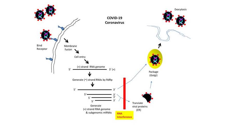
‘RNA interference’ refers to a defense mechanism developed to counter the invading viruses at the RNA level (Agrawal et al., 2003). The post-transcriptional gene silencing mechanism was originally identified in plants and subsequently shown to be operational in various eukaryotes (ex. fungus, vertebrates).  It involves processing a long double stranded RNA (dsRNA) into short 21-25 bp RNA duplexes (with 2 bp overhang at 3’ terminus and phosphorylated 5’ terminus) called ‘small interfering RNAs’ (siRNA) by the enzyme Dicer.  The siRNA duplex is presented by Dicer and TAR RNA binding protein (TRBP) to Argonaute to form the ‘RNA-induced silencing complex’ (RISC) (Wilson et al., 2013).  After unwinding the siRNA duplex, one strand is selected (the other dissociates from RISC) to serve as a guide strand to recognize and cleave target complementary single stranded RNA.  In addition to degrading viral genomic RNA or suppressing the expression of mRNA, RNA interference may function to increase gene expression as targeting a promoter can activate transcription (Li et al., 2008).  

The potential of developing the RNA interference-based therapy for various infectious diseases or genetic disorders (ex. neurodegenerative disorders, cancer) is increasingly being explored.  The siRNA technology involves introducing short dsRNA (~21 bp in length with 2 nucleotide 3’-overhang) into cells.  Upon incorporation into the silencing complex RISC, the sense strand is removed typically.  The remaining antisense strand guides RISC to target mRNA.  Following the cleavage of the target mRNA at a discrete position, the resulting 5’ fragment and 3’ fragment are degraded by exosome and 5'-3' exoribonuclease 1 (XRN1), respectively.  Its catalytic nature allows the degradation process to be repeated with additional mRNA targets.  For pharmacological application, various issues such as stability, delivery, renal clearance and immune response are being addressed.

In 2018, Food and Drug Administration (FDA) approved the first RNA interference inducing drug Onpattro.  Onpattro (also known as Patisiran) was developed by Alnylam Pharmaceuticals, Inc. to treat hereditary transthyretin-mediated amyloidosis (hATTR), a neurodegenerative disorder causing polyneuropathy (dysfunction of peripheral nerves).  This rare but life-threatening disease is caused by the deposition of circulating transthyretin (TTR) amyloid in peripheral nerve, heart, kidney, gastrointestinal tract, etc., causing sensorimotor deterioration and various other symptoms including heart failure.  Most hereditary cases are heterozygous for TTR mutation, and both mutant and normal TTR are found in the amyloid deposits (Coelho et al., 2013). 

Onpattro (Patisiran) is a 21-mer double-stranded small interfering RNA (siRNA) oligonucleotide that targets 3’-UTR (untranslated region) shared by both normal and mutant TTR mRNAs.  It incorporates 2’-O-methylcytidine and 2’-O-methyluridine at specific locations, with 2´-deoxythymidine dinucleotide overhangs at both 3´ ends.  The RNA duplex is encapsulated in a cationic lipid nanoparticle, which is opsonized with ApoE (apolipoprotein E) to facilitate binding to ApoE receptor present on hepatic cell surface for uptake via endocytosis (Coelho et al., 2013).  Phase 3 clinical trial demonstrated that Onpattro improves the symptoms of adult patients suffering from the hereditary transthyretin amyloidosis (Adams et al., 2018).  One caveat is that the prescription cost for Onpattro could be quite high.


These advances have set the stage for the development of RNA interference therapy for other disorders such as cancer, diabetes, cardiac disease, etc.  Additionally, the potential of utilizing siRNA oligonucleotides to treat infectious diseases (ex. hepatitis B virus) has been investigated.  Previously, for SARS coronavirus, siRNAs targeting the regions in the genome encoding spike protein and ORF1b (NSP12) have been designed (Li B et al., 2005).  Another report showed that siRNA targeting the ‘Leader’ sequence could suppress the replication of SARS coronavirus (Li T et al., 2005). 

 With the event of COVID-19 coronavirus pandemic, there is an avid interest in developing siRNA as prophylactic or therapeutic agent.  Alnylam Pharmaceuticals plans to develop siRNA targeting the host factor ACE2 (angiotensin converting enzyme-2; receptor for COVID-19) or TMPRSS2 (transmembrane protease, serine 2; cleaves spike protein to mediate virus entry) to impede the coronavirus infection.  One challenge in developing siRNA therapeutic is the existence of virus encoded proteins that suppress RNA interference as have been documented for influenza A virus, vaccinia virus, Ebola virus, etc.  In the case of SARS coronavirus, its nucleocapsid protein N may repress RNA interference of the host cell (Cul et al., 2015).

 Bio-Synthesis, Inc.—with its capacity to provide siRNA oligonucleotides with unsurpassed specificity and stability--is committed to supporting COVID-19 therapy initiatives. Further, the key to preventing epidemic is the ability to diagnose the infected early to preempt further propagation.  For this, it provides primers and probes (as well as synthetic RNA control) for COVID-19 diagnosis via RT-PCR assay.  Bio-Synthesis, Inc. also specializes in oligonucleotide modification and provides an extensive array of chemically modified nucleoside analogues (over ~200) including bridged nucleic acid (BNA).  A number of options are available to label oligonucleotides (DNA or RNA) with fluorophoreseither terminally or internally as well as conjugate to peptidesIt recently acquired a license from BNA Inc. of Osaka, Japan, for the manufacturing and distribution of BNANC, a third generation of BNA oligonucleotides.  To meet the demands of therapeutic application, its oligonucleotide products are approaching GMP grade.  Bio-Synthesis, Inc. has recently entered into collaborative agreement with Bind Therapeutics, Inc. to synthesize miR-21 blocker using BNA for triple negative breast cancer.  The BNA technology that we offer provides superior, unequalled advantages in base stacking, binding affinity, aqueous solubility and nuclease resistance.  It also improves the formation of duplexes and triplexes by reducing the repulsion between the negatively charged phosphates of the oligonucleotide backbone.  Its single-mismatch discriminating power was especially useful for diagnosis (ex. FISH using DNA probe).  More importantly, BNA oligonucleotide exhibits lesser toxicity than other modified nucleotides for clinical application.


https://www.biosyn.com/oligonucleotide-modification-services.aspx

https://www.biosyn.com/oligo-flourescent-labeling.aspx

https://www.biosyn.com/tew/Speed-up-Identification-of-COVID19.aspx

https://www.biosyn.com/covid-19.aspx

https://www.biosyn.com/sirna-overview.aspx


References

Adams D, Gonzalez-Duarte A, O'Riordan WD, Yang CC, Ueda M, et al.  Patisiran, an RNAi Therapeutic, for Hereditary Transthyretin Amyloidosis.  N Engl J Med. 379:11-21 (2018).  PMID: 29972753 doi: 10.1056/NEJMoa1716153.

Agrawal N, Dasaradhi PV, Mohmmed A, Malhotra P, Bhatnagar RK, Mukherjee SK.  RNA interference: biology, mechanism, and applications.  Microbiol Mol Biol Rev. 67:657-85  (2003).  PMID:14665679  DOI: 10.1128/mmbr.67.4.657-685.2003

Coelho T, Adams D, Silva A, Lozeron P, Hawkins PN, Mant T, et al.  Safety and efficacy of RNAi therapy for transthyretin amyloidosis.  N Engl J Med.  369:819-29 (2013).  PMID: 23984729  doi: 10.1056/NEJMoa1208760.

Cui L, Wang H, Ji Y, Yang J, Xu S, Huang X, Wang Z, et al. The Nucleocapsid Protein of Coronaviruses Acts as a Viral Suppressor of RNA Silencing in Mammalian Cells.  J Virol  89:9029-43 (2015).  PMID: 26085159 PMCID: PMC4524063 DOI: 10.1128/JVI.01331-15

Li BJ, Tang Q, Cheng D, Qin C, Xie FY, Wei Q, et al. Using siRNA in prophylactic and therapeutic regimens against SARS coronavirus in Rhesus macaque.  Nat Med  11:944-51 (2005).  PMID: 16116432  PMCID: PMC7095788 DOI: 10.1038/nm1280 

Li LC.  "Small RNA-Mediated Gene Activation". RNA and the Regulation of Gene Expression: A Hidden Layer of Complexity.  Caister Academic Press.  ISBN 978-1-904455-25-7 (2008).

Li T, Zhang Y, Fu L, Yu C, Li X, Li Y, et al.  siRNA targeting the leader sequence of SARS-CoV inhibits virus replication.  Gene Ther. 12:751-61 (2005).  PMID: 15772689  DOI: 10.1038/sj.gt.3302479

Wilson RC, Doudna JA.  Molecular mechanisms of RNA interference.  Annu Rev Biophys. 42:217-39 (2013).  PMID: 23654304 PMCID: PMC5895182   doi: 10.1146/annurev-biophys-083012-130404. 


Abbreviations commonly used in corona virus research

$
0
0


Abbreviation

 Description


ACE

 Angiotensin-converting enzyme.

ACE2

 Angiotensin-converting enzyme 2.

APN

 Aminopeptidase.

ATP1A1

 ATPase, Na+/K+ transporting, alpha 1 polypeptide.

COPB2

 Coatomer protein complex, subunit beta 2 (beta prime).

CoVs

 Coronaviruses.

CTD

 C-terminal domain.

DDX1

 ATP-dependent RNA helicase.

DPP4

 Dipeptidyl-peptidase 4.

E

 Envelope protein. Approximately 8-12 kDa in size, found in small quantities within the virion. 

ER

 Endoplasmic reticulum.

ERGIC

 Endoplasmic reticulum Golgi intermediate compartment.

ExoN

 Exoribonuclease.

GAPDH

 Glyceraldehyde 3-phosphate dehydrogenase.

HE

 Hemagglutinin-esterase.

HnRNP A1

 Hetrogeneous nuclear ribonucleoprotein A1.

IFITM

 Interferon inducing transmembrane protein.

M

 Membrane protein. Most abundant structural protein in the virion. A small ~25-30 kDa protein with 3 transmembrane   domains giving the virion its shape. Has a small N-terminal glycosylated ectodomain and a larger C-terminal endodomain extending 6-8 mm into the viral particle. Mediates attachment to the host receptor(s).

MADP1

 Zinc Finger CCHC-type and RNA binding motif 1.

MERS-CoV

 Middle Eastern Respiratory Syndrome Coronavirus.

MHV

 Murine hepatitis virus.

Mpro

 Main protease, a serine type protease, encoded by nsp5. Eleven cleavage events.

N

 Nucleocapsid hosphor-protein. Present in the nucleocapsid. Maybe exist as a disulfide linked multimer. Has two separate domains, NYD and CTD. Both bind RNA. Binds viral genome in a beads-on-a-string type conformation. Binds TRS and the genomic packaging signal.

Nsps or NPS

 Non-structural proteins.

NTD

 N-terminal domain.

PABP

 Poly A binding protein.

PCBP1/2

 Poly(rC)-binding protein 1.

Ps

 Packaging signal. A 69-nt stem – loop structure positioned in the 3’-end of ORF1b.

PLpro

 Papain-like protease, encoded within nsp3. PLpro cleaves the nsp1/2, and nsp3/4 boundaries.

RBD

 Receptor Binding Domain.

RdRp

 RNA-dependent RNA polymerase.

RTC

 Replicase-transcriptase complex.

S

 Spike protein. Structural protein. A trimeric glycoprotein. Approximately, 150 kDa in size. Utilizes an N-terminal signal sequence to enter the endoplasmic reticulum (ER), heavily N-linked glycosylated. Homotrimers of the S protein make up the spike structure on the surface of the virus.

SARS-CoV

 Severe Acute Respiratory Syndrome Coronavirus.

sgRNA

 Sub-genomic mRNA.

TRS

 Transcriptional regulatory sequence.

UTR

 Untranslated region.


Bio-Synthesis Inc. is pleased to offer a large variety of oligonucleotides and peptides for a number of research applications, including COVID 19 testing, analysis and vaccine development!

---...---

Efficient delivery of therapeutic RNA

$
0
0

The efficient delivery of RNA molecules into cells or tissue, such as siRNAs, is a necessity for the development of therapeutic RNAs. For many years, the difficulty encountered in efficient and safe in vivodelivery of RNAs prevented the widespread clinical use of siRNA-based therapies until recently.

The first RNA interference (RNAi) drug Onpattro for the treatment of polyneuropathies was approved by the FDA in 2018.


Already, siRNAs for the SARS coronavirus targeting the regions in the genome encoding spike protein and ORF1b (NSP12) have been designed as well, one targeting the ‘Leader’ sequence that could potentially suppress the replication of SARS coronaviruses. 

To improve cellular uptake of small interfering RNAs (siRNAs), Osborn et al. studied the effect of lipid conjugated siRNAs on cellular endogenous lipid transport pathways to determine the biodistribution of conjugated siRNAs.

For the design and synthesis of lipid hydrophobically modified RNA conjugates (hsiRNAs), the researchers used a panel of biologically occurring, but structurally diverse lipids. A clinically validated, chemically altered siRNA scaffold is at the heart of the design. Earlier studies established that cholesterol-conjugated siRNAs have an 8- to 15-fold higher activity when pre-complexed into purified high-density lipoprotein (HDL) than when injected alone. Recent studies indicated that the asialoglycoprotein receptor (ASGPR) contributes to the uptake of unconjugated phosphorothioate-modified antisense oligonucleotides (PS ASOs). The most clinically advanced trivalent N-acetylgalactosamine (GalNac)-siRNA binds to ASGPR on hepatocytes with high selectivity triggering a potent and durable gene silencing in patients. Lipids, when conjugated to siRNA molecules, enhance circulation time, and promote local and systemic delivery and efficacy. For example, cholesterol-modified siRNA silence liver apolipoprotein B (ApoB) expression at high doses.

Lipids, Linkers and Modification for the design of hsiRNA conjugates

Lipids used:


Lithocholic acid (LCA), Docosahexaenoic acid (DHA), Docosanoic acid (DCA), Cholesterol (Chol).


Linker used:  


Triethylene glycol (TEG).


Modifications used: 

2’-Fluoro RNA, 2’-O-Methyl RNA, Cholesterol, Phosphorothioate, TEG linker, 5’-Vinylphosphonate.


Figure 1: Chemical structures of lipids and linker used for conjugation of siRNAs. 

Design of Lipid-hsiRNA conjugates

The scaffold used for the design of lipid-hsiRNA conjugates consists of a hydrophobically modified siRNA scaffold, called hsiRNA. The conjugate contains a 20-nucleotide (nt) antisense or guide strand and a 15-nt sense or passenger strand with alternating 2’-O-methyl and 2’-fluoro sugar modifications. The 5’-end of the antisense strand contains the stable phosphate mimic (E)-vinylphosphonate (VP). The addition of VP to the 5’-end of the guide strand increases siRNA tissue accumulation, extends silencing activity duration, and shields oligonucleotides from exonuclease-mediated degradation, thereby promoting cellular internalization. Because of the rapid degradation and clearance of unmodified oligonucleotides, these modifications stabilize siRNAs and prevent their degradation in cells and tissue. The 3’-end of the sense strand is an optimal position for the attachment of conjugates since it has a minimal effect on siRNA-RISC or intracellular RNA-induced silencing complex loading. The use of linkers, such as the TEG linker, allows attachment of lipid moieties.


Figure 2: Modification pattern of lipid-hsiRNA.

Osborn et al. reported that lipid-conjugated hsiRNA engage distinct lipid transport pathways and elicit silencing in a variety of tissues with minimal systemic toxicity.

Reference

Feingold, K.R., & Grunfeld, C.; Introduction to Lipids and Lipoproteins. NIH ENDOTEXT.

Osborn MF, Coles AH, Biscans A, Haraszti RA, Roux L, Davis S, Ly S, Echeverria D, Hassler MR, Godinho BMDC, Nikan M, Khvorova A. Hydrophobicity drives the systemic distribution of lipid-conjugated siRNAs via lipid transport pathways. Nucleic Acids Res. 2019 Feb 20;47(3):1070-1081. [PMC]


Bio-Synthesis Inc. is pleased to offer a large variety of oligonucleotides and peptides for a number of research applications, including COVID 19 testing, analysis and vaccine development!

---...---

Texan Bats and Coronaviruses

$
0
0

When watching bats in Austin, Texas at the South Congress Bridge, one does not think that these critters carry a coronavirus. The emergence of the Mexico free-tail bats every evening from March to November between 5:30 PM to 6:30 PM is a major spectacle in Austin. Many evenings, bat enthusiasts and watchers wait at lookout spots below and above the South Congress Bridge until the bats emerge in huge flocks, mostly visible as a vibrating cloud. Images of a Townsend's big-eared bat, a horse-shoe bat, and the Mexican free-tailed bat are shown below.


        Townsend's big-eared bat                       Horseshoe bat                         Mexican free-tailed bat

Many viruses are found in bats

Horseshoe bats in China harbor SARS-like coronaviruses. The suggestion that wildlife markets led to the spillover of the newly emerged corona virus SARS-CoV-2 has now given bats a bad reputation. Bats can be carriers of rabies as well as of coronaviruses but are no danger to humans unless people come in contact with their blood or saliva. This is very rare in the western world.

Approximatelly 60 to 80% of emerging infectious diseases in humans originated from wild life. Bats are a natural reservior for a large variety of viruses. The origin and evolution of bat viromes can be studied using modern molecular biology technologies such as the construction of viral nucleic acid libraries, genome analysis via next-generation sequencing, and phylogenetic analysis.

In 1989,
Steece and Altenbach found that a rabies virus infected young Mexican free-tailed bats shortly after birth. However, an immunological study carried out by these two scientists determined that the Mexican free-tailed bats readily recovered from their rabies virus infection. Bats are known to harbor a wide range of human pathogens, including Nipah, Hendra, rabies, Ebola, Marburg, and severe acute respiratory syndrome coronavirus (SARS-CoV). Hence, there is a need to monitor the presence of infectious pathogens, including for the existence of coronaviruses (CoVs).

Anthony et al., in 2013, investigated the presence of CoVs in bats from Mexico and found that an individual bat from the species Lonchorhina aurita or Tomes’s sword-nosed bat testes positive for the novel coronavirus α-CoV Mex-CoV-3. The research group screened 606 bats from 42 different species in Campeche, Chiapas, and Mexico City and identified 13 distinct CoVs. Nine were alpha (α)-CoVs; four wereβ-CoVs.

However, it is essential to remember that most of the viruses carried by bats will not pose any clinical risk. Therefore, bats should not be stigmatized ubiquitously as significant threats to public health.

In recent years, our knowledge of coronaviruses has increased exponentially. Several outbreaks of zoonotic diseases caused by several pathogenic CoVs, including SARS, MERS, and more recently SARS-CoV-2 (COVID-19), stimulated a new era of coronavirus research.

SARS-Cov-2 is the cause of COVID-19. SARS-CoV-2 is a betacoronavirus like MERS-CoV and SARS-CoV. A large family of coronaviruses exists, and many are common in people as well as in different animal species. Sequence analysis suggests that SARS-CoV-2 recently emerged from an animal reservoir in
Wuhan, China

In a dispatch by the CDC, Lau et al. suggested that the genome of SARS-CoV-2 is closest to that detected in a related virus from horseshow bats. Phylogenetic analysis of coronavirus genomes indicated that the genomic region of SARS-CoV-2 is closely related to SARSr-Ra-BatCoV-RaTG13 from an intermediate horseshoe bat in Yunnan. In contrast, its receptor-binding domain (RBD) resembles that of pangolin-SARSr-CoV/MP789/Guangdong/2019 observed in smuggled pangolins in Guangzhou. Identified potential recombination sites around the RBD region, suggest that SARS-CoV-2 maybe a recombinant virus. The genomic backbone appears to have evolved from the Yunnan bat virus-like SARSr-CoVs, and the RBD region came from pangolin virus-like SARSr-CoVs.

The availability of next-generation sequencing (NGS) technologies allows increased surveillance of wild animal species. As of 2019, an enormous number of novel coronaviruses were identified. So far, over 200 novel coronaviruses have been found in bats. Approximately 35% of the sequenced bat virome is from coronaviruses.

Bats are known to be natural reservoirs of a large variety of viruses, including coronaviruses. Severe Acute Respiratory Syndrome Coronavirus (SARS-CoV) and Middle East Respiratory Syndrome Coronavirus (MERS-CoV), are thought to have originated from bats. Various species of horseshoe bats in China harbor genetically diverse SARS-like coronaviruses. Some strains are highly similar to SARS-CoV, even in the spike protein sequence. Some CoVs use the same receptor as SARS-CoV for cell entry. Bat also harbor coronaviruses genetically related to human coronavirus 229E and NL63.


Reference

Anthony, Simon & Ojeda-Flores, Rafael & Rico, Oscar & Navarrete-Macias, Isamara & Zambrana-Torrelio, Carlos & Rostal, Melinda & H. Epstein, Jonathan & Tipps, Teresita & Liang, Eliza & Sanchez-Leon, Maria & Sotomayor-Bonilla, J & Aguirre, A. Alonso & Avila-Flores, Rafael & Medellín, Rodrigo & Goldstein, Tracey & Suzan, Gerardo & Daszak, Peter & Lipkin, W. (2013). Coronaviruses in bats from Mexico.The Journal of general virology. 94. 10.1099/vir.0.049759-0. [PMC]

Banerjee A, Kulcsar K, Misra V, Frieman M, Mossman K. Bats and Coronaviruses. Viruses. 2019 Jan 9;11(1):41. [PMC]

Dominguez SR, O'Shea TJ, Oko LM, Holmes KV. Detection of group 1 coronaviruses in bats in North America. Emerg Infect Dis. 2007 Sep;13(9):1295-300. [PMC]

Ben Hu, Xingyi Ge, Lin-Fa Wang and Zhengli Shi1.; Bat origin of human coronaviruses. Virology Journal (2015) 12:221. [Link]

Joffrin, L., Goodman, S.M., Wilkinson, D.A. et al. Bat coronavirus phylogeography in the Western Indian Ocean. Sci Rep 10, 6873 (2020). [PMC]

Lau SKP, Luk HKH, Wong ACP, Li KSM, Zhu L, He Z, et al. Possible bat origin of severe acute respiratory syndrome coronavirus 2. Emerg Infect Dis. 2020 Jul. [Article]

Quan P. L., Firth C., Street C., Henriquez J. A., Petrosov A., Tashmukhamedova A., Hutchison S. K., Egholm M., Osinubi M. O. V. & other authors (2010). Identification of a severe acute respiratory syndrome coronavirus-like virus in a leaf-nosed bat in Nigeria. MBio 1, e00208–e00210 10.1128/mBio.00208-10 [PMC]

Richard Steece and J. Scott Altenbach; PREVALENCE OF RABIES SPECIFIC ANTIBODIES IN THE MEXICAN FREE-TAILED BAT (TADARIDA BRASILIENSIS MEXICANA) AT LAVA CAVE, NEW MEXICO. Journal of Wildlife Diseases 1989 25:4, 490-496. [Article]


Wu Z, Yang L, Ren X, He G, Zhang J, Yang J, Qian Z, Dong J, Sun L, Zhu Y, Du J, Yang F, Zhang S, Jin Q. Deciphering the bat virome catalog to better understand the ecological diversity of bat viruses and the bat origin of emerging infectious diseases. ISME J. 2016 Mar;10(3):609-20. [PMC]

Bio-Synthesis Inc. is pleased to offer a large variety of oligonucleotides and peptides, modified or unmodified, for a number of research applications, including COVID 19 testing, analysis and vaccine development!

---...---

Endocytic trafficking inhibitors: ongoing clinical trials assess chloroquine’s impact on COVID-19: Bio-Synthesis, Inc. offers CDC-designed Synthetic RNA Control and primers and probes for COVID-19 diagnosis

$
0
0
To date, hundreds of different species of cornaviruses have been identified, which infect a variety of organisms.  For the poultry industry, a significant economic loss has been associated with avian coronavirus IBV, which causes avian infectious bronchitis.  IBV infects a variety of tissues including respiratory tract, digestive system, reproductive system, kidney, etc (Niesters et al., 1986).  Despite the availability of vaccines, its containment is beset by the occurrence of random mutants, which give rise to new variants that escape ‘herd immunity’.

 The current pandemic causing severe respiratory dysfunction is caused by COVID-19 coronavirus.  During infection, spike protein (S) plays a critical role as its interaction with a cellular receptor initiates a series of events leading to its internalization.  For COVID-19, the receptor binding domain located in S1 subunit of S protein may bind to angiotensin-converting enzyme 2 (ACE2) expressed by specific cells in bronchi, lung, tongue, etc. (which is also utilized by SARS and HCoV-NL63 human coronaviruses for cell entry) (Walls et al., 2020).

As a component of the renin-angiotensin system that regulates blood pressure, angiotensin I is converted to angiotensin II by ACE (angiotensin converting enzyme), which constricts blood vessels to reduce flow.  This is countered by ACE2 that hydrolyzes angiotensin II.  The binding of ACE2 by COVID-19 spike protein, was suggested to downregulate ACE2 (Bombardini et al., 2020), which may decrease the flow of pulmonary artery into the lung, hence affecting the level of oxygenated blood.  This may exacerbate the negative effects brought on by cytokine release syndrome (also known as ‘cytokine storm’) due to hyperinflammation, which causes ARDS (acute respiratory distress syndrome), that a minor subset of COVID-19 infected people experience.

 The cell entry of COVID-19 requires the fusion of vial envelope with the cell membrane.  Whether the fusion occurs at the plasma membrane or endosomal membrane following endocytic uptake (or both) has not been clearly delineated.  Following the binding of S protein to ACE2 receptor, it undergoes a proteolytic cleavage to separate the receptor binding domain from the fusion domain located in S2 subunit.  This is thought to be mediated by TMPRRS2 protease (or possibly others, ex. endosomal proteases that are active at lower pH).

Further, an additional cleavage occurs at a distinct site to expose the ‘fusion peptide’ comprised of hydrophobic residues.  The liberated fusion domain inserts into the targeted cellular membrane to form a ‘pre-hairpin’ structure using the fusion peptide.  Then, two heptad repeats (HR1 and HR2) residing in S2 subunit join to form an antiparallel 6-helix bundle, which serves to pull back on the viral and cellular membranes, allowing tem to fuse.  The fusion creates a channel through which the virus capsid (protein sheath containing its RNA genome) can enter the cell interior.


Chloroquine (CQ) has been used extensively to manage malaria though its efficacy is limited by the emergence of resistant strains.  The plasmodium malaria invades red blood cells and catabolizes hemoglobin in its food vacuoles (acidic), with the resultant byproduct (heme) being detoxified.  Chloroquine, which permeates through diffusion, becomes trapped inside the vacuole upon protonation, and disrupts the detoxification of heme, leading to cell lysis (Lin et al., 2015).

Likewise, chloroquine accumulates in late endosomes (or lysosomes) and increases their pH.  This may potentially interfere with the function of endosomal proteases that activate spike proteins for fusion with endosomal membrane for COVID-19 entry.   This view (i.e interference with endocytic uptake) is indirectly supported by the finding that chloroquine prevents the receptor mediated endocytosis of nanoparticles (of similar size as COVID-19 virus) by inhibiting PICALM (phosphatidyl inositol binding clathrin assembly protein) that regulates endocytosis (Hu et al., 2020). To explore, the potential efficacy of chloroquine in preventing COVID-19 pathogenesis is being assessed through numerous (as many as ~50) clinical trials that are currently underway.  Nevertheless, chloroquine may cause side effects (ex. affect vision) and several recent clinical trials examining chloroquine (or its derivative hydroxychloroquine) have reported cardiac problems or deaths. 

Chloroquine may interfere with additional steps in endocytic trafficking.  Of interest is the finding that chloroquine may block the glycosylation of ACE2 receptor to interfere with SARS infection (Vincent et al., 2005).   Endocytic trafficking is also exploited for cell entry by oncogenic viruses such as human papilloma virus (HPV), which causes head and neck cancer, cervical cancer, and other malignancies (~5% of all cancers globally).

 Bio-Synthesis, Inc.—with its capacity to provide siRNA oligonucleotides with unsurpassed specificity and stability--is committed to supporting COVID-19 therapy initiatives. Further, the key to preventing epidemic is the ability to diagnose the infected early to preempt further propagation.  For this, it provides primers and probes (as well as synthetic RNA control) for COVID-19 diagnosis via RT-PCR assay.  Bio-Synthesis, Inc. also specializes in oligonucleotide modification and provides an extensive array of chemically modified nucleoside analogues (over ~200) including bridged nucleic acid (BNA).  A number of options are available to label oligonucleotides (DNA or RNA) with fluorophoreseither terminally or internally as well as conjugate to peptidesIt recently acquired a license from BNA Inc. of Osaka, Japan, for the manufacturing and distribution of BNANC, a third generation of BNA oligonucleotides.  To meet the demands of therapeutic application, its oligonucleotide products are approaching GMP grade.  Bio-Synthesis, Inc. has recently entered into collaborative agreement with Bind Therapeutics, Inc. to synthesize miR-21 blocker using BNA for triple negative breast cancer.  The BNA technology that we offer provides superior, unequalled advantages in base stacking, binding affinity, aqueous solubility and nuclease resistance.  It also improves the formation of duplexes and triplexes by reducing the repulsion between the negatively charged phosphates of the oligonucleotide backbone.  Its single-mismatch discriminating power was especially useful for diagnosis (ex. FISH using DNA probe).  More importantly, BNA oligonucleotide exhibits lesser toxicity than other modified nucleotides for clinical application.


https://www.biosyn.com/oligonucleotide-modification-services.aspx

https://www.biosyn.com/oligo-flourescent-labeling.aspx

https://www.biosyn.com/tew/Speed-up-Identification-of-COVID19.aspx

https://www.biosyn.com/covid-19.aspx

https://www.biosyn.com/sirna-overview.aspx


References

Bombardini T, Picano E. Angiotensin-Converting Enzyme 2 as the Molecular Bridge Between Epidemiologic and Clinical Features of COVID-19.  Can J Cardiol. Mar 29. pii: S0828-282X(20)30299-3.  (2020)  PMID: 32299780  doi: 10.1016/j.cjca.2020.03.026.  

Hu TY, Frieman M, Wolfram J. Insights from nanomedicine into chloroquine efficacy against COVID-19.  Nat Nanotechnol. 15:247-249 (2020). PMID: 32203437  doi: 10.1038/s41565-020-0674-9.

Lin JW, Spaccapelo R, Schwarzer E, Sajid M, Annoura T, Deroost K, et al. Replication of Plasmodium in reticulocytes can occur without hemozoin formation, resulting in chloroquine resistance. J Exp Med.212:893-903 (2015). PMID: 25941254   doi: 10.1084/jem.20141731.

Niesters HG, Lenstra JA, Spaan WJ, Zijderveld AJ, Bleumink-Pluym NM, Hong F, et al.  The peplomer protein sequence of the M41 strain of coronavirus IBV and its comparison with Beaudette strains.  Virus Res. 5:253-63 (1986). PMID: 2429473   PMCID: PMC7134181 DOI: 10.1016/0168-1702(86)90022-5

Vincent MJ, Bergeron E, Benjannet S, Erickson BR, Rollin PE, Ksiazek TG, et al.  Chloroquine is a potent inhibitor of SARS coronavirus infection and spread.  Virol J.  2:69 (2005). PMID: 16115318   doi: 10.1186/1743-422X-2-69.

Walls AC, Park YJ, Tortorici MA, Wall A, McGuire AT, Veesler D. Structure, Function, and Antigenicity of the SARS-CoV-2 Spike Glycoprotein.  Cell 181:281-292.e6. (2020) PMID: 32155444 doi: 10.1016/j.cell.2020.02.058. Epub 2020 Mar 9.


Remdesivir and COVID-19

$
0
0

As of May 04, 2002, according to the John Hopkins University, more than 3.5 million people have tested positive for the coronavirus worldwide, and over 248,000 people have died. Therefore, effective therapeutic drugs for coronavirus disease 2020 (COVID-19) are desperately needed for the control of the current pandemic.

A recent
clinical trial at the NIH now shows that Remsdesivir accelerates recovery from advanced COVID-19. Apparently, according to the latest report, patients treated with Remdesivir recover more quickly in comparison to untreated ones. There is hope.  It is hoped that this treatment helps COVID-19 patients recover with minimal side effects. However, only time will tell how effective Remdesivir is as a treatment of COVID-19.


Figure 1: Remdesivir. (Left) Chemical structure of Remdesivir, or GS 5734. Formula: C27H35N6O8P; 2-ethylbutyl (2S)-2-[[[(2R,3S,4R,5R)-5-(4-aminopyrrolo[2,1-f][1,2,4]triazin-7-yl)-5-cyano-3,4-dihydroxyoxolan-2-yl]methoxy-phenoxyphosphoryl]amino]propanoate; l-alanine, N-((S)-hydroxyphenoxyphosphinyl)-, 2-ethylbutyl ester, 6-ester with 2-C-(4-aminopyrrolo(2,1-f)(1,2,4)triazin-7-yl)-2,5-anhydro-d-altrononitrile. Molecular weight: Average: 602.585; Monoisotopic: 602.225399109.  (Right) Partial structure derived from the structure PDB ID 7BV2. https://pubchem.ncbi.nlm.nih.gov/compound/Remdesivir


Remdesivir, GS-5734, is an antiviral drug developed by Gilead Sciences. Remdesivir is an adenosine triphosphate analog. In 2016 Warren et al. investigated the drug for the treatment of Ebola, and Sheahan et al., in 2017, demonstrated the drug is also active in coronaviruses. A more recent test investigated Remdesivir as a potential drug for the treatment of patients infected with SARS-CoV-2, the coronavirus responsible for COVID-19 (de Wit et al.; Ledford). More recently, in May 2020, the FDA authorized the antiviral drug Remdesivir for the emergency treatment of hospitalized patients with coronavirus infections, as told by the President of the United States of America on Friday, May 01, 2020.

The viral RNA-dependent RNA polymerase (RdRp) is needed for replication of SARS-CoV-2 viral RNA. RdRp is the likely target of the investigational nucleotide analogue Remdesivir. Agostini et al., in 2018, suggested that the Remdesivir nucleoside analog inhibits the viral RNA polymerase. Recently, Gao et al. reported the structure of the COVID-19 virus full-length nsp12 in complex with cofactors nsp7 and nsp8 at 2.9-Å resolution solved by cryo-EM. The structure revealed the presence of the conserved architecture of the polymerase core of the viral polymerase protein family. The non-structural protein nsp12 was found to contain a newly identified β-hairpin domain at its N-terminus. The structural model shows how Remdesivir binds to the polymerase. The structure provides a guiding principle for the design of new antiviral therapeutics targeting viral RdRp.


Figure 2: Complex of nsp12-nsp7-nsp8 bound to the template-primer RNA and triphosphate form of Remdesivir. Cryo EM structure PDB ID 7BV2.  https://www.rcsb.org/structure/7BV2.

The nonstructural protein nsp7 produced by both pp1a and pp1ab forms a hexadecameric complex with nsp8 and is thought to act as a processivity clamp for RNA polymerase. The RNA-dependent RNA polymerase (RdRp), nsp12, is produced by pp1ab only.  


Figure 3: Remdesivir-RNA primer. Extracted from source PDB ID 7BV2. (Left) Stick model with Remdesivir shown as spheres. (Right) Surface model of the RNA with Remdesivir shown as sticks.

Incorporation of the analog into viral RNA prevents the addition of nucleotides leading to the termination of RNA transcription preventing the virus from multiplying. Enzyme kinetic studies performed by Gordon et al., in 2020, indicate that this RdRp efficiently incorporates the active triphosphate form of Remdesivir into RNA. Incorporation of the Remdesivir triphosphate at position i caused termination of RNA synthesis at position i+3. Almost identical results were found for SARS-CoV, MERS-CoV, and SARS-CoV-2 RdRps. A delayed chain-termination is Remdesivir’s plausible mechanism of action. Also, Remdesivir shows broad-spectrum antiviral activity against several RNA viruses, including Ebola virus (EBOV) and Middle East respiratory syndrome coronavirus (MERS-CoV). 


Reference

John Hopkins University coronavirus info:  https://coronavirus.jhu.edu/map.html

Remsdesivir trial:
https://www.cnn.com/2020/04/16/health/coronavirus-remdesivir-trial/index.html.

Agostini ML, Andres EL, Sims AC, Graham RL, Sheahan TP, Lu X, Smith EC, Case JB, Feng JY, Jordan R, Ray AS, Cihlar T, Siegel D, Mackman RL, Clarke MO, Baric RS, Denison MR: Coronavirus Susceptibility to the Antiviral Remdesivir (GS-5734) Is Mediated by the Viral Polymerase and the Proofreading Exoribonuclease. mBio. 2018 Mar 6;9(2). pii: mBio.00221-18. doi: 10.1128/mBio.00221-18. [PubMed]

de Wit E, Feldmann F, Cronin J, Jordan R, Okumura A, Thomas T, Scott D, Cihlar T, Feldmann H: Prophylactic and therapeutic remdesivir (GS-5734) treatment in the rhesus macaque model of MERS-CoV infection. Proc Natl Acad Sci U S A. 2020 Feb 13. pii: 1922083117. doi: 10.1073/pnas.1922083117. [PNAS]

Ledford H: Hopes rise on coronavirus drug remdesivir. Nature. 2020 Apr 29. pii: [PubMed]

Calvin J. Gordon, Egor P. Tchesnokov, Emma Woolner, Jason K. Perry, Joy Y. Feng, Danielle P. Porter, and Matthias Götte; Remdesivir is a direct-acting antiviral that inhibits RNA-dependent RNA polymerase from severe acute respiratory syndrome coronavirus 2 with high potency. JBC April 13, 2020. [JBC]

Warren TK, Jordan R, Lo MK, Ray AS, Mackman RL, Soloveva V, Siegel D, Perron M, Bannister R, Hui HC, Larson N, Strickley R, Wells J, Stuthman KS, Van Tongeren SA, Garza NL, Donnelly G, Shurtleff AC, Retterer CJ, Gharaibeh D, Zamani R, Kenny T, Eaton BP, Grimes E, Welch LS, Gomba L, Wilhelmsen CL, Nichols DK, Nuss JE, Nagle ER, Kugelman JR, Palacios G, Doerffler E, Neville S, Carra E, Clarke MO, Zhang L, Lew W, Ross B, Wang Q, Chun K, Wolfe L, Babusis D, Park Y, Stray KM, Trancheva I, Feng JY, Barauskas O, Xu Y, Wong P, Braun MR, Flint M, McMullan LK, Chen SS, Fearns R, Swaminathan S, Mayers DL, Spiropoulou CF, Lee WA, Nichol ST, Cihlar T, Bavari S: Therapeutic efficacy of the small molecule GS-5734 against Ebola virus in rhesus monkeys. Nature. 2016 Mar 17;531(7594):381-5. doi: 10.1038/nature17180. Epub 2016 Mar 2. [PMC]

Bio-Synthesis Inc. is pleased to offer a large variety of oligonucleotides and peptides, modified or unmodified, for a number of research applications, including COVID 19 testing, analysis and vaccine development!

---...---

The SARS-CoV-2 Envelope Protein

$
0
0

Coronaviruses are known to cross the species barrier and infect humans. The recent outbreak of SARS-CoV-2 (COVID-19) is a vivid example. The outbreaks of SARS in 2003 and, more recently, Middle-East respiratory syndrome (MERS) already demonstrated the lethality of coronaviruses (CoVs).


Since 2003, much progress has been made in understanding aspects of the virus’ life cycle. The CoV envelope (E) protein is a small, integral membrane protein taking part in virus assembly, budding envelope formation, and pathogenesis. Recent biochemical studies increased our knowledge of E protein’s structure, structural motifs, its function as an ion-channeling viroprotein, as well as its interaction with other coronavirus proteins and host cell proteins.

Coronaviral genomes encode four major structural proteins required to produce a structural complete viral particle. The four major structural proteins are the spike or surface protein (S), nucleocapsid (N), membrane (M) protein, and the envelope (E) protein. Some C0Vs do not need the E protein to form a complete, infectious virion.

The E protein is the smallest of the major structural proteins. During the replication cycle, the virus expresses the E protein in high abundance inside the cell. However, only a small portion is incorporated into the virion envelope. Most of the protein is localized at the site of intercellular trafficking, at the endoplasmic reticulum (ER), the Golgi, and the Endoplasmic reticulum Golgi intermediate compartment (ERGIC). The E protein participates in viral assembly and budding. Coronaviruses without the E protein appear to mature much slower than CoVs in which the E protein is present.

The E protein of coronaviruses is a conserved, short, integral membrane protein of 76 to 109 amino acids, ranging in size from 8.4 to 12 kDa. Figure 1 shows the alignment of E protein sequences for MERS, SARS1, SARS-CoV-2, sequences from solved protein structures 5X29, 2MM4, 5XES, and the self-assembly peptide TVYVYSRVK.

Figure 1: Alignment of CoV E proteins for MERS, SARS and SARS CoV-2, and the C-terminal peptide TVYVYSRVK known to self-assemble.

The E protein has a short, hydrophilic amino terminus of 7 to 12 amino acids, followed by a large hydrophobic transmembrane domain (TMD) of 25 amino acids, and a hydrophilic carboxyl terminus (Figure 2). The hydrophobic TMD region has at least one amphipatic α-helix that can oligomerize to form an ion-conductive pore of membranes. Sequence analysis of both, E and S protein for SARS and SARS CoV-2, revealed a triple cysteine motif located after the transmembrane region of the E protein (LCAYCN) and in the C-terminal end of the S protein (SCGSCCK). Wu et al., in 2003, suggested that these two motifs could serve as a structural basis for the association between the E and S protein. However, this may need to be experimentally verified. Synthetic peptides spanning the motif could serve as tools for the study of these motifs.


 Figure 2: Amino acid sequences and domains of the E protein from MERS, SARS and SARS-CoV-2 virus. The E protein contains three domains, the amino (N)-terminal domain, the transmembrane domain (TMD), and the carboxy (C)-terminal domain. The putative interaction peptide and the C-terminal peptide known to self-assemble are also indicated within the sequence.

Li et al., in 2014, reported that the E protein in the severe acute respiratory syndrome virus has only one transmembrane (TM) domain in micelles and that the predicted β-coil-β motif forms a short membrane-bound α-helix connected by a disordered loop to the TM domain. More recently, Surya et al., in 2018, reported the structural model of the SARS coronavirus E channel in lyso-myristoyl phosphatidylglycerol (LMPG) micelles (Figure 3).


Figure 3: NMR model of the SARS coronavirus E channel in LMPG micelles (Surya et al.; PCB ID 5X29). The model of the pentamer was optained by docking the monomeric form of the protein using the software HADDOCK 2.2. In LMPG micelles, the C-terminal tail of SARS-Cov E protein is α-helical. 

Reference

Li Y, Surya W, Claudine S, Torres J. Structure of a conserved Golgi complex-targeting signal in coronavirus envelope proteins. J Biol Chem. 2014 May 2;289(18):12535-49. [PMC]

Schoeman, D., Fielding, B.C. Coronavirus envelope protein: current knowledge. Virol J 16, 69 (2019).

Surya W, Li Y, Torres J. Structural model of the SARS coronavirus E channel in LMPG micelles. Biochim Biophys Acta Biomembr. 2018 Jun;1860(6):1309-1317. [PMC]

Wu Q, Zhang Y, Lü H, Wang J, He X, Liu Y, et al. The E protein is a multifunctional membrane protein of SARS-CoV. Genomics, Proteomics & Bioinformatics. 2003;1(2):131–44. [Article]  


Bio-Synthesis Inc. is pleased to offer a large variety of DNA/RNA oligonucleotides and peptides, modified or unmodified, conjugates, as well as stapled peptides, for a number of research applications, including COVID 19 testing, analysis and vaccine development!


---...---


FDA approves first-ever siRNA oligonucleotide drug for neurodegenerative disorder raising the prospect for cancer or COVID-19 therapy

$
0
0
‘RNA interference’ refers to a defense mechanism developed to counter the invading viruses at the RNA level (Agrawal et al., 2003). The post-transcriptional gene silencing mechanism was originally identified in plants and subsequently shown to be operational in various eukaryotes (ex. fungus, vertebrates).  It involves processing a long double stranded RNA (dsRNA) into short 21-25 bp RNA duplexes (with 2 bp overhang at 3’ terminus and phosphorylated 5’ terminus) called ‘small interfering RNAs’ (siRNA) by the enzyme Dicer.  The siRNA duplex is presented by Dicer and TAR RNA binding protein (TRBP) to Argonaute to form the ‘RNA-induced silencing complex’ (RISC) (Wilson et al., 2013).  After unwinding the siRNA duplex, one strand is selected (the other dissociates from RISC) to serve as a guide strand to recognize and cleave target complementary single stranded RNA.  In addition to degrading viral genomic RNA or suppressing the expression of mRNA, RNA interference may function to increase gene expression as targeting a promoter can activate transcription (Li et al., 2008).  

The potential of developing the RNA interference-based therapy for various infectious diseases or genetic disorders (ex. neurodegenerative disorders, cancer) is increasingly being explored.  The siRNA technology involves introducing short dsRNA (~21 bp in length with 2 nucleotide 3’-overhang) into cells.  Upon incorporation into the silencing complex RISC, the sense strand is removed typically.  The remaining antisense strand guides RISC to target mRNA.  Following the cleavage of the target mRNA at a discrete position, the resulting 5’ fragment and 3’ fragment are degraded by exosome and 5'-3' exoribonuclease 1 (XRN1), respectively.  Its catalytic nature allows the degradation process to be repeated with additional mRNA targets.  For pharmacological application, various issues such as stability, delivery, renal clearance and immune response are being addressed.

In 2018, Food and Drug Administration (FDA) approved the first RNA interference inducing drug Onpattro.  Onpattro (also known as Patisiran) was developed by Alnylam Pharmaceuticals, Inc. to treat hereditary transthyretin-mediated amyloidosis (hATTR), a neurodegenerative disorder causing polyneuropathy (dysfunction of peripheral nerves).  This rare but life-threatening disease is caused by the deposition of circulating transthyretin (TTR) amyloid in peripheral nerve, heart, kidney, gastrointestinal tract, etc., causing sensorimotor deterioration and various other symptoms including heart failure.  Most hereditary cases are heterozygous for TTR mutation, and both mutant and normal TTR are found in the amyloid deposits (Coelho et al., 2013). 

Onpattro (Patisiran) is a 21-mer double-stranded small interfering RNA (siRNA) oligonucleotide that targets 3’-UTR (untranslated region) shared by both normal and mutant TTR mRNAs.  It incorporates 2’-O-methylcytidine and 2’-O-methyluridine at specific locations, with 2´-deoxythymidine dinucleotide overhangs at both 3´ ends.  The RNA duplex is encapsulated in a cationic lipid nanoparticle, which is opsonized with ApoE (apolipoprotein E) to facilitate binding to ApoE receptor present on hepatic cell surface for uptake via endocytosis (Coelho et al., 2013).  Phase 3 clinical trial demonstrated that Onpattro improves the symptoms of adult patients suffering from the hereditary transthyretin amyloidosis (Adams et al., 2018).  One caveat is that the prescription cost for Onpattro could be quite high.


These advances have set the stage for the development of RNA interference therapy for other disorders such as cancer, diabetes, cardiac disease, etc.  Additionally, the potential of utilizing siRNA oligonucleotides to treat infectious diseases (ex. hepatitis B virus) has been investigated.  Previously, for SARS coronavirus, siRNAs targeting the regions in the genome encoding spike protein and ORF1b (NSP12) have been designed (Li B et al., 2005).  Another report showed that siRNA targeting the ‘Leader’ sequence could suppress the replication of SARS coronavirus (Li T et al., 2005). 

 With the event of COVID-19 coronavirus pandemic, there is an avid interest in developing siRNA as prophylactic or therapeutic agent.  Alnylam Pharmaceuticals plans to develop siRNA targeting the host factor ACE2 (angiotensin converting enzyme-2; receptor for COVID-19) or TMPRSS2 (transmembrane protease, serine 2; cleaves spike protein to mediate virus entry) to impede the coronavirus infection.  One challenge in developing siRNA therapeutic is the existence of virus encoded proteins that suppress RNA interference as have been documented for influenza A virus, vaccinia virus, Ebola virus, etc.  In the case of SARS coronavirus, its nucleocapsid protein N may repress RNA interference of the host cell (Cul et al., 2015).

 Bio-Synthesis, Inc.—with its capacity to provide siRNA oligonucleotides with unsurpassed specificity and stability--is committed to supporting COVID-19 therapy initiatives. Further, the key to preventing epidemic is the ability to diagnose the infected early to preempt further propagation.  For this, it provides primers and probes (as well as synthetic RNA control) for COVID-19 diagnosis via RT-PCR assay.  Bio-Synthesis, Inc. also specializes in oligonucleotide modification and provides an extensive array of chemically modified nucleoside analogues (over ~200) including bridged nucleic acid (BNA).  A number of options are available to label oligonucleotides (DNA or RNA) with fluorophoreseither terminally or internally as well as conjugate to peptidesIt recently acquired a license from BNA Inc. of Osaka, Japan, for the manufacturing and distribution of BNANC, a third generation of BNA oligonucleotides.  To meet the demands of therapeutic application, its oligonucleotide products are approaching GMP grade.  Bio-Synthesis, Inc. has recently entered into collaborative agreement with Bind Therapeutics, Inc. to synthesize miR-21 blocker using BNA for triple negative breast cancer.  The BNA technology that we offer provides superior, unequalled advantages in base stacking, binding affinity, aqueous solubility and nuclease resistance.  It also improves the formation of duplexes and triplexes by reducing the repulsion between the negatively charged phosphates of the oligonucleotide backbone.  Its single-mismatch discriminating power was especially useful for diagnosis (ex. FISH using DNA probe).  More importantly, BNA oligonucleotide exhibits lesser toxicity than other modified nucleotides for clinical application.


https://www.biosyn.com/oligo-flourescent-labeling.aspx

https://www.biosyn.com/tew/Speed-up-Identification-of-COVID19.aspx

https://www.biosyn.com/covid-19.aspx

https://www.biosyn.com/sirna-overview.aspx


References

Adams D, Gonzalez-Duarte A, O'Riordan WD, Yang CC, Ueda M, et al.  Patisiran, an RNAi Therapeutic, for Hereditary Transthyretin Amyloidosis.  N Engl J Med. 379:11-21 (2018).  PMID: 29972753 doi: 10.1056/NEJMoa1716153.

Agrawal N, Dasaradhi PV, Mohmmed A, Malhotra P, Bhatnagar RK, Mukherjee SK.  RNA interference: biology, mechanism, and applications.  Microbiol Mol Biol Rev. 67:657-85  (2003).  PMID:14665679  DOI: 10.1128/mmbr.67.4.657-685.2003

Coelho T, Adams D, Silva A, Lozeron P, Hawkins PN, Mant T, et al.  Safety and efficacy of RNAi therapy for transthyretin amyloidosis.  N Engl J Med.  369:819-29 (2013).  PMID: 23984729  doi: 10.1056/NEJMoa1208760.

Cui L, Wang H, Ji Y, Yang J, Xu S, Huang X, Wang Z, et al. The Nucleocapsid Protein of Coronaviruses Acts as a Viral Suppressor of RNA Silencing in Mammalian Cells.  J Virol  89:9029-43 (2015).  PMID: 26085159 PMCID: PMC4524063 DOI: 10.1128/JVI.01331-15

Li BJ, Tang Q, Cheng D, Qin C, Xie FY, Wei Q, et al. Using siRNA in prophylactic and therapeutic regimens against SARS coronavirus in Rhesus macaque.  Nat Med  11:944-51 (2005).  PMID: 16116432  PMCID: PMC7095788 DOI: 10.1038/nm1280 

Li LC.  "Small RNA-Mediated Gene Activation". RNA and the Regulation of Gene Expression: A Hidden Layer of Complexity.  Caister Academic Press.  ISBN 978-1-904455-25-7 (2008).

Li T, Zhang Y, Fu L, Yu C, Li X, Li Y, et al.  siRNA targeting the leader sequence of SARS-CoV inhibits virus replication.  Gene Ther. 12:751-61 (2005).  PMID: 15772689  DOI: 10.1038/sj.gt.3302479

Wilson RC, Doudna JA.  Molecular mechanisms of RNA interference.  Annu Rev Biophys. 42:217-39 (2013).  PMID: 23654304 PMCID: PMC5895182   doi: 10.1146/annurev-biophys-083012-130404. 


Endocytic trafficking inhibitors: ongoing clinical trials assess chloroquine’s impact on COVID-19

$
0
0
To date, hundreds of different species of cornaviruses have been identified, which infect a variety of organisms.  For the poultry industry, a significant economic loss has been associated with avian coronavirus IBV, which causes avian infectious bronchitis.  IBV infects a variety of tissues including respiratory tract, digestive system, reproductive system, kidney, etc (Niesters et al., 1986).  Despite the availability of vaccines, its containment is beset by the occurrence of random mutants, which give rise to new variants that escape ‘herd immunity’.

 The current pandemic causing severe respiratory dysfunction is caused by COVID-19 coronavirus.  During infection, spike protein (S) plays a critical role as its interaction with a cellular receptor initiates a series of events leading to its internalization.  For COVID-19, the receptor binding domain located in S1 subunit of S protein may bind to angiotensin-converting enzyme 2 (ACE2) expressed by specific cells in bronchi, lung, tongue, etc. (which is also utilized by SARS and HCoV-NL63 human coronaviruses for cell entry) (Walls et al., 2020).

As a component of the renin-angiotensin system that regulates blood pressure, angiotensin I is converted to angiotensin II by ACE (angiotensin converting enzyme), which constricts blood vessels to reduce flow.  This is countered by ACE2 that hydrolyzes angiotensin II.  The binding of ACE2 by COVID-19 spike protein, was suggested to downregulate ACE2 (Bombardini et al., 2020), which may decrease the flow of pulmonary artery into the lung, hence affecting the level of oxygenated blood.  This may exacerbate the negative effects brought on by cytokine release syndrome (also known as ‘cytokine storm’) due to hyperinflammation, which causes ARDS (acute respiratory distress syndrome), that a minor subset of COVID-19 infected people experience.

 The cell entry of COVID-19 requires the fusion of vial envelope with the cell membrane.  Whether the fusion occurs at the plasma membrane or endosomal membrane following endocytic uptake (or both) has not been clearly delineated.  Following the binding of S protein to ACE2 receptor, it undergoes a proteolytic cleavage to separate the receptor binding domain from the fusion domain located in S2 subunit.  This is thought to be mediated by TMPRRS2 protease (or possibly others, ex. endosomal proteases that are active at lower pH).

Further, an additional cleavage occurs at a distinct site to expose the ‘fusion peptide’ comprised of hydrophobic residues.  The liberated fusion domain inserts into the targeted cellular membrane to form a ‘pre-hairpin’ structure using the fusion peptide.  Then, two heptad repeats (HR1 and HR2) residing in S2 subunit join to form an antiparallel 6-helix bundle, which serves to pull back on the viral and cellular membranes, allowing tem to fuse.  The fusion creates a channel through which the virus capsid (protein sheath containing its RNA genome) can enter the cell interior.


Chloroquine (CQ) has been used extensively to manage malaria though its efficacy is limited by the emergence of resistant strains.  The plasmodium malaria invades red blood cells and catabolizes hemoglobin in its food vacuoles (acidic), with the resultant byproduct (heme) being detoxified.  Chloroquine, which permeates through diffusion, becomes trapped inside the vacuole upon protonation, and disrupts the detoxification of heme, leading to cell lysis (Lin et al., 2015).

Likewise, chloroquine accumulates in late endosomes (or lysosomes) and increases their pH.  This may potentially interfere with the function of endosomal proteases that activate spike proteins for fusion with endosomal membrane for COVID-19 entry.   This view (i.e interference with endocytic uptake) is indirectly supported by the finding that chloroquine prevents the receptor mediated endocytosis of nanoparticles (of similar size as COVID-19 virus) by inhibiting PICALM (phosphatidyl inositol binding clathrin assembly protein) that regulates endocytosis (Hu et al., 2020). To explore, the potential efficacy of chloroquine in preventing COVID-19 pathogenesis is being assessed through numerous (as many as ~50) clinical trials that are currently underway.  Nevertheless, chloroquine may cause side effects (ex. affect vision) and several recent clinical trials examining chloroquine (or its derivative hydroxychloroquine) have reported cardiac problems or deaths. 

Chloroquine may interfere with additional steps in endocytic trafficking.  Of interest is the finding that chloroquine may block the glycosylation of ACE2 receptor to interfere with SARS infection (Vincent et al., 2005).   Endocytic trafficking is also exploited for cell entry by oncogenic viruses such as human papilloma virus (HPV), which causes head and neck cancer, cervical cancer, and other malignancies (~5% of all cancers globally).

 Bio-Synthesis, Inc.—with its capacity to provide siRNA oligonucleotides with unsurpassed specificity and stability--is committed to supporting COVID-19 therapy initiatives. Further, the key to preventing epidemic is the ability to diagnose the infected early to preempt further propagation.  For this, it provides primers and probes (as well as synthetic RNA control) for COVID-19 diagnosis via RT-PCR assay.  Bio-Synthesis, Inc. also specializes in oligonucleotide modification and provides an extensive array of chemically modified nucleoside analogues (over ~200) including bridged nucleic acid (BNA).  A number of options are available to label oligonucleotides (DNA or RNA) with fluorophoreseither terminally or internally as well as conjugate to peptidesIt recently acquired a license from BNA Inc. of Osaka, Japan, for the manufacturing and distribution of BNANC, a third generation of BNA oligonucleotides.  To meet the demands of therapeutic application, its oligonucleotide products are approaching GMP grade.  Bio-Synthesis, Inc. has recently entered into collaborative agreement with Bind Therapeutics, Inc. to synthesize miR-21 blocker using BNA for triple negative breast cancer.  The BNA technology that we offer provides superior, unequalled advantages in base stacking, binding affinity, aqueous solubility and nuclease resistance.  It also improves the formation of duplexes and triplexes by reducing the repulsion between the negatively charged phosphates of the oligonucleotide backbone.  Its single-mismatch discriminating power was especially useful for diagnosis (ex. FISH using DNA probe).  More importantly, BNA oligonucleotide exhibits lesser toxicity than other modified nucleotides for clinical application.


https://www.biosyn.com/oligonucleotide-modification-services.aspx

https://www.biosyn.com/oligo-flourescent-labeling.aspx

https://www.biosyn.com/tew/Speed-up-Identification-of-COVID19.aspx

https://www.biosyn.com/covid-19.aspx


References

Bombardini T, Picano E. Angiotensin-Converting Enzyme 2 as the Molecular Bridge Between Epidemiologic and Clinical Features of COVID-19.  Can J Cardiol. Mar 29. pii: S0828-282X(20)30299-3.  (2020)  PMID: 32299780  doi: 10.1016/j.cjca.2020.03.026.  

Hu TY, Frieman M, Wolfram J. Insights from nanomedicine into chloroquine efficacy against COVID-19.  Nat Nanotechnol. 15:247-249 (2020). PMID: 32203437  doi: 10.1038/s41565-020-0674-9.

Lin JW, Spaccapelo R, Schwarzer E, Sajid M, Annoura T, Deroost K, et al. Replication of Plasmodium in reticulocytes can occur without hemozoin formation, resulting in chloroquine resistance. J Exp Med.212:893-903 (2015). PMID: 25941254   doi: 10.1084/jem.20141731.

Niesters HG, Lenstra JA, Spaan WJ, Zijderveld AJ, Bleumink-Pluym NM, Hong F, et al.  The peplomer protein sequence of the M41 strain of coronavirus IBV and its comparison with Beaudette strains.  Virus Res. 5:253-63 (1986). PMID: 2429473   PMCID: PMC7134181 DOI: 10.1016/0168-1702(86)90022-5

Vincent MJ, Bergeron E, Benjannet S, Erickson BR, Rollin PE, Ksiazek TG, et al.  Chloroquine is a potent inhibitor of SARS coronavirus infection and spread.  Virol J.  2:69 (2005). PMID: 16115318   doi: 10.1186/1743-422X-2-69.

Walls AC, Park YJ, Tortorici MA, Wall A, McGuire AT, Veesler D. Structure, Function, and Antigenicity of the SARS-CoV-2 Spike Glycoprotein.  Cell 181:281-292.e6. (2020) PMID: 32155444 doi: 10.1016/j.cell.2020.02.058. Epub 2020 Mar 9.


SARS-CoV-2 (COVID-19) FRET Peptides

$
0
0

Fluorescence‐based peptide assays allow screening of protease inhibitor compounds in a high throughput format. Fluorescence resonance energy transfer (FRET) peptides are useful tools for the study of protease and peptidase specificities. FRET peptides enable continuous monitoring of cleavage reactions as well as the determination of enzyme activities.

In general, FRET peptides allow investigation of any biochemical reaction causing a change in the physical distance between donor and acceptor molecule.

Typical applications for FRET peptides are

(i)     the functional characterization of peptidases, proteases, kinases, or phosphatases,
(ii)    kinetic characterization of peptidases, proteases, kinases, or phosphatases,
(iii)   screening and detection of new proteolytic enzymes, or
(iv)   conformational investigation of peptide folding.

Hydrolysis of a peptide bond between a donor-acceptor pair generates fluorescence. FRET peptides allow enzyme activity studies at nanomolar concentrations. A FRET event is the transfer of energy from an initially excited donor molecule, a dye (Dye 1), to an acceptor molecule, also a dye (Dye 2). FRET is a distant dependent dipole-dipole interaction without the emission of a photon. A fluorescent donor group attached to one of the amino acid residues of the peptide transfers energy to a quenching acceptor group attached to another residue in the sequence. Theodor Foerster explained the mechanism of this energy migration or transfer in 1948. When the emission spectrum of the fluorophore overlaps with the absorption spectrum of the acceptor, FRET occurs.

Efficient FRET requires

(i)    the proximity between donor and acceptor, usually in the range of 10 to 100 Ångström,

(ii)   the overlap of the absorption spectrum of acceptor with the emission spectrum of the donor, and

(iii)   transition dipole orientation of donor and acceptor to be approximately parallel.

Energy transfer in FRET can happen in two ways

(i) conversion of energy transfer into molecular vibration (in the case of dark quencher groups), or

(ii) emission of transferred energy as light of longer wavelength (in the case is the acceptor molecule is fluorescent).

The physical separation of the two dyes generates a flurescent signal. Flurescent signals differ for different dye pairs.


Figure 1:  Schematic representation of a FRET peptide and FRET mechanism. Upon cleavage of any peptide bond within the amino acid sequence, fluorescence occurs.  P1, P2, P3, and P4 are designated for amino acid residues in the N-terminal direction from the cleavage site or cleaved peptide bond. P1’, P2’, P3’, and P4’ designate amino acids in the C-terminal direction. The terminology used is the one, according to Schechter & Berger (1967).

Typically, FRET peptides use ortho-aminobenzoic acid (Abz) or 5-((2-aminoethyl)amino)naphthalene-1-sulfonic acid (EDANS) as the fluorescent group and 2,4-dinitrophenyl (DNP), 4-dimethylaminobenzene-4’-sulfonyl chloride (DAPSYL) or N-(2,4-dinitrophenyl)ethylenediamine (EDDnP) as the quencher. Many more dye and quencher pairs are now commercially available.

Proteolytic enzymes are fundamental in regulating biological processes and are often associated with pathological conditions. Viruses, such as coronaviruses, including SARS-CoV-2 (COVID-19), utilize proteases for their viral life cycle.

The 3C-like (3CL) protease is essential for the life cycle of acute severe respiratory syndrome-coronavirus (SARS-CoV). This protease is a key target for antiviral drugs or agents. Hsu et al., in 2004, reported virus inhibitory effects for compounds phenylmercuric acetate, hexachlorophene, and thimerosal, when studying virus-infected Vero E6 cells. Phenylmercuric acetate and thimerosal are widely used as antimicrobial preservatives in parenteral and topical pharmaceutical formulations. Phenylmercuric acetate is used in cosmetics, as an antimicrobial preservative, as a bactericide in parenterals, and eye‐drops, and as a spermicide. Hexachlorophene is commonly found in soaps and scrubs and is known as a cholinesterase inhibitor. The metals Hg2+, Zn2+, and Cu2+ as well as 1‐hydroxypyridine‐2‐thione zinc, and zinc salts such as zincum gluconicum (Zenullose) also showed inhibitory effects in the study. Zinc is also known as an effective treatment for the common cold.

Coronaviruses belong to a family of positive-stranded RNA viruses containing one of the largest viral RNA genomes. The SARS coronavirus replicase gene produces two overlapping translation products, polyproteins 1a (∼450 kDa) and 1ab (∼750 kDa). Polyprotein sequences are highly conserved within other coronaviruses.

The internally encoded 3C-like proteinase processes polyproteins 1a and 1ab by cleaving at specific sites. The processing event releases functional proteins necessary for virus replication.

The SARS 3C-like proteinase is highly conserved among all the released SARS coronavirus genomes and also homologous to other coronavirus 3C-like proteinases. Fan et al. reported 11 cleavage sites of the 3C-like proteinase on the SARS polyprotein, as revealed by sequence analysis. SARS 3C-like proteinase cleaves 11 peptides containing all the 11 cleavage sites in the viral polyprotein sequence with different efficiency. SARS 3C-like proteinase tolerates Phe, Val, and Met residues at P2 position. However, mainly P1, P2, and P1′ positions determine the specificity of the protease.

For the study of substrate specificity of SARS 3C-like proteinase, Fan et al. cloned, expressed, and purified the protein. The 11 peptides covering the 11 cleavage sites on the virus polyprotein were studied. 
The cloned and purified SARS 3C-like proteinase cleaved the substrate peptides containing the polyprotein cleavage sites. FRET peptides using these sequences allow the study of coronavirus SARS 3C-like proteinases.

Table 1: Coronavirus Cleavage Site Peptides with focus on SARS-CoV-2

Polyprotein 1ab

SEQUENCE

AAs

Origin

Orf1ab


CS 1:  3257 to 3271

 Itsavlq/sgfrkmAF

15

SARS-CoV-2

CS 1:  3258 to 3268

TSAVLQ/SGFRK

11

SARS-CoV-2

CS 1:  2897 to 2911

Svnstlq/sglrkmAQ

15

FIPV

CS 1:  3266 to 3281

mfgvnlq/sgkttsmf

15

hCoV-229E

CS 1:  3385 to 3403

vstsflq/sgivkmVS

15

CoV HKU1; ABD75543.1

CS 2:  3560 to 3574

QCSGVTFQ/SAVKRTI

15

SARS-CoV-2

CS 2:  3541 to 3551

SGVTFQ/GKFKK

11

SARS CoV TW11

CS 3:  3854 to 3864

KVATVQ/SKMSD

11

SARS-CoV-2

CS 4:  3937 to 3947

NRATLQ/AIASE

11

SARS-CoV-2

CS 5:  4135 to 4145

SAVKLQ/NNELS

11

SARS-CoV-2

CS 6:  4248 to 4258

ATVRLQ/AGNAT

11

SARS-CoV-2

CS 7:  4364 to 4374

REPLMQ/SADAS

11

SARS-CoV; ACZ72150.1

CS 7:  4387 to 4396

REPMLQ/SADAQ

11

SARS-CoV-2

CS 7:  4389 to 4403

PMLQ/SADAQ/SFLNRV

15

SARS-CoV-2

CS 8:  5319 to 5329

PHTVLQ/AVGAC

11

SARS-CoV-2

CS 9:  5920 to 5930

NVATLQ/AENVT

11

SARS-CoV-2

CS 10: 6447 to 6457

TFTRLQ/SLENV

11

SARS-CoV-2

CS 11:  6793 to 6803

FYPKLQ/SSQAW

11

SARS-CoV-2

CS 11: 6770 to 6780

FYPKLQ/ASQAW

11

SARS-CoV


 CS = cleavage site. PP1ab FIPV = polyprotein 1ab: SARS [Feline infectious peritonitis virus, FIPV]


The relative cleavage efficiencies of the SARS 3C-like proteinase for the 11 peptides representing the eleven cleavage sites in SARS coronavirus polyprotein as determined by Fan et al. (2004) are illustrated in Figure 2. The SARS 3C-like proteinase used for the studied was a cloned and purified SARS-CoV 3C-like proteinase. A peptide cleavage assay using synthetic peptides containing the cleavage sites in combination with HPLC allowed determination of the relative enzyme activity.


Figure 2: Relative cleavage efficiencies for eleven peptides (Source: Fan et al. 2004).


FRET oligonucleotides are also available for biochemical studies. Similar dye pairs enable the design of FRET oligonucleotides.


Reference


Amos CARMEL and Arieh YARON; An Intramolecularly Quenched Fluorescent Tripeptide as a Fluorogenic Substrate of Angiotensin-I-Converting Enzyme and of Bacterial Dipeptidyl Carboxypeptidase. Eur. J. Biochem. 87, 265-273 (1978). [PubMed]

Carmona Adriana K., Juliano Maria Aparecida, Juliano Luiz. The use of Fluorescence Resonance Energy Transfer (FRET) peptidesfor measurement of clinically important proteolytic enzymes. An. Acad. Bras. Ciênc. 2009  Sep;  81( 3 ): 381-392. [PubMed]

Dye and quencher pairs:
https://www.biosyn.com/faq/dye-and-quencher-pair.aspx#!

Fan K., Wei P., Feng Q., Chen S., Huang C., Ma L., Lai B., Pei J., Liu Y., Chen J., Lai L. Biosynthesis, purification, and substrate specificity of severe acute respiratory syndrome coronavirus 3C-like proteinase. J. Biol. Chem. 2004;279:1637–1642. [PubMed]

Förster, Th.; 1948. Intermolecular energy migration and fluorescence. Ann. Phys. 2: 55-75. [PubMed]

Hsu JT, Kuo CJ, Hsieh HP, Wang YC, Huang KK, Lin CP, Huang PF, Chen X, Liang PH. Evaluation of metal-conjugated compounds as inhibitors of 3CL protease of SARS-CoV. FEBS Lett. 2004 Sep 10;574(1-3):116-20. [PMC]

Kuo CJ, Chi YH, Hsu JT, Liang PH. Characterization of SARS main protease and inhibitor assay using a fluorogenic substrate. Biochem Biophys Res Commun. 2004 Jun 11;318(4):862-867. [PMC]

Schechter I, Berger A.; On the size of the active site in proteases. I. Papain.  Biochem Biophys Res Commun. 1967 Apr 20;27(2):157-62. [PubMed]

Sekar RB, Periasamy A. Fluorescence resonance energy transfer (FRET) microscopy imaging of live cell protein localizations. J Cell Biol. 2003 Mar 3;160(5):629-33. [PMC]


Bio-Synthesis Inc. is pleased to offer a large variety of bioconjugates, oligonucleotides and peptides, modified and unmodified, for a number of research applications, including COVID 19 testing, analysis and vaccine development!

---...---


Therapeutic strategies to counter COVID-19 coronavirus or cancer though vaccination

$
0
0
Recent works have allowed a greater advancement in our understanding of the mechanism regulating immune response at the molecular level.  This, in turn, has led to improved modulation of the immunological parameters to develop an intervention strategy to counter difficult-to-treat disorders. A case in point concerns the delineation of the regulatory mechanism that inhibits the activation of immune response (dubbed ‘immune checkpoint’) by J. Allison of Univ. of Texas M. D. Anderson Cancer Center (Nobel prize, 2018), whose blocking led to the suppression of cancer in a subset of melanoma patients.  Though the reason why only a subset of melanoma patients has responded is not clear, its therapeutic efficacy has been partly attributed to the presence of ‘professional antigen presenting cells’ (ex. dendritic cells) in the skin (Akinleye et al., 2019).                                                                                       

Vaccination to counter infectious diseases caused by virus has become an integral part of modern medicine.  Amongst the notable achievements was the successful development of vaccine against poliovirus in the 1950s.  Poliovirus is a nonenveloped virus containing single-stranded positive-sense RNA genome.  Though most healthy individuals develop minor symptoms following the infection, in approximately 0.1-0.5% of infected cases, it causes poliomyelitis, resulting in paralysis due to weakened muscles.  The development of inactivated polio vaccine by J. Salk and the oral attenuated polio vaccine by A. Sabin was instrumental in suppressing a number of cases (Baicus, 2012).  According to WHO (World Health Organization), polio has been largely eradicated globally due to these two vaccines.

Consequently, on the immunological front, a high level of optimism lies in developing vaccines that could neutralize COVID-19 coronavirus.  Recently, an experimental support for this view was gained by the investigators at the University of Washington (United States), who showed that antibodies directed against SARS coronavirus could recognize COVID-19 coronavirus.  Consistently, treating COVID-19 with the plasma containing antibodies (from mice immunized with SARS coronavirus) blocked the virus from entering VeroE6 cells (derived from kidney epithelial cells of African green monkey) in the laboratory (Walls et al., 2020).  Nonetheless, FDA has yet to approve a vaccine for human coronaviruses including SARS.


The attempt to develop anti-COVID-19 vaccine is being pursued at multiple universities, research institutes and pharmaceutical industries.  Notable among the current approaches is the initiative to inoculate (using electric current) DNA plasmids encoding the suspected epitopes of COVID-19 protein to stimulate immune response.   Previously, a similar strategy was used to transfer DNA (via gene gun) encoding tumor specific antigens (that are mutated or overexpressed in cancer) into the skin (Norell et al., 2020).  Upon expression, the antigen is proteolytically cleaved into peptides, some of which are presented by MHC (major histocompatibility complex) molecules of target cells to circulating T cells for recognition and immune stimulation.  One hurdle with the above approach has been an inadequate expression of the antigen by the injected cells. 

Similarly, injecting mRNA (encoding spike protein) for vaccination is being attempted as it is safer than using infectious agents—though this approach had difficulty advancing to phase III clinical trial in the past.  Continuing on this theme, the possibility of transferring mRNA enclosed in a lipid nanoparticle is also being explored as a potential COVID-19 vaccine (Johns Hopkins, 2020).

Previously, various viral vectors have been used to express foreign genes in vivo for gene therapy, ex. blood clotting factor VIII for haemophilia A patients, Rb or p53 gene for cancer patients (Rangarajan et al., 2017; Zhang & Roth., 1997).  Consequently, one strategy is to utilize recombinant adenovirus Ad26 to express a suspected immunogenic protein of COVID-19 coronavirus.  Aside from non-replicating viruses, replicating types such as weakened measles virus are being considered as alternative viral vectors to express COVID-19 immunogens. 

Protein-based approaches for vaccination include the direct injection of protein(s) derived from COVID-19 coronavirus (with or without adjuvants).  Alternatively, introducing the COVID-19 virus shell devoid of the genetic material (to avoid infectivity) is being considered (Callaway, 2020).  Another approach is to use live attenuated influenza virus to express the antigenic regions of SARS coronavirus to induce cross-reactive immunity against COVID-19 (Johns Hopkins, 2020).

 The key to preventing epidemic is the ability to diagnose the infected early to preempt further propagation.  For this, Bio-Synthesis, Inc. provides primers and probes (as well as synthetic RNA control) for COVID-19 diagnosis via RT-PCR assay. Antibody purification, characterization/quantification, modification and labeling are also offered. It specializes in oligonucleotide modification and provides an extensive array of chemically modified nucleoside analogues (over ~200) including bridged nucleic acid (BNA).  A number of options are available to label oligonucleotides (DNA or RNA) with fluorophoreseither terminally or internally as well as conjugate to peptidesIt recently acquired a license from BNA Inc. of Osaka, Japan, for the manufacturing and distribution of BNANC, a third generation of BNA oligonucleotides.  To meet the demands of therapeutic application, its oligonucleotide products are approaching GMP grade.  Bio-Synthesis, Inc. has recently entered into collaborative agreement with Bind Therapeutics, Inc. to synthesize miR-21 blocker using BNA for triple negative breast cancer.  The BNA technology provides superior, unequalled advantages in base stacking, binding affinity, aqueous solubility and nuclease resistance.  It also improves the formation of duplexes and triplexes by reducing the repulsion between the negatively charged phosphates of the oligonucleotide backbone.  Its single-mismatch discriminating power is especially useful for diagnosis (ex. FISH using DNA probe).  For clinical application, BNA oligonucleotide exhibits lesser toxicity than other modified nucleotides.

https://www.biosyn.com/oligo-flourescent-labeling.aspx

https://www.biosyn.com/tew/Speed-up-Identification-of-COVID19.aspx

https://www.biosyn.com/covid-19.aspx


References

Akinleye A, Rasool Z.  Immune checkpoint inhibitors of PD-L1 as cancer therapeutics.  J Hematol Oncol. 12:92 (2019). PMID: 31488176   doi: 10.1186/s13045-019-0779-5.

Baicus A.  History of polio vaccination.  World J Virol  1:108-14 (2012).  PMID: 24175215  doi: 10.5501/wjv.v1.i4.108.

Callaway E.  The race for coronavirus vaccines: a graphical guide.  Nature 580:576-577 (2020). PMID: 32346146  doi: 10.1038/d41586-020-01221-y

Johns Hopkins Center for Health Security, Vaccines in Development to Target COVID-19 Disease. April 20, 2020. [centerforhealthsecurity.org]

Norell H, Poschke I, Charo J, Wei WZ, Erskine C, Piechocki MP, et al. Vaccination with a plasmid DNA encoding HER-2/neu together with low doses of GM-CSF and IL-2 in patients with metastatic breast carcinoma: a pilot clinical trial.  J Transl Med 8:53 (2010).  PMID: 20529245  doi: 10.1186/1479-5876-8-53.

Rangarajan S, Walsh L, Lester W, Perry D, Madan B, Laffan M, et al.  AAV5-Factor VIII Gene Transfer in Severe Hemophilia A. N Engl J Med 377:2519-2530 (2017). PMID: 29224506 doi: 10.1056/NEJMoa1708483

Walls AC, Park YJ, Tortorici MA, Wall A, McGuire AT, Veesler D. Structure, Function, and Antigenicity of the SARS-CoV-2 Spike Glycoprotein.  Cell 181:281-292.e6 (2020). PMID: 32155444  doi: 10.1016/j.cell.2020.02.058

Zhang WW, Roth JA. Methods for cancer gene therapy using tumor suppressor genes.  Methods Mol Med. 7:403-18 (1997). PMID: 24493444  doi: 10.1385/0-89603-484-4:403

Potential application of minor groove binders for cancer therapy or COVID-19 diagnosis via RT-PCR using TaqMan-MGB probe

$
0
0
 Given the expanding scope of infection, the origin of recently emerged COVID-19 coronavirus remains a topic of interest to both scientists and the public at large.  Identifying the origin of COVID-19 could be important as it may provide an opportunity to intervene.  By targeting the source of the zoonotic transfer, transmission from the animal host to humans may be abrogated.  In the case of SARS coronavirus, civet cat was identified as a potential intermediate host between bats and humans.  Subsequently, their elimination was ordered in hopes of removing the reservoir host (Parry, 2004).

From a pharmaceutical perspective, spike protein (S) has been the focus of research for both vaccine-based approaches as well as molecularly targeted approaches.  For COVID-19 coronavirus, the gene encoding S protein is under intense evolutionary pressure.  To adapt to a new host, spike protein must evolve to optimize its binding efficacy to human receptor(s).  Being exposed at the virus exterior, spike protein is often the target of humoral response involving antibodies.  To avoid viral elimination, the S gene must evolve further though mutation to escape recognition by the host immune system.  Ultimately, these changes may have contributed to the emergence of a novel strain capable of the zoonotic transfer.

Previous works have identified 5 key residues in S protein of coronaviruses that are critical for binding to receptor (Wan et al., 2020).  Subsequently, when the amino acids at the corresponding locations (plus one other residue) of COVID-19 (L455, F486, Q493, S494, N501 and Y505) and SARS coronavirus (Y442, L472, N479, D480, T487 and Y491) were compared, researchers at the Scripps Research Institute (United States) found only one of the six residues identical (Andersen et al., 2020).  With Bat–RaTG13 coronavirus, which represents the closest relative (96% homology based on genomic sequence) (Zhou et al., 2020), COVID-19 also shared one identical amino acid (distinct from the above residue).  Intriguingly, all six residues of S protein were found to be identical between COVID-19 and a coronavirus infecting Malayan pangolins (Manis javanica, a mammal with protective keratin scale) that may have been imported into Guangdong province, China.  Hence, the possibility of genetic recombination occurring between Bat–RaTG13 coronavirus and the pangolin infecting strain was suggested.

However, the above explanation alone may not suffice.  Spike protein is comprised of S1 subunit (containing the receptor binding domain) and S2 subunit that contains heptad repeats HR1 and HR2 (which mediate the fusion of viral membrane with cell membrane for the entry of virus capsid containing its RNA genome).  At the junction of S1 and S2 subunits of COVID-19, 4 novel residues (PRRA, proline-arginine-arginine-alanine) were inserted, which were missing in all other strains examined (SARS, Bat–RaTG13, pangolin-infecting coronaviruses, 2 other Bat-SARS related coronaviruses).  With the presence of R (arginine) residue located adjacent to the inserted residues, it generated RRAR (argine-arginine-alanine-arginine), a polybasic cleavage site of furin.  Furin is a serine endoprotease that cleaves precursor polypeptides (ex. pro-parathyroid hormone, pro-albumin, TGF beta-1 precursor) at the paired basic residues. To explain, the researchers posited that the insertion may have occurred during the human-to-human transfer.
                     

Evident from the above analysis is the need to discriminate between closely related strains of coronaviruses with a high degree of accuracy.  This is especially the case for determining COVID-19 infection status in individuals for epidemiological anlaysis.  Different techniques can be deployed to diagnose COVID-19 at the RNA or protein level.  For specificity, RT-PCR remains the ‘gold standard’ as it interrogates the genomic sequence albeit after reverse transcription.  A commonly prescribed RT-PCR assay developed by CDC (Center for Disease Control) employs oligonucleotide primers and a TaqMan probe (dual-labeled with FAM and BHQ-1 fluorescent labels).

Of interest is a modification that provides even greater sensitivity to the above protocol.  DNA binding proteins can be classified into ‘major groove binders’ versus ‘minor groove binders’.  In general, proteins that interact with specific DNA sequences (ex. transcription factor) bind at the major groove to access the nucleotide bases.  Minor groove binders (MGB) include Hoechst dye or DAPI (commonly used for DNA labeling) and the antibiotics (netropsin, distamycin).  Duocarmycin is another minor groove binder that alkylates DNA (adenine) resulting in cell death, whose cytotoxicity has been exploited for cancer therapy (Tietze et al., 2009). 

For RT-PCR, the minor groove binder, dihydrocyclopyrroloindole tripeptide (DPI3), has been used in TaqMan-MGB probe as its incorporation elevates Tm value, allowing for shorter probe (Kutyavin et al., 2000). Upon hybridization, the crescent shaped MGB (DPI3) folds back into the minor groove of the duplex formed by the terminal 5-6 bp of the oligonucleotide probe and the target DNA, which is primarily stabilized by van der Waal’s force (Kumar et al, 1998).  Using TaqMan-MGB real-time RT-PCR assay, nearly 100-fold increase in sensitivity was achieved for diagnosing Muscovy duck reovirus (Zheng et al., 2020).  TaqMan-MGB probe has been used for virus detection (ex. equine herpes virus 5, avian paramyxovirus type 1, infectious bursal disease virus, avian influenza virus) as well as SNP (single nucleotide polymorphism) genotyping (Zhang et al., 2017; Fratnik et al, 2010; Akkutay et al., 2014).

The key to preventing epidemic is the ability to diagnose the infected early to preempt further propagation.  For this, Bio-Synthesis, Inc. provides primers and probes (as well as synthetic RNA control) for COVID-19 diagnosis via RT-PCR assay. Antibody purification, characterization/quantification, modification and labeling are also offered. It specializes in oligonucleotide modification and provides an extensive array of chemically modified nucleoside analogues (over ~200) including bridged nucleic acid (BNA).  A number of options are available to label oligonucleotides (DNA or RNA) with fluorophoreseither terminally or internally as well as conjugate to peptidesIt recently acquired a license from BNA Inc. of Osaka, Japan, for the manufacturing and distribution of BNANC, a third generation of BNA oligonucleotides.  To meet the demands of therapeutic application, its oligonucleotide products are approaching GMP grade.  Bio-Synthesis, Inc. has recently entered into collaborative agreement with Bind Therapeutics, Inc. to synthesize miR-21 blocker using BNA for triple negative breast cancer.  The BNA technology provides superior, unequalled advantages in base stacking, binding affinity, aqueous solubility and nuclease resistance.  It also improves the formation of duplexes and triplexes by reducing the repulsion between the negatively charged phosphates of the oligonucleotide backbone.  Its single-mismatch discriminating power is especially useful for diagnosis (ex. FISH using DNA probe).  For clinical application, BNA oligonucleotide exhibits lesser toxicity than other modified nucleotides.


https://www.biosyn.com/oligo-flourescent-labeling.aspx

https://www.biosyn.com/tew/Speed-up-Identification-of-COVID19.aspx

https://www.biosyn.com/covid-19.aspx

https://www.biosyn.com/tew/Minor-Groove-Binders-or-MGBs.aspx#!

https://www.biosyn.com/tew/Minor-Groove-Binder-Phosphoramidites.aspx#!


References

Andersen KG, Rambaut A, Lipkin WI, Holmes EC, Garry RF.Andersen KG, et al. The proximal origin of SARS-CoV-2.  Nat Med. 26:450-452 (2020). PMID: 32284615  doi: 10.1038/s41591-020-0820-9

Kumar S, Reed MW, Gamper HB Jr, Gorn VV, Lukhtanov EA, Foti M, et al. Solution structure of a highly stable DNA duplex conjugated to a minor groove binder.  Nucleic Acids Res. 26:831-8 (1998). PMID: 9443977  doi: 10.1093/nar/26.3.831.

 Kutyavin I, Afonina IA, Mills A, Hedgpeth J. 3’-minor groove binder-DNA probes increase sequence specificity at PCR extension temperatures. Nucleic Acids Research. 2000; 28:655–61. PMID: 10606668  DOI:  10.1093/nar/28.2.655  

Parry J. WHO queries culling of civet cats.  BMJ 328(7432): 128 (2004). PMCID: PMC1150312  PMID: 14726333  doi: 10.1136/bmj.328.7432.128-b

Tietze LF, Krewer B.Tietze LF, et al. Novel analogues of CC-1065 and the duocarmycins for the use in targeted tumour therapies.  Anticancer Agents Med Chem. 9:304-25 (2009). PMID: 19275523  doi: 10.2174/1871520610909030304

Wan Y, Shang J, Graham R, Baric RS, Li F.Wan Y, et al. Receptor Recognition by the Novel Coronavirus from Wuhan: an Analysis Based on Decade-Long Structural Studies of SARS Coronavirus.  J Virol  94:e00127-20 (2020). PMID: 31996437  doi: 10.1128/JVI.00127-20.

Zhang Z, Liu D, Sun W, Liu J, He L, Hu J, et al. Multiplex one-step Real-time PCR by Taqman-MGB method for rapid detection of pan and H5 subtype avian influenza viruses.  PLoS One  12:e0178634. PMID: 28575115  doi: 10.1371/journal.pone.0178634. eCollection 2017.

 Zheng M, Chen X, Wang S, Wang J, Huang M, et al. A TaqMan-MGB Real-Time RT-PCR Assay With an Internal Amplification Control for Rapid Detection of Muscovy Duck Reovirus.  Mol Cell Probes  (2020) Apr 17;101575.  doi: 10.1016/j.mcp.2020.101575.  (Online ahead of print)

Zhou P, Yang XL, Wang XG, Hu B, Zhang L, Zhang W, et al.  A pneumonia outbreak associated with a new coronavirus of probable bat origin.  Nature  579:270-273 (2020). PMID: 32015507  doi: 10.1038/s41586-020-2012-7.


siRNAs or interference RNA for COVID-19

$
0
0

RNA interference as a technology allows knocking down genes after transcription. RNA interference can potentially allow selective knocking down or silencing of targeted SARS-CoV-2 (COVID-19) genes. Packaging RNA interference drugs for COVID-19 using synthetic RNA as nasal sprays may offer an effective way to treat COVID-19 patients. Also, carrier molecules are known to increase half-lives, chemical stability, and prevent degradation of RNA by nucleases useful for the delivery of RNA into cells. However, for efficient gene-silencing, it is essential to select the correct double-stranded RNA molecules.

Small interfering RNA (siRNA), also known as short interfering RNA or silencing RNA, is known to regulate gene expression. This type of regulation is also known as RNA interference (RNAi).

Andrew Z. Fire and Craig C. Mello received the Nobel Prize for Physiology or Medicine in 2006 for their discovery of RNA interference – gene silencing by double-stranded RNA.  Fire and Mello found that double-stranded RNA can silence genes. The two scientists showed that RNAi is specific for the gene whose code matches the injected RNA molecule.


Figure 1: RNAi, the process in which small RNA molecules activate the cellular response to destroy specific RNA molecules such as mRNAs (Source: Wiki Commons).

siRNAs are a class of double-stranded non-coding RNA molecules, usually 20 to 25 base pairs in length. siRNA based therapeutics have already been developed and implemented as anticancer and antiviral drugs, including drugs for the treatment of genetic diseases.

The outbreak of the novel coronavirus Severe Acute Respiratory Syndrome 2 (SARS-CoV-2 – COVID-19) pandemic in December 2019, started testing many drugs for the treatment of the disease. Candidates for testing are the antiviral drugs remdesivir, favipiravir, lopinavir, ritonavir, and arbidol.  Also, candidates for clinal trials are the antimalarial drug hydroxychloroquine, and anticancer agents interferon-alpha 2b.  However, the efficacy of these drugs against SARS-CoV-2 has yet to be proven.

In that light, siRNA based treatments may also provide a therapeutic solution. A few studies have already demonstrated that selected siRNA candidates have the potential to be effective against the outbreak of SARS and Middle-East Respiratory Syndrome (MERS). Several siRNA-related patents already were issued: CN1548054, WO2005019410, CN101173275, CN101113158, CN1010085986, and US865352.

Earlier studies showed that siRNAs targeting sequences coding for several viral genes of various SARS viruses decreased the viral load between 50 to 95 %. siRNAs targeting the N protein gene appeared to work the best. Zhang et al., in 2004, demonstrated that the expression of SARS-CoV spike protein is silenced in cultured cells using RNA interference. For expression the spike protein in cultured cells, the research group used an expression vector with a cytomegalovirus (CMV) promoter.  Tagging the spike protein with hemagglutinin allowed the monitoring of its expression. Also, Vero e6 cells allowed the propagation of the SARS-CoV strain investigated. A vector containing a U6 promotor enabled the construction of a 22 base pair hairpin siRNAs using the sequence UUCAAGAGA for the hairpin loop.

Figure 2:  Diagram of predicted structure of siRNAs used by Zhang et al. in 2004.

Reference

RNA delivery;  https://www.biosyn.com/tew/Nanocarriers-for-RNA-delivery.aspx

Zhang Y, et al. Silencing SARS-CoV spike protein expression in cultured cells by RNA interference. FEBS Lett. 2004;560:141–146. doi: 10.1016/S0014-5793(04)00087-0. [
PMC] [PubMed]


---...---

Standards for Industry and Research

$
0
0

GLP, GMP, cGMP, ISO 2001:2015 and ISO 13485


Business technologies usually adhere to a set of standards and best practices. International organizations such as the International Organization for Standardization (ISO), the Industry Standards and Technology Organization (ISTO), the Institute of Electrical and Electronics Engineers (IEEE), or manufacturers of products publish guidelines for best practices used by various business technologies.

Best Practices

Best practices are methods or technics generally accepted as superior to alternatives. Best practices produce better results than those achieved by other means. Best practices maintain quality and are a feature of accredited management standards such as ISO 9000 and ISO 14001 family. [Best Practice].

GLP

GLP is a quality system concerned with organizational processes and conditions for planning health and environmental safety studies, design, performance, monitoring, recording, achieving, and reporting.

To ensure uniformity, consistency, reliability, quality, and the integrity of experimental results and products, research laboratories, and organizations developed quality systems of management controls. One of these is good laboratory practice or GLP. GLP applies to non-clinical studies conducted for the assessment of the safety of products in development. Also, the Organization for Economic Co-operation and Development (OECD) developed internationally accepted guidelines for the testing of chemicals. [GLP; OECD; Testing].

GMP

Good manufacturing practices (GMP) are guidelines recommended by agencies for practices controlling the authorization and licensing of manufacture and sale of food and beverages, cosmetics, pharmaceutical products, dietary supplements, and medical devices. These guidelines describe minimal requirements a manufacturer must meet to ensure that their products are consistently high.


The main goal of GMP is to prevent harm to the end-user. The main principles of GMP ensure that the end product is free from contamination; the manufacture is consistent and well documented; all personnel is well trained and that the product has been checked for quality more than just at the end phase. A well-designed quality management system ensures this. [GMP].


cGMP

Current good manufacturing practice (cGMP) refers to the Current Good Manufacturing Practise regulations enforced by the FDA. cGMP provides systems that assure proper design, monitoring, and control of manufacturing processes and facilities. Following cGMP guidelines ensures the identity, strength, quality, and purity of drug products.  [cGMP facts; Regulatory Info]
.

Quality Management System (QMS)
  
A quality management system or QMS focusses on consistently meeting customer requirements and enhancing their satisfaction. QMS is a formalized system documenting processes, procedures, and responsibilities for archiving quality policies and objectives. A well designed QMS coordinates activities of an organization to meet customer and regulatory requirements. Also, QMS continuously improves an organization’s effectiveness. The international standard specifying requirements for a QMS is ISO-9001:2015. [QMS].

ISO = International Organization for Standardization

ISO 9001:2015


ISO 9001 refers to the international standard for a quality management system (QMS), the most noted standard concerning quality management. The ISO certification creates trust between customers, partners, and suppliers. Also, to get recertified, a company is continuously asked to improve. Continuous improvements assure that customers receive products and services that meet their requirements delivered consistently. ISO 9001 ensures that customers’ needs are satisfied and that internally, an organization profits from increased job satisfaction, improved morale, and improved operational results.

The ISO 9001 Standard sets forth the requirements for a company to follow, based on several quality management principles. These principles include a strong focus on customers, leadership motivation, and implication of top management, process-based approach, system approach to management, factual approach to decision making, and continual improvement. ISO 9001 ensures that customers receive consistent, high-quality products and services.

The QMS defines planning, control, assurance, and improvement activities to direct and control an organization concerning quality. An ISO-based QMS creates confidence in processes
and products, providing the basis for continual improvement that ultimately leads to customer satisfaction and mutual success.


Figure 1:  Example of the ISO 9001 QMS model. The ISO 9001 model is structured in five (5) major sections: 1. Quality Management System 2. Management Responsibility 3. Resource Management 4.  Product Realization 5. Measurement, Analysis and Improvement.


The ISO 9001:2015 based QMS helps with the overall improvement and performance of an organization. When integrated, the QMS becomes a sustainable part of the company. Following the standard, organizations demonstrate their ability to consistently provide products and services that meet customer and regulatory requirements. Continuous improvements in products and services are also part of the standard. Implementation of a quality management system (QMS) ensures an organization’s sustained success. Further, the QMS increases the confidence of the organization or company in their ability to provide products and services consistently.

ISO 9001 requires certification. The current version is ISO 9001:2015. The “ISO 9001 Certificate” indicates that an organization has met the requirements in ISO 9001 that define the ISO 9001 Quality Management System (QMS). ISO 9001 evaluates and verifies if the QMS used is appropriate and effective, and the organization or company also identifies and implements improvements.

ISO 9001:2015 certification requires control of documents, control of records, internal audits, monitoring of non-conforming products and services, corrective actions, and preventive actions. Internal audits performed by the organization checks that its quality management system is working. Organizations can decide to invite an independent certification body for verifying their conformity to the standard. Alternatively, a client audit of the quality system is possible as well.

BSI ISO 9001:2015 Certificate

ISO 13485

ISO 13485, as an internationally agreed standard, defines requirements for a quality management system specific to the medical devises industry. ISO 13485 is modelled on ISO 9001 but focusses on components and products included in a finished medical device.

The regulatory standard ISO 13485 focuses on meeting customer requirements, regulatory requirements, and maintaining the effectiveness of the QMS. ISO 13485 differs from ISO 9001:2000 by concentrating on meeting customer requirements and the maintenance of QMS effectiveness. ISO 13485 demonstrates to cusomers that quality systems are properly implemented and maintained. Furthermore, the ISO 13485  regulatory standard requires more documented procedures.


Figure 2: Example of the ISO 9001:2015-ISO 13485 QMS model. ISO 13485 adds value to the ISO 9011 QMS system.

Critical requirements of ISO 13485 are:

  • Risk management at all stages during product development and production.

  • Training and supervision of staff
  • Prevention of contamination.
  • Strict documentation requirements.
  • Monitoring if customer requirements are met.
  • Maintaining continued suitability and effectiveness of the QMS.

A third party acting as an auditor confirms if the standards are met. A certificate is issued after a successful external audit.

BSI ISO 13485:2016 Certificate


---...---

Vaccination with neoantigenic peptides or RNA may extend the survival of melanoma patients: implications for COVID-19 therapy

$
0
0
The recent years have seen the development of multiple innovative treatments for cancer to complement standard therapies consisting of surgery, radiotherapy and chemotherapy.  These include targeted therapy, anti-hormone therapy, etc., which may prolong the lifespan of patients (though less as effective in reducing mortality for advanced stage cancers).  Nevertheless, for the year 2020, National Cancer Institute predicts ~1.8 million people will be expected to be diagnosed with cancer—with ~606,000 deaths—in the U. S. alone, a sobering figure given the steady decline in cancer mortality rate observed since 1991.

In clinical oncology, there has been a resurgent interest in immunotherapy.  For cytokine immunotherapy, the use of IL-2 (interleukin 2) was approved by FDA (Food and Drug Administration) for the treatment of metastatic melanoma and renal cell carcinoma in 1990s.  Since then, various antibodies (monoclonal or humanized type) recognizing antigenic molecules expressed on the surface of cancer cells have been FDA approved.  These include antibodies targeting CD20 (leukemia), PD-1 (melanoma), Her2 (breast cancer) and EGF receptor (non-small cell lung cancer)

For cellular immunotherapy, currently available methods include vaccination with dendritic cells that have been incubated with tumor associated proteins or T cells engineered to express T cell receptors recognizing tumor specific antigens, i.e. CAR-T (chimeric antigen receptor) therapy.   The recognition of cancer target by these modified cells is mediated through the interaction of T cell receptor with the MHC (major histocompatibility complex) molecule presenting a peptide derived from tumor specific antigen. 

Tumor specific antigens’ can be immunogenic domains of normal proteins aberrantly expressed in tumor cells (ex. via gene amplification, promoter mutation) or mutant proteins exclusively expressed by cancer cells.  ‘Neoantigens’ refer to the latter type and are derived from mutant genes, which may play a role in cancer development.  Targeting neoantigens is ideal as it avoids inducing autoimmunity against normal tissues.  It may allow greater target-binding affinity as neoantigen-activated T cells may escape clearance (removal of self-reactive T cells) through programmed cell death.
                    
 Identification of neoantigens can be challenging.  In addition to being mutated, other criteria must be met: (i) translation into protein, (ii) proteolytic degradation into peptides; (iii) high affinity to MHC; (iv) strong binding of mutant peptide-MHC complex to T cell receptor.  To identify, whole-exon sequencing is performed on cancer cell’s DNA using next generation sequencing, followed by comparison with the corresponding normal sequences to identify candidate neoantigens.  It takes into account of the binding pattern of individual MHC alleles—a considerable feat given that ~5000 class I MHC alleles exist and each patient may express 3 to 6 alleles.  To assist, various bioinformatics software have been developed to predict neoantigens (Peng et al., 2019), with some incorporating artificial intelligence for machine learning (Ott el al., 2017).  Yet, it has been a difficult endeavor as very few candidates were found to be expressed by tumors or capable of triggering immune response.  Nonetheless, in 2017, several reports described that vaccination with long synthetic neoantigenic peptides or RNA-based poly-neo-epitope could extend the survival of a limited number of melanoma patients (Ott el al., 2017; Sahin et al., 2017).

Similarly, an immunological solution to the current pandemic caused by COVID-19 coronavirus is being sought.  Recently, the investigators at Oregon Health & Science University (USA) performed an in silico (computer modeling-based) study to characterize potential peptides for vaccination (Ngyuen et al., 2020).  They identified three HLA alleles that may bind to highly conserved peptides, which suggested that prior exposure to other coronaviruses (OC43, HKU1, NL63, and 229E) may endow cross-protection (T cell-based immunity) against COVID-19. They also found correlation between a HLA allele with fewest binding peptides (of COVID-19) and the vulnerability of individuals expressing this specific allele to SARS coronavirus.  Hence, conducting HLA typing concomitantly with COVID-19 testing was suggested.

The key to preventing epidemic is the ability to diagnose the infected early to preempt further propagation.  For this, Bio-Synthesis, Inc. provides primers and probes (as well as synthetic RNA control) for COVID-19 diagnosis via RT-PCR assay.  For medicinal chemistry, it specializes in peptide synthesis, characterization, modification, purification to generate various peptide-based building blocks as well as pharmaceutical intermediates.  Antibody purification, characterization/quantification, modification and labeling are also offered.  It specializes in oligonucleotide modification and provides an extensive array of chemically modified nucleoside analogues (over ~200) including bridged nucleic acid (BNA).  A number of options are available to label oligonucleotides (DNA or RNA) with fluorophoreseither terminally or internally as well as conjugate to peptidesIt recently acquired a license from BNA Inc. of Osaka, Japan, for the manufacturing and distribution of BNANC, a third generation of BNA oligonucleotides.  To meet the demands of therapeutic application, its oligonucleotide products are approaching GMP grade.  Bio-Synthesis, Inc. has recently entered into collaborative agreement with Bind Therapeutics, Inc. to synthesize miR-21 blocker using BNA for triple negative breast cancer.  The BNA technology provides superior, unequalled advantages in base stacking, binding affinity, aqueous solubility and nuclease resistance.  It also improves the formation of duplexes and triplexes by reducing the repulsion between the negatively charged phosphates of the oligonucleotide backbone.  Its single-mismatch discriminating power is especially useful for diagnosis (ex. FISH using DNA probe).  For clinical application, BNA oligonucleotide exhibits lesser toxicity than other modified nucleotides.

https://www.biosyn.com/oligo-flourescent-labeling.aspx

https://www.biosyn.com/tew/Speed-up-Identification-of-COVID19.aspx

https://www.biosyn.com/covid-19.aspx

https://www.biosyn.com/peptide-synthesis.aspx

https://www.biosyn.com/tew/Potential-Peptide-Targets-for-a-COVID-19-Vaccine.aspx


References

Nguyen A, David JK, Maden SK, Wood MA, Weeder BR, Nellore A, et al. Human Leukocyte Antigen Susceptibility Map for Severe Acute Respiratory Syndrome Coronavirus 2.  J Virol  94:e00510-20 (2020).  PMID: 32303592

Ott PA, Hu Z, Keskin DB, Shukla SA, Sun J, Bozym DJ, et al.  An immunogenic personal neoantigen vaccine for patients with melanoma.  Nature 547:217-221 (2017). PMID: 28678778

Peng M, Mo Y, Wang Y, Wu P, Zhang Y, Xiong F, Guo C, et al.  Neoantigen vaccine: an emerging tumor immunotherapy.  Mol Cancer 18:128 (2019).  PMID: 31443694  

Sahin U, Derhovanessian E, Miller M, Kloke BP, Simon P, Löwer M, et al. Personalized RNA mutanome vaccines mobilize poly-specific therapeutic immunity against cancer.  Nature 547:222-226 (2017).  PMID: 28678784

Very Fast CRISPR using Caged Deoxythymidine

$
0
0

A very fast CRISPR method employing caged RNA (vfCRISPR) allows Cas9 to bind DNA without cleaving the targeted DNA until activated by light.

Liu et al. recently designed a Cas9 CRISPR system that allows genome-editing manipulations in seconds. The key to this method is the partially chemicallycaged guide RNA allowing the Cas9-guide RNA complex to bind to a specific genomic location without cleavage. Following light activation, vfCRISPR creates double-strand breaks at a submicrometer resolution within seconds.

The discovery of RNA-guided DNA targeting using CRISPR-Cas9 in 1993 revolutionized modern gene editing. Precise genome-editing utilizes the repair of sequence-specific nuclease-induced DNA nicking or double-strand breaks (DSBs) by homology-directed repair (HDR). However, nonhomologous end-joining (NHEJ) is error-prone. NHEJ acts simultaneously to HDR and reduces the rate of high-fidelity edits. To avoid off-target effects, CRISPR based gene editing methods need to be highly precise and specific.

Liu et al. showed that light-induced synchronized cleavage using vfCRISPR enables kinetic analysis of DNA repair. Using this method, the research group revealed how cells respond to Cas9-induced double-strand breaks (DSBs) within minutes and retain the double-strand break repair protein MRE11 after DNA ligation.

The vfCRISPR approach utilizes the Streptococcus pyogenes Cas9 (Cas9) cleavage mechanism. The protospacer adjacent motif (PAM) is a 9 to 10 base pair region of the guide RNA (gRNA) that governs Cas9 binding to its target DNA. However, additional base pairing at the PAM-distal region, approximately 10 to 20 base pairs in length, is required for cleavage.

The presence of mismatches in the PAM-distal region prevents full unwinding of target DNA and conformational changes of the HNH domain required for cleavage. Based on the CRISPR system's mechanics, the researchers replaced two to three uracils at the PAM-distal region of crRNA with light-sensitive, 6-nitropiperonyloxymethyl (NPOM)–modified deoxythymidine caged nucleotides. Hybridization of crRNA to wild-type transactivating CRISPR RNA (tracrRNA) resulted in a caged guide RNA (cgRNA).

TheCas9/cgRNA complex can bind its target DNA but cannot cleave because the steric hindrance of the caging groups prevents full DNA unwinding and nuclease activation. Stimulation with light at 365 or 405 nm removes the caging groups. The pre-bound, now-activated Cas9/cgRNA complex rapidly cleaves target DNA.

The HNH domain contains approximately 30 amino acids with two conserved histidines and one asparagine. For more detail, please review the structure of the HNH homing endonuclease I-Hmul PDB ID 1U3E.

Photocaging is an attractive approach for the control and study of complex biological processes. The term “caging” is used to describe the attachment of a photolabile protecting group to a biologically active molecule. When placed at specific locations of the selected molecule, the biological function is blocked, and the molecule is inactivated. Uncaging restores the biological function. Photoremovable protecting groups allow spatial and temporal control over the release of various biologic active molecules. Examples are ATP, neurotransmitters and cell-signaling molecules, acids, bases, calcium ions, oxidants, insecticides, pheromones, and many others. Liu et al. used the light-sensitive, 6-nitropiperonyloxymethyl (NPOM)–modified deoxynucleotide thymine (dT) caged nucleotide for vfCRISPR. The structure of the NPOM modified dT and its uncaging reaction is illustrated in figure 1.

Figure 1: NPOM modified dT. The presence of the photolabile 6-nitropiperonyloxymethyl (NPOM) group at the N3 position of thymidine inhibits base-pairing and prevents hybridization. Caging refers to the attachment of a photolabile protecting group to a biologically active molecule, such as an oligonucleotide. Irradiation of the caged compound using light at the wavelength required to remove the protecting group ‘uncages” the caged compound and restores its biological function. Caged nucleoside phosphoramidites allow the synthesis of caged oligonucleotides. Caged oligonucleotides are versatile tools for PCR, the study of polymerase activity, antisense and gene silencing, regulation of restriction endonuclease activity, enzyme-free mutagenesis, and activation and deactivation of DNAzyme, as well as photochemical control of DNA function or regulation. Placing NPOM-caged dT in oligonucleotides at every five or six bases inhibit hybridization to their complementary strands. However, incorporating a single NPOM-dT residue into an oligonucleotide may not be enough to inhibit hybridization. Photo-uncaging is carried out with UV light at 365 nm for seconds or minutes. A UV transilluminator, a hand-held UV light, or a fluorescence microscope allows the uncaging of the oligonucleotide in a specific location within a cell.

Figure 2: Illustration of Cas9 activation (Modified after Liu et al. 2020). The caged guide RNA hybridizes to the seed region in the Cas9-cgRNA complex, where the caged part of the guide creates a roadblock preventing the cleavage of the DNA strand. Stimulation with UV light at 365 or 406 nm removes the caging groups and activates the Cas9-cgRNA complex. Once activated, the Cas9-cgRNA complex cleaves the DNA strand within seconds.

According to Liu et al. vfCRISPR, combined with time-resolved biochemical, sequencing, and imaging readouts allow systematic studies of DNA damage responses. The research group suggests that the combination of vfCRISPR with subcellular photoactivation will allow precise genome editing with single-allele specificity and the elimination of off-target activity. 


Reference

A general design for caging groups

Petr Klán, Tomᚊolomek, Christian G. Bochet, Aurélien Blanc, Richard Givens, Marina Rubina, Vladimir Popik, Alexey Kostikov, and Jakob Wirz; Photoremovable Protecting Groups in Chemistry and Biology: Reaction Mechanisms and Efficacy. Chemical Reviews 2013, 113, 119-191. [PMC]

Lander ES; The Heroes of CRISPR. Cell. 2016;164(1-2):18-28. [Pubmed]

Yang Liu, Roger S. Zou, Shuaixin He, Yuta Nihongaki, Xiaoguang Li, Shiva Razavi, Bin Wu,Taekjip Ha; Very fast CRISPR on demand. Science  12 Jun 2020:Vol. 368, Issue 6496, pp. 1265-1269. DOI: 10.1126/science.aay8204. [Science]

MRE11 protein

NPOM dT phosphoramidite

Stracker TH, Petrini JH. The MRE11 complex: starting from the ends. Nat Rev Mol Cell Biol. 2011 Feb;12(2):90-103. [PMC]

Zuo, Z., Liu, J. Structure and Dynamics of Cas9 HNH Domain Catalytic State. Sci Rep 7, 17271 (2017). [Scientific Reports

---...---


What is a carrier molecule?

$
0
0

A carrier molecule is typically involved in the transport of other biological compounds such as proteins, DNA or RNA, electrons, or protons including ions. For example, carrier proteins can transport other molecules such as ions, sugar, fat, or peptides through the cell membrane.

For the isolation of
DNA and RNA using mini purification kits, the use of carrier DNA is recommended when expected yields are low, for example, below ten (10) ng.

DNA carrier molecules such as poly-dA (
poly-(A)), poly-dT (poly-(T)) or poly-dA:dT (poly-(AT)) should be used for RT-PCR. Other carriers such as herring sperm DNA may interfere with PCR by possibly nonspecific binding of primers.

Poly-(A) may interfere with oligo-dT primers. In this case, use a different carrier. 

Poly-(A) refers to the poly-(A) tail at the 3'-end of oligonucleotides consisting of a repetitive sequence of adenine nucleotides. The 3'-end of nearly all eukaryotic mRNAs include a string of 50 to 250 adenylate residues, called poly-A tail.

Typical concentrations of carriers are >10 μg/ml. The size distribution of the carrier molecules ranges from 100 base pairs ten (10) kilobases (kb). But optimal amounts used need to be experimentally determined for each application.

Beránek et al., in 2016, investigated the effect of carrier molecules on the extraction yields of circulating tumor DNA (ctDNA) from plasma samples. Their results revealed that poly-(A) worked best and therefore is the carrier molecule of choice when extracting low levels of DNA or RNA from biological samples.

Alternatively, a linear polyacrylamide (LPA) is also an option for DNA and RNA template coprecipitation. Gaillard and Stauss precipitated DNA with ethanol in the presence of LPA to improve yields. Sachdeva and Simm demonstrated the precipitation effect of LPA during their isolation and characterization of X-DING-CD4 cDNA using touch-down PCR.


Carrier molecules used for immunization are:

  • Protein carries: KLH, BSA, Thyroglobulin, OVA, Tetanus and Diptheria Toxoids
  • Liposome-oligonucleotide conjugates
  • Synthetic or natural polymers (dextran, agarose, poly-L-lysine), or
  • Synthetically designed organic molecules (dendrimers).
The primary criterion for a carrier molecule used for immunization is the potential for immunogenicity. A suitable functional group for conjugation with oligonucleotides, DNA, RNA, nucleic acids, proteins, or peptides should be available for conjugation as well.

Reference

Beránek M, Sirák I, Vošmik M, Petera J, Drastíková M, Palička V. Carrier molecules and extraction of circulating tumor DNA for next generation sequencing in colorectal cancer. Acta Medica (Hradec Kralove). 2016;59(2):54-58. doi:10.14712/18059694.2016.54. [Pubmed]

Claire Gaillard, François Strauss, Ethanol precipitation of DNA with linear polyacrylamide as carrier, Nucleic Acids Research, Volume 18, Issue 2, 25 January 1990, Page 378.[Link]

Poly-(A); Poly-A

Sachdeva R, Simm M. Application of linear polyacrylamide coprecipitation of denatured templates for PCR amplification of ultra-rapidly reannealing DNA. Biotechniques. 2011 Apr;50(4):217-9. [PMC]

Contact us for more info


---...---

Viewing all 591 articles
Browse latest View live