Quantcast
Channel: Latest Articles of Bio-Synthesis Inc.
Viewing all 591 articles
Browse latest View live

A highly sensitive digoxigenin-labeled probe generated using BNA primer for direct measurement of telomeric length in scarce human tumor tissues

$
0
0
A highly sensitive digoxigenin-labeled probe generated using BNA primer for direct measurement of telomeric length in scarce human tumor tissues


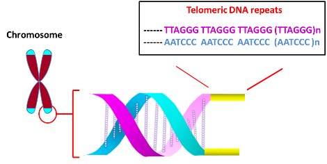
The main function of telomere is to protect chromosome ends from degradation and avoid genetic instability arising from the fusion with other chromosomes at the termini.  Telomeres at the ends of chromosomes are comprised of numerous repeats of simple DNA sequence such as TTAGGG in the case of humans.  To prevent DNA repair/recombination from occurring at the chromosome ends, the DNA sequence repeats and a single stranded 3’-overhang of telomere fold back on itself to form a loop (T loop) with the single strand invading the duplex to anneal with one of the two strands (D loop) (Xu et al., 2016).  This ‘capping’ function of telomere is mediated by Shelterin proteins, which incudes TRF1, TRF2, POT1, RAP1 and others.


In most organisms, the length of telomere decreases after each round of DNA replication in normal cells.  The loss of telomere occurs as the terminal DNA sequence occupied by the RNA primer towards the end of the lagging strand (Richter et al., 2007) cannot be replicated by DNA polymerase (though other factors such as oxidative stress may also contribute).  However, the progressive shortening of telomeres can be reversed through telomerase, which regenerates repeat sequences.  Telomerase is comprised of telomerase RNA (TERC), telomerase reverse transcriptase (TERT) and several associating proteins (Venteicher et al., 2008; Xu et al., 2016).  To extend shorter telomeres, TERT uses TERC’s template to add sequence repeats at the 3' end.  Whereas most normal somatic cells express little telomerase, TERT activity is up-regulated in continuously dividing stem cells and ectopically expressing hTERT immortalizes non-stem cells, indicating that telomeres may affect cell’s lifespan.  Consistently, an elevated telomerase activity was observed in most cancer specimens (Kim et al., 1994).


Several highly sensitive assays have been developed to diagnose the telomere status.  First, to survey the altered level of telomerase activity in cancer versus normal cells, Telomere Repeat Amplification Protocol (TRAP) assay was developed (Kim et al. 1994).  It involves extending the substrate oligonucleotide by telomerase, followed by PCR amplification using alternate primers, and separation by gel electrophoresis to visualize the extended products (Mender et al 2015a).  


Second, the loss of telomere function due to defective shelterin proteins (see above) or very short telomeric repeats elicits DNA damage response associated with double stranded DNA breaks.  The response is typified by the accumulation of the DNA damage checkpoint and DNA repair associated proteins such as ATM, Mre11, Rad17, gH2AX, and p53BP1 at the uncapped telomeres.  Telomere dysfunction-Induced Foci (TIFs) assay was developed to detect the DNA damage foci at uncapped telomeres (Mender et al., 2015b).  The assay relies on the co-localization of antibodies recognizing proteins associated with DNA damage response (ex. gH2AX) and telomere (ex. TRF2). 


Third, to determine the telomere length of individual chromosomes, Fluorescence In Situ Hybriization (FISH) was performed. Investigators at the British Columbia Cancer Agency (Canada) analyzed human metaphase or interphase chromosomes in fetal liver, adult bone marrow and chronic myeloid leukemia (CML) cells using a PNA (peptide nucleic acid)-based oligonucleotide probe recognizing the TTAGGG repeats.  The observed fluorescence intensities of sister chromatids’ telomeres were comparable, indicative of similar number of the repeats.  The mean telomerase fluorescence intensity correlated with the mean size of the ‘terminal restriction fragment’ (see below) (Lansdorp et al.., 1996).  Quenching by red blood cells (RBC) or autofluorescing molecules in granulocytes may affect the fluorescence level, however (Baerlocher et al., 2002).


Terminal Restriction Fragment (TRF) represents an alternate method to directly measure the length of telomeres (Mender et al 2015c).  After cleaving the genomic DNA with a restriction endonuclease, the resultant fragments (~800 bp) were subjected to Southern blot analysis.  To increase detection sensitivity for analyzing small amounts of tumor DNA and avoid hazards associated with radioactive labels, a modified TRF assay utilizing digoxigenin (DIG)-labeled probes was developed at the University of Texas Southwestern Medical Center (Lai et al., 2016).  To increase signal, the probe was labeled internally.  To incorporate multiple DIG labels, a template oligonucleotide encoding the complementary sequence of telomeric repeats was prepared, to which a primer was hybridized to extend using a mixture of DIG-dUTP and dNTPs.  After the elongation/modification, the resultant DNA was treated (5’ à 3’) with exonuclease to yield single stranded DIG-labeled probe.  To prevent degradation, bridged nucleic acid (BNA) analogues were incorporated at the 3’ terminus of the primer, which was used to generate the probe.  The enhanced sensitivity of the method was demonstrated using human cervical cancer-derived HeLa cells.


Bio-Synthesis, Inc. provides extensive options for the application of various modified nucleosides for research or therapy purposes.  It specializes in oligonucleotide modification and provides an extensive array of chemically modified nucleoside analogues (over ~200).  For instance, we provide digoxigenin oligonucleotide labeling services at 5', 3' and internally using NHS ester cross-linking chemistry that reacts with an amine group to form an amide.  The digoxigenin-labeld probes could be used for in situ hybridization, Northern or Southern blot analysis.  For bridged nucleic acid (BNA), it has recently acquired a license from BNA Inc. of Osaka, Japan, for the manufacturing and distribution of BNANC, a third generation of BNA oligonucleotides.  Bio-Synthesis, Inc. has recently entered into collaborative agreement with Bind Therapeutics, Inc. to synthesize miR-21 blocker using BNA.  The BNA technology that we offer provides superior, unequalled advantages in base stacking, binding affinity, aqueous solubility and nuclease resistance.  More importantly, BNA oligonucleotide exhibits lesser toxicity than other modified nucleotides for clinical application.

https://www.biosyn.com/digoxigenin-oligo-labeling.aspx#!


References

Baerlocher GM, Mak J, Tien T, Lansdorp PM. Telomere length measurement by fluorescence in situ hybridization and flow cytometry: tips and pitfalls.  (2002) Cytometry. 47:89-99.  PMID: 11813198

Kim NW, Piatyszek MA, Prowse KR, Harley CB, West MD, Ho PL, Coviello GM, Wright WE, Weinrich SL, Shay JW.  Specific association of human telomerase activity with immortal cells and cancer.  (1994). Science  266:2011-5.  PMID: 7605428  DOI: 10.1126/science.7605428

Lai TP, Wright WE, Shay JW.  Generation of digoxigenin-incorporated probes to enhance DNA detection sensitivity.  (2016)  Biotechniques. 60:306-9. doi: 10.2144/000114427.  PMID: 27286808

Lansdorp PM, Verwoerd NP, van de Rijke FM, Dragowska V, Little MT, Dirks RW, Raap AK, Tanke HJ. Heterogeneity in telomere length of human chromosomes.  (1996) Hum Mol Genet. 5:685-91.  PMID: 8733138

Mender I, Shay JW. Telomerase Repeated Amplification Protocol (TRAP). (2015a)  Bio Protoc.  5(22). pii: e1657.  PMID: 27182535

Mender I, Shay JW.  Telomere Dysfunction Induced Foci (TIF) Analysis.  (2015b)  Bio Protoc.  5(22). pii: e1656.  PMID: 27500188

Mender I, Shay JW.   Telomere Restriction Fragment (TRF) Analysis. (2015c)  Bio Protoc. 5(22). pii: e1658.  PMID: 27500189

Richter T, von Zglinicki T.  A continuous correlation between oxidative stress and telomere shortening in fibroblasts.  (2007)  Exp Gerontol. 42:1039-42. PMID: 17869047 DOI: 10.1016/j.exger.2007.08.005

Venteicher A.S., Meng Z., Mason P.J., Veenstra T.D., Artandi S.E. Identification of APTases pontin and reptin as telomerase components essential for holoenzyme assembly. (2008). Cell 132:945–957.   PMID: 18358808  PMCID: PMC2291539   DOI: 10.1016/j.cell.2008.01.019

Xu Y,  Goldkorn A.  Telomere and telomerase therapeutics in cancer. (2016). Genes (Basel) 7: 22.  PMCID: PMC4929421   PMID: 27240403


TRAIP and DNA Repair

$
0
0

Cellular Genomes

Cellular genomes
are very vulnerable to damage during chromosomal DNA replicationcan cause mutations as a result of the incorporation of incorrect bases during DNA replication. For the maintenance of genomic integrity, cells have several mechanisms to repair these damages. For example, the reversal of chemical reactions that caused DNA breakage and the removal of damaged bases followed by their replacement with newly synthesized DNA. Cells also have other means to cope with damaged DNA.

The protein proliferating cell nuclear antigen (PCNA) initially identified as an antigen in the nuclei of DNA synthesizing cells, is a DNA clampessential for DNA repair. PCNA is a homotrimer protein complex that encircles DNA.  Bound to DNA, it functions as a scaffold for the recruitment of proteins needed for the replication of DNA, remodeling of chromatin as well as for epigenetic modifications. Many of proteins required for DNA repair interact with PCNA via the PCNA-interacting motif called PCNA-interacting peptide box (PIP) or the AlkB homolog 2 PCNA interacting motif (APIM). PCNA can function as a sliding clamp during DNA synthesis, a polymerase switch factor, or as a recruitment factor. Interactions of PCNA with various proteins together with post-translational modifications of PCNA control the regulation of the cell cycle and chromatid cohesion.

Wu et al. recently identify the E3 ubiquitin ligase TRAIP (TRAF-interacting protein) as a factor located at active and stressed replication forks. TRAIP directly interacts with PCNA via a conserved PCNA-interacting peptide (PIP) box motif. TRAIP promotes ATR-dependent checkpoint signaling in human cells via the generation of replication protein A (RPA)-bound single-stranded DNA regions. Replication stress requires its E3 ligase activity, which is potentiated by the PIP box. Wu et al. report that the loss of TRAIP leads to chromosomal instability and decreased cell survival after replication stress. TRAIP appears to be a PCNA-binding ubiquitin ligase protecting genome integrity after difficult DNA replication.

RPA is the major eukaryotic ssDNA-binding protein with essential roles in genome maintenance. RPA binds to ssDNA through multiple dynamic modes. RPA can alternate between different binding ways modifying ssDNA structures. However, many of these interactions remain unknown.

TRAIP and DNA repair

Cellular genomes are very vulnerable to damage during chromosomal DNA replication. Alterations of genomic DNA can cause mutations as a result of the incorporation of incorrect bases during DNA replication. For the maintenance of genomic integrity, cells have several mechanisms to repair these damages. For example, the reversal of chemical reactions that caused DNA breakage and the removal of damaged bases followed by their replacement with newly synthesized DNA. Cells also have other means to cope with damaged DNA.

The protein proliferating cell nuclear antigen (PCNA), initially identified as an antigen in the nuclei of DNA synthesizing cells, is a DNA clamp essential for DNA repair. PCNA is a homotrimer protein complex that encircles DNA.  Bound to DNA, it functions as a scaffold for the recruitment of proteins needed for the replication of DNA, remodeling of chromatin as well as for epigenetic modifications. Many of proteins required for DNA repair interact with PCNA via the PCNA-interacting motif called PCNA-interacting peptide box (PIP) or the AlkB homolog 2 PCNA interacting motif (APIM). PCNA can function as a sliding clamp during DNA synthesis, a polymerase switch factor, or as a recruitment factor. Interactions of PCNA with various proteins together with post-translational modifications of PCNA control the regulation of the cell cycle and chromatid cohesion.

Wu et al. recently identify the E3 ubiquitin ligase TRAIP
(TRAF-interacting protein) as a factor located at active and stressed replication forks. TRAIP directly interacts with PCNA via a conserved PCNA-interacting peptide (PIP) box motif. TRAIP promotes ATR-dependent checkpoint signaling in human cells via the generation of replication protein A (RPA)-bound single-stranded DNA regions. Replication stress requires its E3 ligase activity, which is potentiated by the PIP box. Wu et al. report that the loss of TRAIP leads to chromosomal instability and decreased cell survival after replication stress. TRAIP appears to be a PCNA-binding ubiquitin ligase protecting genome integrity after difficult DNA replication.

RPA is the major eukaryotic ssDNA-binding protein with essential roles in genome maintenance. RPA binds to ssDNA through multiple dynamic modes. RPA can alternate between different binding ways modifying ssDNA structures. However, many of these interactions remain unknown.

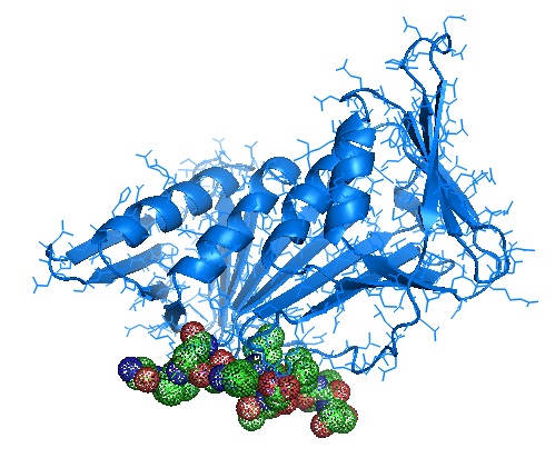
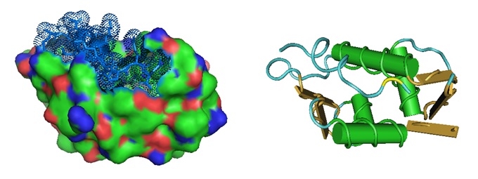
Crystal structure of human PCNA in complex with a TRAIP peptide.

Monomer structure of PCNA in complex with Ala-phe-gln-ala-lys-leu-asp-thr-phe-leu-trp-ser and Ala-gly-ala-gly-ala [4ZTD].


Sequence alignment of the peptide sequence with the protein sequence shows that the interacting peptide is located at the C-terminal end of E3 ubiquitin-protein ligase TRAIP.

E3 ubiquitin-protein ligase TRAIP [Homo sapiens]

>NP_005870.2 E3 ubiquitin-protein ligase TRAIP [Homo sapiens]

MPIRALCTICSDFFDHSRDVAAIHCGHTFHLQCLIQWFETAPSRTCPQCRIQVGKRTIINKLFFDLAQEE

ENVLDAEFLKNELDNVRAQLSQKDKEKRDSQVIIDTLRDTLEERNATVVSLQQALGKAEMLCSTLKKQMK

YLEQQQDETKQAQEEARRLRSKMKTMEQIELLLQSQRPEVEEMIRDMGVGQSAVEQLAVYCVSLKKEYEN

LKEARKASGEVADKLRKDLFSSRSKLQTVYSELDQAKLELKSAQKDLQSADKEIMSLKKKLTMLQETLNL

PPVASETVDRLVLESPAPVEVNLKLRRPSFRDDIDLNATFDVDTPPARPSSSQHGYYEKLCLEKSHSPIQ

DVPKKICKGPRKESQLSLGGQSCAGEPDEELVGAFPIFVRNAILGQKQPKRPRSESSCSKDVVRTGFDGL

GGRTKFIQPTDTVMIRPLPVKPKTKVKQRVRVKTVPSLFQAKLDTFLWS

Earlier in 2017, Klattenhoff et al. showed that the protein Nei endonuclease VIII-like 3 (NEIL3) is also localized at DNA double-strand break (DSB) sites during oxidative DNA damage and replication stress.

Loss of NEIL3 significantly increased spontaneous replication-associated DSBs and recruitment of replication protein A (RPA). In contrast, we observed a marked decrease in Rad51 on nascent DNA strands at the replication fork, suggesting that HR-dependent repair is compromised in NEIL3-deficient cells. Interestingly, NEIL3-deficient cells were sensitive to ataxia–telangiectasia and Rad3 related protein (ATR) inhibitor alone or in combination with PARP1 inhibitor. This study elucidates the mechanism by which NEIL3 is critical to overcome oxidative and replication-associated genotoxic stress.

Reference

DNA repair: https://www.ncbi.nlm.nih.gov/books/NBK9900/

https://en.wikipedia.org/wiki/Proliferating_cell_nuclear_antigen

https://www.ncbi.nlm.nih.gov/books/NBK9900/

Deeba N. Syed, Mohammad Imran Khan, Maria Shabbir, Hasan Mukhtar; MicroRNAs in Skin Response to UV Radiation.  Current Drug Targets Volume 14 , Issue 10 , 2013. DOI :
10.2174/13894501113149990184. [https://www.ncbi.nlm.nih.gov/pmc/articles/PMC3985496/]

Klattenhoff AW, Thakur M, Chu CS, Ray D, Habib SL, Kidane D. Loss of NEIL3 DNA glycosylase markedly increases replication associated double strand breaks and enhances sensitivity to ATR inhibitor in glioblastoma cells. Oncotarget. 2017 Dec 4;8(68):112942-112958. doi: 10.18632/oncotarget.22896. PMID: 29348879; PMCID: PMC5762564. [https://www.ncbi.nlm.nih.gov/pubmed/29348879]

Strzalka W, Ziemienowicz A. Proliferating cell nuclear antigen (PCNA): a key factor in DNA replication and cell cycle regulation. Ann Bot. 2011 May;107(7):1127-40. doi: 10.1093/aob/mcq243. Epub 2010 Dec 17. PMID: 21169293; PMCID: PMC3091797. [https://www.ncbi.nlm.nih.gov/pmc/articles/PMC3091797/]

Qing-Man Wang, Yan-Tao Yang, Yi-Ran Wang, Bo Gao, Xuguang Xi and Xi-Miao Hou; Human Replication protein A induces dynamic changes in single-stranded DNA and RNA structures. The Journal of Biological Chemistry294, 13915-13927. [
http://www.jbc.org/content/early/2019/07/26/jbc.RA119.009737.abstract]

Wu RA, Semlow DR, Kamimae-Lanning AN, Kochenova OV, Chistol G, Hodskinson MR, Amunugama R, Sparks JL, Wang M, Deng L, Mimoso CA, Low E, Patel KJ, Walter JC. TRAIP is a master regulator of DNA interstrand crosslink repair. Nature. 2019 Mar;567(7747):267-272. doi: 10.1038/s41586-019-1002-0. Epub 2019 Mar 6. PMID: 30842657; PMCID: PMC6417926.
https://www.ncbi.nlm.nih.gov/pmc/articles/PMC6417926.


Octaneuropeptide or ODN protects brain cells

$
0
0


The octadecaneuropeptide (ODN), QATVGDVNTDRPGLLDLK, C81H138N24O29, Mw: 1912.1 g/mol, protects both neurons and astrocytes from oxidative stress-induced cell death.


ODN is a neuropeptide similar to other neuropeptides for example, the nerve growth factor (NGF) that exhibit cytoprotective activities, and some also induce cell differentiation.

ODN is highly expressed in the developing nervous system where it has cytoprotective and antioxidant effects in cultured murine astrocytes and cerebellar granule neurons in vitro and dopaminergic neurons in vivo.


ODN is a peptide derived from diazepam-binding inhibitor. ODN is also known to reduce food intake in goldfish as well as in rodents, and possibly humans.

In humans “this gene encodes a diazepam binding inhibitor, a protein that is regulated by hormones and is involved in lipid metabolism and the displacement of beta-carbolines and benzodiazepines, which modulate signal transduction at type A gamma-aminobutyric acid receptors located in brain synapses. The protein is conserved from yeast to mammals, with the most highly conserved domain consisting of seven contiguous residues that constitute the hydrophobic binding site for medium- and long-chain acyl-Coenzyme A esters. Diazepam binding inhibitor is also known to mediate the feedback regulation of pancreatic secretion and the postprandial release of cholecystokinin, in addition to its role as a mediator in corticotropin-dependent adrenal steroidogenesis. Three pseudogenes located on chromosomes 6, 8 and 16 have been identified. Multiple transcript variants encoding different isoforms have been described for this gene. [provided by RefSeq, Jul 2008]” [Source: https://www.ncbi.nlm.nih.gov/gene/1622]

In neurodegenerative diseases such as Alzheimer’s disease, Parkinson’s disease, and multiple sclerosis by neuronal cell death is caused in part by inflammatory processes, mitochondrial alterations, and an elevation of oxidative stress. Oxidative stress induces imbalance in ROS generation, impairs cellular antioxidant defenses and can trigger both neurons and astroglial cell death via apoptosis. Astrocytes synthesize and release endozepines, a family of regulatory peptides, including ODN.

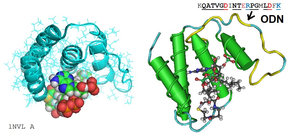
1NVL: RDC-refined NMR structure of bovine Acyl-coenzyme A Binding Protein, ACBP, in complex with palmitoyl-coenzyme A

Alignment of selected Octaneuropeptides


ODN was originally characterized as an endogenous ligand for central-type benzodiazepine receptors. ODN is known to increase intracellular calcium concentration ([Ca2+]i) in rat astroglial cells.

Recently, using a peptidomic approach, two N-terminal fragments of acyl-Co-A-binding protein were identified in yeast, a single N-terminal fragment was identified in human cells, and a number of N-terminal peptides were identified in mouse tissues along with a couple of internal peptides. In addition, ODN prevents toxicity induced by 6-hydroxydopamine in cultured rat astrocyte. Apparently ODN triggers or stimulates the endogenous antioxidant systems and the intrinsic apoptotic pathway.


Reference

Dasgupta S, Yang C, Castro LM, Tashima AK, Ferro ES, Moir RD, Willis IM, Fricker LD. Analysis of the Yeast Peptidome and Comparison with the Human Peptidome. PLoS One. 2016 Sep 29;11(9):e0163312. doi: 10.1371/journal.pone.0163312. PMID: 27685651; PMCID: PMC5042401.  https://www.ncbi.nlm.nih.gov/pmc/articles/PMC5042401/

Givalois L, Grinevich V, Li S, Garcia-De-Yebenes E, Pelletier G.;
The octadecaneuropeptide-induced response of corticotropin-releasing hormone messenger RNA levels is mediated by GABA(A) receptors and modulated by endogenous steroids. Neuroscience. 1998 Jul;85(2):557-67.

Hamdi Y, Kaddour H, Vaudry D, Leprince J, Zarrouk A, Hammami M, Vaudry H, Tonon MC, Amri M, Masmoudi-Kouki O.
Octadecaneuropeptide ODN prevents hydrogen peroxide-induced oxidative damage of biomolecules in cultured rat astrocytes. Peptides. 2015 Sep;71:56-65. doi: 10.1016/j.peptides.2015.06.010. Epub 2015 Jul 2. Review. https://doi.org/10.1101/266379.

https://www.biorxiv.org/content/10.1101/266379v1.full

Hamdi Y, Kaddour H, Vaudry D, Bahdoudi S, Douiri S, Leprince J, Castel H, Vaudry H, Tonon MC, Amri M, Masmoudi-Kouki O.; The octadecaneuropeptide ODN protects astrocytes against hydrogen peroxide-induced apoptosis via a PKA/MAPK-dependent mechanism. PLoS One. 2012;7(8):e42498. doi: 10.1371/journal.pone.0042498. Epub 2012 Aug 21.

Kaddour H, Hamdi Y, Vaudry D, Basille M, Desrues L, Leprince J, Castel H, Vaudry H, Tonon MC, Amri M, Masmoudi-Kouki O.;
The octadecaneuropeptide ODN prevents 6-hydroxydopamine-induced apoptosis of cerebellar granule neurons through a PKC-MAPK-dependent pathway. J Neurochem. 2013 May;125(4):620-33. doi: 10.1111/jnc.12140. Epub 2013 Feb 19.

Masmoudi-Kouki O, Hamdi Y, Ghouili I, Bahdoudi S, Kaddour H, Leprince J, Castel H, Vaudry H, Amri M, Vaudry D, Tonon MC.;
Neuroprotection with the Endozepine Octadecaneuropeptide, ODN. Curr Pharm Des. 2018;24(33):3918-3925. doi: 10.2174/1381612824666181112111746.

Namsi A, Nury T, Khan AS, Leprince J, Vaudry D, Caccia C, Leoni V, Atanasov AG, Tonon MC, Masmoudi-Kouki O, Lizard G.;

Octadecaneuropeptide (ODN) Induces N2a Cells Differentiation through a PKA/PLC/PKC/MEK/ERK-Dependent Pathway: Incidence on Peroxisome, Mitochondria, and Lipid Profiles. Molecules. 2019 Sep 11;24(18). pii: E3310. doi: 10.3390/molecules24183310.

ODN:  https://www.ncbi.nlm.nih.gov/pubmed/?term=Octadecaneuropeptide

---…---


Bio-Synthesis, Inc. offers LNA or 2',4'-BNANC based oligonucleotides for basic research, genome editing, diagnosis or therapy of multiple human disorders including cancer

$
0
0
 Bio-Synthesis, Inc. offers LNA or 2',4'-BNANC based oligonucleotides for basic research, genome editing, diagnosis or therapy of multiple human disorders including cancer

The successful development of synthetic chemistry methodology allowed the production of high-quality oligonucleotides that are cost effective.  The availability of the synthetic oligonucleotides opened up multiple opportunities for novel basic research, diagnostic and therapeutic applications.  With increasing utility, came the demand for further modification to enhance oligonucleotide’s binding affinity to various targets including DNA, RNA and protein.  Equally significant is the chemical modification to block degradation by nucleases in vivo.  For clinical application, oligonucleotide drugs must meet additional criteria including delivery, potency, toxicity, off-target activity, etc.

Xeno nucleic acids collectively refer to nucleic acid analogues that have been chemically modified.    Among them is locked nucleic acid (LNA), representing modified RNA nucleotide with an extra bridge linking 2' oxygen and 4' carbon of the sugar moiety.  By locking the ribose in the 3'-endo conformation, the bridge reduces the entropic cost for transitioning from single stranded state to A-form helix (Julien et al., 2008). This entropic advantage translates into significantly higher binding affinity to target complementary strand, promoting hybridization and augmenting Tm.  Hence, a significant increase in the stability of duplex (RNA:DNA, DNA:DNA) or triplex can be achieved by incorporating even a smaller number of LNAs.

This, in turn, inspired the development of 2'-O,4'-aminoethylene bridged nucleic acid (2',4'-BNANC), the third-generation bridged nucleic acid containing a six-member bridged structure with an N-O linkage.  It was developed along the same general principle to enhance the binding affinity.  The 2',4'-BNANC modification provides greater binding affinity, better single-mismatch discrimination, enhanced RNA specificity, stronger/selective triplex-forming properties, and considerably higher nuclease resistance (Rahman et al., 2008; Miyashita et al., 2007).  Antisense oligonucleotides containing 2',4'-BNANC are more stable and better tolerated than LNA modified oligonucleotides in the murine models (Rahman et al., 2008;  Yamamoto et al., 2012).  Further, 2',4'-BNANC modified oliigonucleotides induce lesser caspase activity, explaining its reduced toxicity (Manning et al., 2017).  These attributes make 2',4'-BNANC ideal for clinical application.


A wide range of applications are possible with conformationally restricted nucleoside analogues, which includes basic science research.  For the structure-function relationship study concerning the role of ribose structure and dynamics in RNA function, LNA was used to probe the site-specific conformational/energetic properties in ribozyme (Julien et al., 2008).  For genome editing, incorporating LNA or BNANC at specific points in CRISPR-RNAs (crRNAs) decreased off-target DNA cleavage by Cas9, improving specificity (Cromwell et al., 2018).  The potential use of BNANC to cap aptamers using terminal transferase to increase nuclease resistance has been described (Kim et al., 2015).  Other potential uses include modulating mRNA splicing by oligonucleotides with such modification.

For diagnosis, LNA or 2',4'-BNANC incorporated into oligonucleotide probes greatly improved the efficacy of SNP genotyping, allelic discrimination, real time PCR amplification, qPCR, hybridization probe or FISH analysis.  As LNA bases render the probe greater duplex stability than single MGB (minor grove binders) at the 3’ end, it provides an ideal substitute for TaqMan MGB probes.  Using bead-based suspension assay with BNANC modified hybridization probes, specific mutation in DNMT3A gene was quantified in hematological malignancies, for which BNANC probes performed better than LNA probes (Shivarov et al., 2014).  For detecting single T790M mutation in EGFR, Bio-Synthesis, Inc. developed a highly effective method using 2',4'-BNANC modified clamp for real-time PCR (Kim et al., 2015).

The utility of bridged nucleic acids for therapy is increasingly being recognized.  For gene knockdown, antisense oligonucleotides have been used to inhibit gene expression by triggering RNase-H mediated cleavage of targeted mRNAs or through hybrid-arrest of binding/scanning by mRNA translation complexes.  These include antisense LNA oligodeoxynucleotides that bind to complementary H-Ras mRNA or miR-17-5p microRNA (miRNA) to inhibit cancer progression (Fluiter et al., 2005).  The antisense oligonucleotides with BNANC targeting proprotein convertase subtilisin/kexin type 9 (PCSK9), which is involved in LDL receptor regulation, lowered the cholesterol level (Yamamoto et al., 2012).  BNANC modified antisense oligonucleotide targeting aac(6′)-Ib gene conjugated to a permeabilizing peptide increased susceptibility to the antibiotic amikacin in the pathogen Acinetobacter baumannii (Lopez et al., 2015).  A gapmer incorporating BNANC efficiently degraded CUG expanded repeat RNA, which causes myotonic dystrophy (Manning et al., 2017).  For RNA interference, the LNA modification endowed greater functionality/stability to siRNA therapeutics (Elmén et al., 2005). 

Bio-Synthesis, Inc. specializes in oligonucleotide modification and provides an extensive array of chemically modified nucleoside analogues (over ~200) including bridged nucleic acid (BNA). It recently acquired a license from BNA Inc. of Osaka, Japan, for the manufacturing and distribution of BNANC, a third generation of BNA oligonucleotides.  To meet the demands of therapeutic application, its oligonucleotide products are approaching GMP grade.  Bio-Synthesis, Inc. has recently entered into collaborative agreement with Bind Therapeutics, Inc. to synthesize miR-21 blocker using BNA for triple negative breast cancer.  The BNA technology that we offer provides superior, unequalled advantages in base stacking, binding affinity, aqueous solubility and nuclease resistance.  It also improves the formation of duplexes and triplexes by reducing the repulsion between the negatively charged phosphates of the oligonucleotide backbone.  Its single-mismatch discriminating power was especially useful for diagnosis (ex. FISH using DNA probe).  More importantly, BNA oligonucleotide exhibits lesser toxicity than other modified nucleotides for clinical application.


https://www.biosyn.com/oligonucleotide-modification-services.aspx


References

Cromwell CR, Sung K, Park J, Krysler AR, Jovel J, Kim SK, Hubbard BP.  Incorporation of bridged nucleic acids into CRISPR RNAs improves Cas9 endonuclease specificity. (2018) Nat Commun. 9:1448. doi: 10.1038/s41467-018-03927-0.

Elmén J, Thonberg H, Ljungberg K, Frieden M, Westergaard M, Xu Y, Wahren B, Liang Z, Ørum H, Koch T, Wahlestedt C.  Locked nucleic acid (LNA) mediated improvements in siRNA stability and functionality. (2005).  Nucleic Acids Res. 33:439-47.  PMID: 15653644

Fluiter K, Frieden M, Vreijling J, Rosenbohm C, De Wissel MB, Christensen SM, Koch T, Ørum H, Baas F.  On the in vitro and in vivo properties of four locked nucleic acid nucleotides incorporated into an anti-H-Ras antisense oligonucleotide. (2005).Chembiochem. 6:1104-9. PMID: 4244378 PMCID: PMC3824000 DOI: 10.1371/journal.pone.0078863

Julien KR, Sumita M, Chen PH, Laird-Offringa IA, Hoogstraten CG. Conformationally restricted nucleotides as a probe of structure-function relationships in RNA. (2008)  RNA. 14:1632-43. PMID: 18596252   doi: 10.1261/rna.866408.

Kim S-K, Liu X, Castro A, Loredo L, Castro M. An effective detection method for the EGFR single mutation T790M using BNA-NC clamping real-time PCR. (2015)  Proc Am Assoc Cancer Res 56:1197–1198.

Lopez C, Arivett BA, Actis LA, Tolmasky ME. Inhibition of AAC(6')-Ib-mediated resistance to amikacin in Acinetobacter baumannii by an antisense peptide-conjugated 2',4'-bridged nucleic acid-NC-DNA hybrid oligomer. (2015)  Antimicrob Agents Chemother.  59:5798-803  doi: 10.1128/AAC.01304-15.

Manning KS, Rao AN, Castro M, Cooper TA.   BNANC gapmers revert splicing and reduce RNA foci with low toxicity in myotonic dystrophy cells. (2017). ACS Chem Biol. 12:2503-2509.  PMID: 28853853 PMCID: PMC5694563  doi: 10.1021/acschembio.7b00416.

Miyashita, K., Rahman, S. M., Seki, S., Obika, S., and Imanishi, T. N-Methyl substituted 2′,4′- BNANC: a highly nuclease resistant nucleic acid analogue with high-affinity RNA selective hybridization. (2007) Chem. Commun. (Cambridge, U. K.), 3765−3767.

Rahman SM, Seki S, Obika S, Yoshikawa H, Miyashita K, Imanishi T. Design, synthesis, and properties of 2',4'-BNA(NC): a bridged nucleic acid analogue.  (2008)  J Am Chem Soc. 130:4886-96. PMID: 18341342  doi: 10.1021/ja710342q.

Shivarov V, Ivanova M, Naumova E. Rapid detection of DNMT3A R882 mutations in hematologic malignancies using a novel bead-based suspension assay with BNA(NC) probes. (2014)  PLoS One. 9:e99769.   doi: 10.1371/journal.pone.0099769.

Yamamoto, T., Harada-Shiba, M., Nakatani, M., Wada, S., Yasuhara, H., Narukawa, K, et al. Cholesterol-lowering action of BNA-based antisense Ooonucleotides targeting PCSK9 in atherogenic diet-induced hypercholesterolemic mice. (2012)  Mol. Ther.– Nucleic Acids 1, e22.


Antisense oligonucleotides slow the progression of the pediatric neurodegenerative disorder Batten’s disease by correcting mRNA splicing, paving the way for personalized medicine

$
0
0
Antisense oligonucleotides slow the progression of the pediatric neurodegenerative disorder Batten’s disease by correcting mRNA splicing, paving the way for personalized medicine

Neuronal ceroid lipofuscinoses (NCLs) refer to a group of neurodegenerative disorders, which negatively impacts vision, behavior, learning, speech, etc., causing seizure, impaired motor skill, reduced intellectual capacity and early death.  The NCLs have been categorized based on the age of onset for symptoms (i.e. infantile, late infantile, juvenile, and adult) and the identity of the predisposing gene (Chabrol et al., 2013).  ‘Batten’s disease’ selectively refers to juvenile NCL (JNCL) form; however, some have used the term to refer to all NCL forms collectively.  The neurodegenerative disorder, which is progressive and can be fatal, typically occurs during childhood and there is no cure presently.  Approximately 2-4 per 100,000 children are affected in the U. S. while the frequency may be higher (~1/12,500 people) in some populations.

To date, 14 genetically distinct NCL forms have been identified (Chabrol et al., 2013; Anu et al., 2009).  ‘Infantile neuronal ceroid’ (INCL) is associated with CLN gene encoding PPT1 (palmitoyl-protein thioesterase 1) that functions as a lysosomal enzyme.  Late infantile NCL (LINCL) with the life expectancy of 8-12 yrs is linked to CLN2 gene, which encodes the lysosomal enzyme TPP1 (tripeptidyl-peptidase 1).  Variant of the late infantile NCL is caused by the disruption of CLN6 gene, which encodes the transmembrane protein of endoplasmic reticulum CLN6 (linclin).  Another variant of late infantile NCL is associated with the CLN7 gene encoding MFSD8 (‘major facilitator superfamily domain containing eight’), a lysosomal transmembrane protein.  ‘Juvenile NCL’ (JNCL) occurs due to the inactivation of CLN3 gene, which encodes a lysosomal transmembrane protein similar to SLC (solute carrier) transporters.  Accurate diagnosis of NCL requires the determination of the mutated gene and the resultant dysfunctional protein (for genetic counseling).

Most NCLs exhibit autosomal recessive mode of inheritance.  The disease is manifested if both alleles of the predisposing gene inherited from parents are defective.  Though a carrier may not develop symptoms, the individual is capable of passing the defective allele to a descendent.  An exception may be found in Kufs disease, a distinct form of NCL, whose symptoms appear around age 30 with a limited survival of less than 15 years after the onset.  Its type A is linked to PPT1 and CLN6 genes, while type B (autosomal dominant) is associated with CTSF (cathepsin F) and DNAJC5 (‘DnaJ homolog subfamily C member 5’) genes (Benitez et al., 2011).  Cathepsin is involved in proteolytic degradation in lysosomes.

Batten’s disease belongs to a group of disorders known as ‘lysosomal storage disease’, which is comprised of ca. 50 distinct autosomal recessively inherited genetic disorders.  The disease occurs due to the dysfunctional lysosomes, resulting in the intracellular accumulation of excess amount of undegraded substrates.  The dysfunction is caused by a defect or insufficient level of the lysosomal enzymes (ex. cathepsin, alpha-glucosidase, acid phosphatase) for the degradation of carbohydrates, lipids, proteins, nucleic acids, etc.   The failure to breakdown into smaller components could lead to the death of the affected cells (ex. neurons) eventually.  Lysosomes are also involved in autophagy (wherein autophagnosomes fuse with lysosomes to generate autolysosomes to degrade defective intracellular proteins, damaged organelles, pathogenic microbes, etc.), which has been associated with aging-associated disorders, heart disease, cancer, etc.

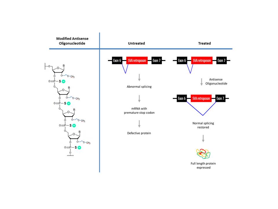
Batten’s disease remains a terminal disease with limited treatment options.  In 2017, FDA approved the enzyme replacement therapy, Brineura, which involves intraventricularly administering recombinant human TPP1, the missing enzyme for NCL patients with defective CLN2 gene (Johnson et al., 2019).  More recently, an mRNA-modulating treatment utilizing antisense oligonucleotides was reported though applicable for only a single patient (Kim et al., 2019).  A 6-year-old girl with worsening symptoms, which included seizures, ataxia, developmental regression, deteriorating vision with mild cerebral/cerebellar atrophy, was diagnosed with Batten’s disease.  Genetic analysis revealed that she was heterozygous for a pathogenic mutation in the MFSD8 (CLN7) gene.  The whole genome sequencing showed that the other MFSD8 allele (both the patient and mother) contains the SVA (a composite of SINE-VNTR-Alu) retroposon inserted in intron 6.  The retroposon caused exon 6 to mis-splice into the cryptic splice-acceptor site, ‘i6.SA’, located upstream of the retroposon, resulting in a faulty transcript with premature translational termination. 

To correct the altered splicing, investigators at the Harvard University-affiliated Boston Children’s Hospital designed Milasen, a 22 bp antisense oligonucleotide, to block the aberrant ‘i6.SA’ splice acceptor site or exonic splicing enhancer (ESE) elements nearby.  To increase stability, the oligonucleotide consisted of 2′-O-methoxyethyl (MOE) ribonucleotides with phosphorothioate internucleotide linkages.  How the modified oligonucleotides were uptaken by neural cells is not clear though phsophorothioate-modified oligonucleotide is known to interact with several receptors (ex. stabilin receptor in the liver) (Miller et al., 2017).  Milasen increased the ratio of normal-to-mutant splicing 2.5 to 3-fold in patient derived fibroblasts. The investigational drug Milasen was administered intrathecally to distribute to the brain via cerebrospinal fluid.  During the treatment (~1 year), no serious adverse events occurred according to the authors.  Though it did not cure, a notable reduction in the intensity and frequency of seizures was reported (Kim et al., 2019). 

Critical to the above undertaking was the ability to synthesize oligonucleotides expeditiously.  Bio-Synthesis, Inc. specializes in oligonucleotide modification and provides an extensive array of chemically modified nucleoside analogues (over ~200) including bridged nucleic acid (BNA). It recently acquired a license from BNA Inc. of Osaka, Japan, for the manufacturing and distribution of BNANC, a third generation of BNA oligonucleotides.  To meet the demands of therapeutic application, its oligonucleotide products are approaching GMP grade.  Bio-Synthesis, Inc. has recently entered into collaborative agreement with Bind Therapeutics, Inc. to synthesize miR-21 blocker using BNA for triple negative breast cancer.  The BNA technology that we offer provides superior, unequalled advantages in base stacking, binding affinity, aqueous solubility and nuclease resistance.  It also improves the formation of duplexes and triplexes by reducing the repulsion between the negatively charged phosphates of the oligonucleotide backbone.  Its single-mismatch discriminating power was especially useful for diagnosis (ex. FISH using DNA probe).  More importantly, BNA oligonucleotide exhibits lesser toxicity than other modified nucleotides for clinical application.


https://www.biosyn.com/oligonucleotide-modification-services.aspx


References

Anu J,Thomas B. "Neuronal ceroid lipofuscinoses". (2009).  Biochimica et Biophysica Acta (BBA) - Molecular Cell Research  1793: 697–709.  PMID 19084560    doi:10.1016/j.bbamcr.2008.11.004.

Benitez BA, Alvarado D, Cai Y, Mayo K, Chakraverty S, Norton J, Morris JC, Sands MS, Goate A, Cruchaga C.  Exome-sequencing confirms DNAJC5 mutations as cause of adult neuronal ceroid-lipofuscinosis.  (2011)  PLoS One. 6(11):e26741.   PMID: 22073189  doi: 10.1371/journal.pone.0026741

Chabrol B, Caillaud C, Minassian B.  Neuronal ceroid lipofuscinoses.  (2013)  Handbk Clin Neurol. 113:1701-6.  PMID: 23622391   doi: 10.1016/B978-0-444-59565-2.00038-1.

Johnson TB, Cain JT, White KA, Ramirez-Montealegre D, Pearce DA, Weimer JM.  Therapeutic landscape for Batten disease: current treatments and future prospects.  (2019) Nat Rev Neurol. 15:161-178.   doi: 10.1038/s41582-019-0138-8.

Kim J, Hu C, Moufawad El Achkar C, Black LE, Douville J, Larson A et al.  Patient-Customized Oligonucleotide Therapy for a Rare Genetic Disease. (2019)  N Engl J Med.  381:1644-1652. PMID:31597037  doi: 10.1056/NEJMoa1813279. Epub 2019 Oct 9.

Miller CM,  Tanowitz M, Donner AJ, Prakash TP, Swayze EE, Harris EN, Seth PP.  Receptor-mediated uptake of phosphorothioate antisense oligonucleotides in different cell types of the liver.  (2018)  Nucleic Acid Ther.  28:119-127.  PMID: 29425080  doi: 10.1089/nat.2017.0709


Co-RNA FISH assay employing fluorescently labeled oligonucleotides detects a long noncoding RNA targeting glutaminase to maintain redox balance in mutant K-RAS driven human pancreatic ductal adenocarcinoma

$
0
0
 Pancreatic cancer is the 4th leading cause of cancer deaths in the U. S. and may claim ~43,000 lives in this year.  Typically, it occurs at ages >45, irrespective of gender.  Pancreatic cancer is difficult to diagnose due to the lack of specific symptoms, thus the majority of the diagnosed present advanced-stage disease.  In early-stage pancreatic cancer, surgical resection may increase 5-year survival rate to ~20% (Aslan et al., 2018).  For all-stage pancreatic cancers, 5-year survival rate remains ~7%.  Chemotherapy, i.e. 5-flourouracil, nab-paclitaxel (albumin–packaged Taxol), gemcitabine or topoisomerase inhibitor, may increase overall survival moderately (~8.5 months following gemcitabine and nab-paclitaxel treatment), and combining with radiotherapy exacerbates side effects.  The efficacy of targeted therapies inhibiting growth signaling pathways (or stroma) and immunotherapies is being explored.

The pancreas located behind the stomach plays a critical role in digestion.  Its exocrine cells secrete enzymes to break down lipids, carbohydrates and proteins while endocrine cells release hormones (ex. insulin to regulate sugar level).  Nearly 85% of all pancreatic cancers occur in exocrine cells, and pancreatic ductal adenocarcinoma (PDAC) rising in the cells lining the pancreatic ducts represents the most aggressive type.  Pancreatic neuroendocrine tumor (PanNET) originating in the endocrine cells represents a minority.  Most of pancreatic cancers harbor chromosomal alterations (ex. translocation, inversion, deletion) and KRAS (>95%), p53, CDKN2A and SMAD4 genes are frequently mutated (BRCA1/BRCA2 is mutated in a subset) (Cicenas et al., 2017).

In normal cells, glucose is metabolized (by glycolysis) to pyruvate, which is converted to acetyl-CoA to enter tricarboxylic acid cycle (TCA) and produce NADH to drive oxidative phosphorylation and generate the maximal amount of ATP in mitochondria.  In cancer cells, however, this pathway is bypassed in favor of increased glycolysis (‘Warburg effect’ by O. Warburg, Nobel prize 1931) to generate glycolytic intermediates to support their uncontrolled growth (Warburg, 1956).  In PDAC cells, constitutively activated signaling by mutant K-RAS oncoprotein facilitates glycolysis by modulating transporters to increase glucose uptake.  It also facilitates shunting of the glycolysis intermediates to ‘pentose phosphate pathway’ to generate ribose 5’-phosphate necessary for DNA replication of rapidly dividing cancer cells (Bryant et al., 2014).

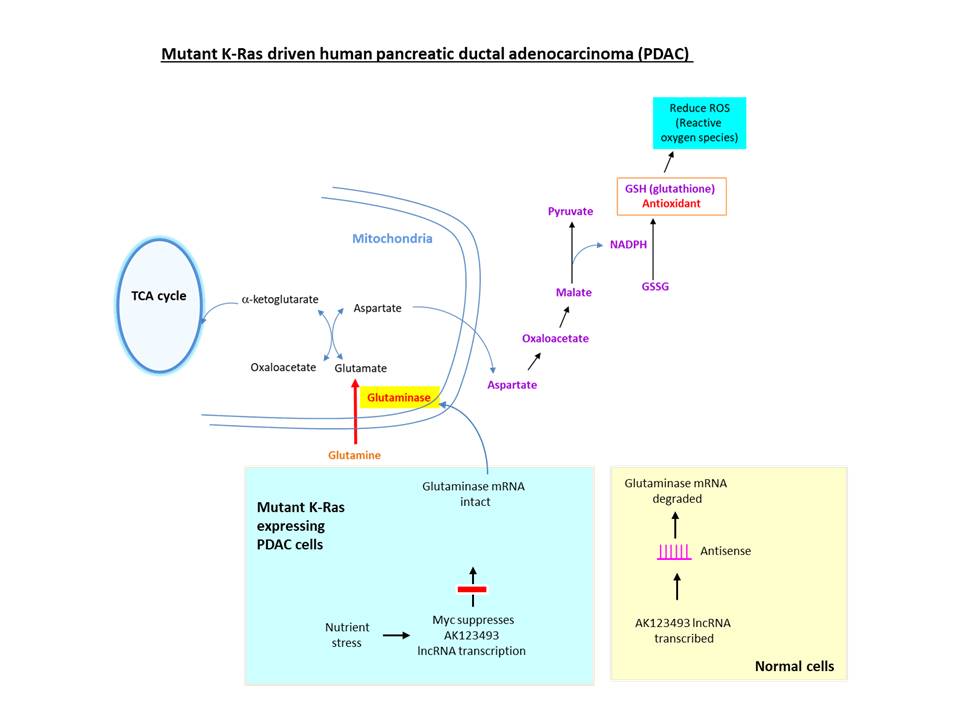
To maintain redox balance, proliferative cells consume greater amounts of glutamine (Eagle, 1955).  In K-RAS driven PDAC cells, instead of converting glutamate (derived from glutamine) to a-ketoglutarate (an intermediate of TCA cycle), it is converted to malate, which is metabolized to yield NADPH necessary for generating the antioxidant glutathione.  The hydrolysis of glutamine to glutamate is catalyzed by the enzyme glutaminase, which is overexpressed in PDAC (Chakrabarti et al., 2015), breast cancer, prostate cancer, etc. 


Long coding RNA (lncRNA) refers to transcripts >200 bp to distinguish from ‘small noncoding RNA’ (ex. siRNA, microRNA, small nucleolar RNA) and >270,000 lncRNA transcripts (Ma et al., 2019) may exist in humans.  Most lncRNAs have similar genomic structure (~42% consist of 2 exons) as protein-coding genes, transcribed as independent transcription unit from promoters epigenetically regulated through histone modification, undergo splicing (Derrien et al., 2012), and exhibit reduced/tissue-specific expression.  They include intergenic lncRNA, antisense lncRNA, sense lncRNA (located in intron, share exon or overlap exons of protein-coding gene).  LncRNA may affect gene transcription directly (as co-factor or via RNA-DNA triplex formation) or indirectly, regulate post-transcriptionally via hybridizing to mRNA to alter splicing/translation, or induce RNA interference for degradation.  Consistent with their presence in chromatin, lncRNAs are involved in epigenetically regulating genes during embryogenesis, imprinting, X chromosome inactivation and telomere protection.

To understand the molecular mechanism regulating glutaminase, the investigators at Huazhong University of Science and Technology (China) examined long noncoding RNAs (lncRNAs) dysregulated in PDAC.  AK123493 is an antisense lncRNA located in intron 17 of human glutaminase gene, which is underexpressed in PDAC.  A ‘co-RNA FISH’ assay revealed that both RNAs are co-localized in the same nuclear foci, indicating that AK123493 lncRNA may hybridize to GLS mRNA (Deng et al., 2019).  For the assay, fluorescently labeled RNA probes were generated by incorporating modified UTP (with NH2 group linked to the base) through in vitro transcription, followed by conjugation with amine-reactive fluorescent dyes.  This, and other data, suggested that AK123493 lncRNA targets glutaminase mRNA to reduce its expression via inducing RNA interference.  Further, they suggested that nutrient stress (caused by glutamine depletion) may activate Myc oncoprotein to inhibit the transcription of AK123493 lncRNA to increase the glutaminase level.

Bio-Synthesis, Inc. specializes in oligonucleotide modification and provides an extensive array of chemically modified nucleoside analogues (over ~200) including bridged nucleic acid (BNA).  A number of options are available to label oligonucleotides (DNA or RNA) with fluorophoreseither terminally or internally as well as conjugate to peptidesIt recently acquired a license from BNA Inc. of Osaka, Japan, for the manufacturing and distribution of BNANC, a third generation of BNA oligonucleotides.  To meet the demands of therapeutic application, its oligonucleotide products are approaching GMP grade.  Bio-Synthesis, Inc. has recently entered into collaborative agreement with Bind Therapeutics, Inc. to synthesize miR-21 blocker using BNA for triple negative breast cancer.  The BNA technology that we offer provides superior, unequalled advantages in base stacking, binding affinity, aqueous solubility and nuclease resistance.  It also improves the formation of duplexes and triplexes by reducing the repulsion between the negatively charged phosphates of the oligonucleotide backbone.  Its single-mismatch discriminating power was especially useful for diagnosis (ex. FISH using DNA probe).  More importantly, BNA oligonucleotide exhibits lesser toxicity than other modified nucleotides for clinical application.


https://www.biosyn.com/oligonucleotide-modification-services.aspx

https://www.biosyn.com/tew/Specific-labeling-of-RNA.aspx

https://www.biosyn.com/labeledrnaprobes.aspx#!


References

Aslan M, Shahbazi R, Ulubayram K, Ozpolat B.  Targeted Therapies for Pancreatic Cancer and Hurdles Ahead.  (2018).  Anticancer Res  38:6591-6606.  PMID: 30504367  doi: 10.21873/anticanres.13026.

Bryant KL, Mancias JD, Kimmelman AC, Der CJ.  KRAS: feeding pancreatic cancer proliferation.  (2014) Trends Biochem Sci.  39:91-100.  PMID: 24388967   doi: 10.1016/j.tibs.2013.12.004

Chakrabarti G, Moore ZR, Luo X, Ilcheva M, Ali A, Padanad M, et al.  Targeting glutamine metabolism sensitizes pancreatic cancer to PARP-driven metabolic catastrophe induced by ß-lapachone.  (2015) Cancer Metab 3:12.  PMID: 26462257   doi: 10.1186/s40170-015-0137-1. eCollection 2015.

Cicenas J, Kvederaviciute K, Meskinyte I, Meskinyte-Kausiliene E, Skeberdyte A, Cicenas J.   KRAS, TP53, CDKN2A, SMAD4, BRCA1, and BRCA2 Mutations in Pancreatic Cancer.   (2017)  Cancers (Basel).  9(5). pii: E42.  PMID: 28452926  doi: 10.3390/cancers9050042.

Deng SJ, Chen HY, Zeng Z, Deng S, Zhu S, Ye Z, et al.  Nutrient Stress-Dysregulated Antisense lncRNA GLS-AS Impairs GLS-Mediated Metabolism and Represses Pancreatic Cancer Progression.  (2019)  Cancer Res.  79:1398-1412.  PMID: 30563888   doi: 10.1158/0008-5472.CAN-18-0419.

Derrien T, Johnson R, Bussotti G, Tanzer A, Djebali S, Tilgner H, et al.  The GENCODE v7 catalog of human long noncoding RNAs: analysis of their gene structure, evolution, and expression.  (2012) Genome Res 22:1775-89.  PMID: 22955988   doi: 10.1101/gr.132159.111.

Eagle H.  Nutrition needs of mammalian cells in tissue culture. (1955)  Science 122:501-14.  PMID: 13255879   DOI: 10.1126/science.122.3168.501

Ma L, Cao J, Liu L, Du Q, Li Z, Zou D, Bajic VB, Zhang Z.   LncBook: a curated knowledgebase of human long non-coding RNAs. (2019)   Nucleic Acids Res  47:2699.  PMID: 30715521   doi: 10.1093/nar/gkz073

Warburg O.  On the origin of cancer cells.  (1956)  Science  123:309-14.  PMID: 13298683


Single-molecule real-time sequencing (SMRT-seq) with fluorescent phospho-linked nucleotides helps to identify extrachromosomal circular DNA containing N-myc gene as a diagnostic marker for neuroblastoma

$
0
0
Neuroblastoma represents the most common cancer for infants less than 1 year old and ~90% are diagnosed at younger than 5 years of age.  In humans, nervous system is comprised of ‘central nervous system’ (brain, spinal cord) and ‘peripheral nervous system’ (ganglia, nerves located externally to spinal cord or brain).  The peripheral system is composed of ‘somatic nervous system’ (cranial nerves originating from brainstem that controls the head and neck anatomical structures) and ‘autonomic nervous system’ (ganglionic neurons connected to brain that involuntarily controls physiological functions by heart, bladder, etc). The autonomic nervous system is divided into ‘sympathetic nervous system’ (controls “fight or flight” response) and ‘parasympathetic nervous system’ (controls salivation, digestion, heart rate, etc).  Neuroblastoma occurs in the nerve tissues of sympathetic nervous system (i.e. paraspinal ganglia, adrenal medulla) (Maris, 2010).  Consequently, the lesions can appear as a mass in various regions (ex. neck, abdomen, chest, etc.) with wide-ranging symptoms, making the diagnosis of neuroblastoma difficult.

For neuroblastoma, the overall 5-year survival rate remains <40% (Eposito et al, 2017).  In another report, a moderate increase in the survival rates (52% for 1975-77 to to 74% for 1999-2005) was documented—primarily attributed to the increased cure rate for the benign type (Maris, 2010).  Nearly half of all diagnosed are classified as ‘high risk’ group for recurrence.  Depending on the type of risk group to which a neuroblastoma patient is assigned, different treatment may follow.  For the ‘low risk’ group with localized tumor, surgery is recommended, whereas for the ‘high risk’ group involving metastasis to bone (or bone marrow), treatments may include surgery, dose-intensive chemotherapy with hematopoietic stem cell transplantation, radiotherapy and immunotherapy.  As such, the ability to diagnose accurately is critical as it directly impacts risk stratification as well as therapy.

In trying to classify patients into different risk groups (low, high, intermediate, ultra-high), multiple prognostic factors are considered, which include the age of patient, clinical stage, tumor differentiation, and tumor histology.  To help identify the stage, ‘Omics’ data (i.e. proteomics, genomics, metabolomics) are increasingly being utilized.  The ability to sequence the entire genome has provided insight on additional layer of information, ex. gene copy number variation, amplification, deletion, genetic variant (ex. single nucleotide polymorphism).  Through high-throughput Omics analysis, other relevant information such as mRNA/protein expression level, post-translational modification and metabolites can be acquired.

Neuroblastoma is thought to be a disease of developing tissues that arise from the precursor cells (Hoehner et al, 1996).  Genetic analysis has shown that hereditary neuroblastoma, which exhibits autosomal dominant mode of inheritance, is associated with the activating mutation in ALK (ana-plastic lymphoma kinase) oncogene (Mossé et al., 2008) and inactivation of homeobox gene PHOX2B (Trochet et al., 2004).  Genome wide association study (GWAS) has implicated several other genes (ex. FLJ22536, BARD1).  In ~20% of the cases, N-myc gene is amplified, which is associated with advanced stage neuroblastoma (Brodeur et al., 1984; Lee et al., 1984).  N-myc belongs to a family of human proto-oncogenes related to v-myc—i.e. c-myc, N-myc, l-myc.  V-myc is an oncogene encoded by avian myelocytomatosis virus (MC29), which causes neoplasm in chickens (myelocytomas, tumors of kidney and liver).  In patients with Burkitt lymphoma, a chromosomal translocation places c-myc gene under the control of immunoglobulin promoter, resulting in elevated expression.


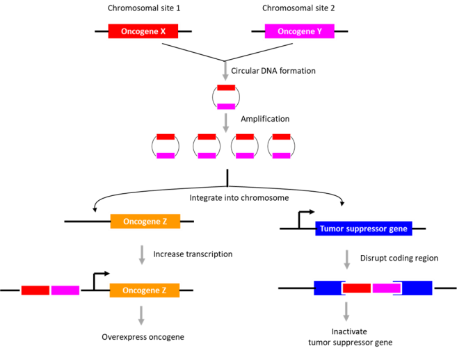
In human tissues, three different types of circular DNAs have been identified, i.e. ring chromosome, large extrachromosomal circular DNA (ecDNA), and small extrachromosomal circular DNA (eccDNA).  The presence of circular DNA in 93 neuroblastoma samples was computationally inferred by applying algorithms to detect circularity in genome sequencing data (Koche et al., 2019).  To increase the sensitivity of detection, the investigators at Sloan Kettering Cancer Center first depleted genomic DNA via nuclease treatment before conducting ‘circle sequencing’ (Circle-seq).  Circle sequencing utilizes circularized DNA template, which yields tandem repeats of sequences that are then aligned to determine the consensus sequence.  With ‘SMRT-seq’ (single molecule real time sequencing) using fluorescent phospho-linked nucleotides, the sequences could be obtained in real time.

In neuroblastoma, no ring chromosomes were found although ecDNA (mean size 680 kbp; ~0.8 copies per tumor) and eccDNA (mean size 2.4 kbp; ~5600 copies per tumor) were detected.  Whereas ecDNA may contain the entire genes, eccDNAs generally contains partial genes, with DNA circularization occurring in both coding and noncoding regions.  Intriguingly, the chromosomal region encompassing N-myc gene was highly circularized; other genes circularized include proto-oncogene JUN or MDM2 and transcription factor SOX11 or TAL2.

The potential role of DNA circularization in tumorigenesis was investigated.  Examination of circle junction sequence revealed microhomologies (minimally 5 bp), indicating that the DNA circles may have arisen during ‘microhomology-mediated DNA repair’.  The data suggested that DNA circularization may be necessary but not sufficient for genomic amplification.  Also, its role in gene overexpression appeared unlikely.   As an alternative, its role in genome remodeling was examined.  The extrachromosomal circular DNAs were found to be chimeras consisting of genomic sequences derived from distinct chromosomes (~2.2 chimeric segments for eccDNA; ~4.8 chimeric segments for ecDNA).  Further, they described an eccDNA (containing a region in chromosome 2 encompassing the N-myc gene), which is partly integrated into chromosome 13, disrupting DLCK1 gene.  Thus, the authors suggest that the extrachromosomal circular DNAs may integrate into various sites in the genome to disrupt tumor suppressor gene function or enhance proto-oncogene expression.  The presence of “circle-derived rearrangements” correlated with poor survivability, indicative of its potential use as a diagnostic marker for neuroblastoma.

 Bio-Synthesis, Inc. specializes in oligonucleotide modification and provides an extensive array of chemically modified nucleoside analogues (over ~200) including bridged nucleic acid (BNA).  A number of options are available to label oligonucleotides (DNA or RNA) with fluorophoreseither terminally or internally as well as conjugate to peptidesIt recently acquired a license from BNA Inc. of Osaka, Japan, for the manufacturing and distribution of BNANC, a third generation of BNA oligonucleotides.  To meet the demands of therapeutic application, its oligonucleotide products are approaching GMP grade.  Bio-Synthesis, Inc. has recently entered into collaborative agreement with Bind Therapeutics, Inc. to synthesize miR-21 blocker using BNA for triple negative breast cancer.  The BNA technology that we offer provides superior, unequalled advantages in base stacking, binding affinity, aqueous solubility and nuclease resistance.  It also improves the formation of duplexes and triplexes by reducing the repulsion between the negatively charged phosphates of the oligonucleotide backbone.  Its single-mismatch discriminating power was especially useful for diagnosis (ex. FISH using DNA probe).  More importantly, BNA oligonucleotide exhibits lesser toxicity than other modified nucleotides for clinical application.

https://www.biosyn.com/oligonucleotide-modification-services.aspx


References

Brodeur GM, Seeger RC, Schwab M, Varmus HE, Bishop JM. Amplification of N-myc in untreated human neuroblastomas correlates with advanced disease stage. (1984)  Science. 224:1121–4.   PMID: 6719137 DOI: 10.1126/science.6719137

Esposito MR, Aveic S, Seydel A, Tonini GP.  Neuroblastoma treatment in the post-genomic era.  (2017).  J Biomed Sci.  24:14.   PMID: 28178969  doi: 10.1186/s12929-017-0319-y

Hoehner JC, Gestblom C, Hedborg F, Sandstedt B, Olsen L, Pahlman S. A developmental model of neuroblastoma: differentiating stroma-poor tumors’ progress along an extra-adrenal chromaffin lineage. (1996)  Lab Invest.  75:659–75.   PMID: 8941212

Koche RP, Rodriguez-Fos E, Helmsauer K, Burkert M, MacArthur IC, Maag J, et al.  Extrachromosomal circular DNA drives oncogenic genome remodeling in neuroblastoma. (2019)  Nat Genet.  PMID: 31844324   DOI: 10.1038/s41588-019-0547-z

Lee WH, Murphree AL, Benedict WF.  Expression and amplification of the N-myc gene in primary retinoblastoma.  (1984)  Nature  309:458-60.  PMID: 6728001 DOI: 10.1038/309458a0

Maris JM.  Recent advances in neuroblastoma. (2010)  N Engl J Med.  362:2202-11.  PMID: 20558371  doi: 10.1056/NEJMra0804577

Mossé YP, Laudenslager M, Longo L, et al.  Identification of ALK as a major familial neuroblastoma predisposition gene. Nature. (2008)  455:930–5.   PMID: 18724359 PMCID: PMC2672043 DOI: 10.1038/nature07261 

Trochet D, Bourdeaut F, Janoueix-Lerosey I, et al. Germ-line mutations of the paired-like homeobox 2B (PHOX2B) gene in neuroblastoma. (2004)  Am J Hum Genet.  74:761–4.   PMID: 15024693 PMCID: PMC1181953 DOI: 10.1086/383253


Amino Acid Hydrophobicity for Structural Prediction of Peptides and Proteins

$
0
0

Hydrophobicity refers to the physical property of molecules that repel water. Hydrophobic molecules tend to be nonpolar and therefore prefer to interact other neutral and nonpolar solvents. In water, hydrophobic molecules often cluster together to form micelles. The hydrophobicity of molecules such as amino acids can be experimentally measured as reported by Richard Wolfenden.

Physiochemical properties routinely allow the characterization of peptide or protein sequences of known or unknown function. “Peptide Property Calculators,” such as the one found at BSIs website for the structural prediction or detection of membrane-associated or embedded β-sheets or α-helices, mostly present in larger peptides or proteins, are based on the Kyte-Doolittle Hydrophobicity scale. Several experimentally defined hydrophobicity parameters for amino acids now allow the calculation of peptide properties. In addition, many different scales for calculating peptide properties using hydrophobicity parameters have been developed in recent decades. However, the Kyte-Doolittle Hydrophobicity scale appears to be used the most in peptide calculators.

Often, to achieve a more accurate prediction for the peptide or protein structure of unknowns, five different hydrophobicity parameters are employed:    

1) the overall hydrophobicity, 

2) the hydrophobic moment for detection of α-helical and β-sheet membrane segments, 

3) the alternating hydrophobicity, 

4) the alternating hydrophobicity, and 

5) the exact β-strand score. 

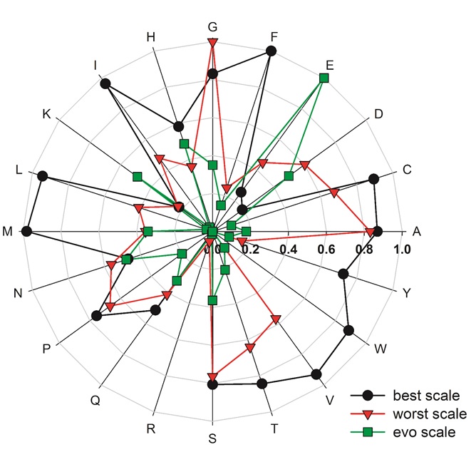
Simm et al. in 2016 reported that most scales allow discriminating between transmembrane α-helices and transmembrane β-sheets. However, using the five different hydrophobicity parameters enables the assignment of peptides to pools of soluble peptides of different secondary structures. But using alternating hydrophobicity is not significantly beneficial. All scales appear to have limitations in separation capacity. Simm et al. observed that the scales derived from the evolutionary approach performed best in separating different peptide pools. The 98 hydrophobicity scales known today have differently defined hydrophobicity values for the 20 amino acids. Unfortunately, because different experimental approaches were used for their definition, a high variance between different scales is observed. Importantly, Simm et al. found that the scale, as defined by Naderi-Manesh, developed in 2001, performed somewhat better than the other hydrophobicity scales. Therefore Simm et al. proposed a rule of thumb for the use of a hydrophobicity scale for the identification of peptides with transmembrane segments from a pool of peptides. Place the hydrophobicity value of arginine (Arg, R) and tyrosine (Tyr, Y) most distant from the value for glutamate (Glu, E). Select the hydrophobicity values for asparagine (Asn, N), aspartate (Asp, D), histidine (His, H), and lysine (Lys, K) such that they are in the center of the scale.

{Simm S, Einloft J, Mirus O, Schleiff E. 50 years of amino acid hydrophobicity scales: revisiting the capacity for peptide classification. Biol Res. 2016 Jul 4;49(1):31. doi: 10.1186/s40659-016-0092-5. PMID: 27378087; PMCID: PMC4932767. https://www.ncbi.nlm.nih.gov/pubmed/27378087}.

Figure 1: Hydrophobicity scale as defined by Naderi-Manesh.

{Naderi-Manesh H, Sadeghi M, Arab S, Moosavi Movahedi AA. Prediction of protein surface accessibility with information theory. Proteins. 2001;2001(42):452–459. doi: 10.1002/1097-0134(20010301)42:4<452::AID-PROT40>3.0.CO;2-Q.[PubMed] [CrossRef] [Google Scholar]}


Selected sources for hydrophobicity scales


http://www.genome.jp/aaindex/
Kawashima S, Pokarowski P, Pokarowska M, Kolinski A, Katayama T, Kanehisa M. AAindex: amino acid index database, progress report 2008. Nucleic Acids Res. 2008;2008(36):D202–D205. [PMC free article] [PubMed] [Google Scholar].

http://split4.pmfst.hr/split/scales.htmlJuretić D. Protein secondary structure conformations and associated hydrophobicity scales. J Math Chem. 1993;1993(14):35–45. doi: 10.1007/BF01164453. [CrossRef] [Google Scholar].

http://web.expasy.org/protscale/;Gasteiger E, Hoogland C, Gattiker A, Duvaud S, Wilkins MR, Appel RD, Bairoch A. Protein identification and analysis tools on the ExPASy server. In: Walker JM, editor. The proteomics protocols handbook. Totowa: Humana Press Inc.; 2005. pp. 571–607. [Google Scholar].

Table 1: Hydrophobicity Scales commonly used

Residue Type

kdHydrophobicitya

wwHydrophobicityb

hhHydrophobicityc

Ile

4.5

0.31

-0.60

Val

4.2

-0.07

-0.31

Leu

3.8

0.56

-0.55

Phe

2.8

1.13

-0.32

Cys

2.5

0.24

-0.13

Met

1.9

0.23

-0.10

Ala

1.8

-0.17

0.11

Gly

-0.4

-0.01

0.74

Thr

-0.7

-0.14

0.52

Ser

-0.8

-0.13

0.84

Trp

-0.9

1.85

0.30

Tyr

-1.3

0.94

0.68

Pro

-1.6

-0.45

2.23

His

-3.2

-0.96

2.06

Glu

-3.5

-2.02

2.68

Gln

-3.5

-0.58

2.36

Asp

-3.5

-1.23

3.49

Asn

-3.5

-0.42

2.05

Lys

-3.9

-0.99

2.71

Arg

-4.5

-0.81

2.58

 a A simple method for displaying the hydropathic character of a protein. Kyte J, Doolittle RF. J Mol Biol. 1982 May 5;157(1):105-32.

 b Experimentally determined hydrophobicity scale for proteins at membrane interfaces. Wimley WC, White SH. Nat Struct Biol. 1996 Oct;3(10):842-8.

 c https://www.cgl.ucsf.edu/chimera/docs/UsersGuide/midas/hydrophob.html#cnote


Amino Acid Hydrophobicity Scale used in Chimera, a modelling software from UCSF

UCSF Chimera is an example of a software package that allows visualization and analysis of molecular structures and related data. In Chimera, amino acid residues are automatically assigned an attribute named kdHydrophobicity, with values according to the hydrophobicity scale of Kyte and Doolittle.
The other scales in the following table are not assigned automatically, but input files to assign them with Define Attribute are linked below. A simple text format allows users to create custom attributes with ease.

Table 2: Chimera Amino Acid Hydrophobicity Scale

Residue Type

kdHydrophobicitya

wwHydrophobicityb

hhHydrophobicityc

mfHydrophobicityd

ttHydrophobicitye

Ile

4.5

0.31

-0.60

-1.56

1.97

Val

4.2

-0.07

-0.31

-0.78

1.46

Leu

3.8

0.56

-0.55

-1.81

1.82

Phe

2.8

1.13

-0.32

-2.20

1.98

Cys

2.5

0.24

-0.13

0.49

-0.30

Met

1.9

0.23

-0.10

-0.76

1.40

Ala

1.8

-0.17

0.11

0.0

0.38

Gly

-0.4

-0.01

0.74

1.72

-0.19

Thr

-0.7

-0.14

0.52

1.78

-0.32

Ser

-0.8

-0.13

0.84

1.83

-0.53

Trp

-0.9

1.85

0.30

-0.38

1.53

Tyr

-1.3

0.94

0.68

-1.09

0.49

Pro

-1.6

-0.45

2.23

-1.52

-1.44

His

-3.2

-0.96

2.06

4.76

-1.44

Glu

-3.5

-2.02

2.68

1.64

-2.90

Gln

-3.5

-0.58

2.36

3.01

-1.84

Asp

-3.5

-1.23

3.49

2.95

-3.27

Asn

-3.5

-0.42

2.05

3.47

-1.62

Lys

-3.9

-0.99

2.71

5.39

-3.46

Arg

-4.5

-0.81

2.58

3.71

-2.57

 a A simple method for displaying the hydropathic character of a protein. Kyte J, Doolittle RF. J Mol Biol. 1982 May 5;157(1):105-32.

 b Experimentally determined hydrophobicity scale for proteins at membrane interfaces. Wimley WC, White SH. Nat Struct Biol. 1996 Oct;3(10):842-8. Attribute assignment file wwHydrophobicity.txt.

 c Recognition of transmembrane helices by the endoplasmic reticulum translocon. Hessa T, Kim H, Bihlmaier K, Lundin C, Boekel J, Andersson H, Nilsson I, White SH, von Heijne G. Nature. 2005 Jan 27;433(7024):377-81, supplementary data. Attribute assignment file hhHydrophobicity.txt. In this scale, negative values indicate greater hydrophobicity.

 d Side-chain hydrophobicity scale derived from transmembrane protein folding into lipid bilayers. Moon CP, Fleming KG. Proc Natl Acad Sci USA. 2011 Jun 21;108(25):10174-7, supplementary data. Attribute assignment file mfHydrophobicity.txt. In this scale, negative values indicate greater hydrophobicity.

e An amino acid “transmembrane tendency” scale that approaches the theoretical limit to accuracy for prediction of transmembrane helices: relationship to biological hydrophobicity. Zhao G, London E. Protein Sci. 2006 Aug;15(8):1987-2001. Attribute assignment file ttHydrophobicity.txt (contributed by Shyam M. Saladi).

Table 3: Distribution Coefficients of the 19 Amino Acid Side Chains

Amino acid

RH (side chain)

v>wa

c>wb

VHc

CHd

GUe

WWf

WWth g


Least polar


ILE

n-butane

2.15

4.92

−0.60

0.24

2.04

2.16

−1.12

LEU

isobutane

2.28

4.92

−0.55

−0.02

1.76

2.29

−1.25

PHE

toluene

−0.76

2.98

−0.32

0.00

2.09

2.68

−1.71

VAL

propane

1.99

4.04

−0.31

0.09

1.18

1.61

−0.46

CYS

methanethiol

−1.24

1.28

−0.13

0.00

ND

1.23

−0.02

MET

methylethylsulfide

−1.48

2.35

−0.10

−0.24

1.32

1.71

−0.67

ALA

methane

1.94

1.81

0.13

−0.29

0.52

0.87

0.50

TRP

3-methylindole

−5.88

2.33

0.30

−0.59

2.51

2.96

−2.09

THR

ethanol

−4.88

−2.57

0.52

−0.71

0.27

0.95

0.25

TYR

4-methylphenol

−6.11

−0.14

0.68

−1.02

1.63

1.67

−0.71

GLY

hydrogen

2.39

0.94

0.74

−0.34

0.00

1.01

1.15

SER

methanol

−5.06

−3.40

0.84

−0.75

0.04

0.85

0.46

ASN

acetamide

−9.68

−6.64

2.05

−1.18

−0.01

0.30

0.85

HIS

4-methylimidazole

−10.27

−4.66

2.06

−0.94

0.95

0.92

2.33

GLN

propionamide

−9.38

−5.54

2.36

−1.53

−0.07

0.30

0.77

ARG

N-propylguanidine

−19.92

−14.92

2.58

−2.71

−1.32

2.99

1.81

GLU

propionic acid

−10.24

−6.81

2.68

−0.90

−0.79

−2.53

3.63

LYS

n-butylamine

−9.52

−5.55

2.71

−2.05

0.08

2.49

2.80

ASP

acetic acid

−10.95

−8.72

3.49

−1.02

ND

−2.46

3.64


Most polar


r2 versus VH h

0.73

0.83

(1.0)

0.66

0.59

0.30

0.77


r2 versus CH h

0.82

0.80

0.66

(1.0)

0.48

0.00

0.35


p versus VHi

0.000001

<0.0000001


0.00003

0.003


Distribution coefficients of the 19 amino acid side chains (RH) at pH 7, expressed in kcal/mol at 25°C, sorted according to their decreasing tendencies (VH) to be found in a transmembrane helix (Hessa et al., 2005). Experimental scales are shown in bold, theoretical scale in normal type.

aSide-chain Kd values for side-chain transfer from vapor to water (Wolfenden et al., 1981) ;  bSide-chain Kd values for transfer from cyclohexane to water (Radzicka and Wolfenden, 1988);  cTendency of amino acid residue to be found in a transmembrane helix (Hessa et al., 2005);  dTendency of amino acid residue to be buried in the interior of a globular protein (Chothia, 1976);  eAmino acid side-chain Kd values for transfer from wet octanol to water (Guy, 1985);  fPentapeptide Kd values for transfer from water to wet octanol (Wimley et al., 1996);  gTheoretical pentapeptide Kd values for transfer from water to wet octanol, after adjustment for the estimated effects of occlusion by neighboring residues (Wimley et al., 1996, Table II, column 3);  hValue of the correlation coefficient (r) obtained by linear regression of distribution coefficients against the VH or the CH scales;  iProbability that this experimental scale is not related to the VH scale, i.e., that the null hypothesis is true.

{Source: Wolfenden R. Experimental measures of amino acid hydrophobicity and the topology of transmembrane and globular proteins. J Gen Physiol. 2007 May;129(5):357-62. doi: 10.1085/jgp.200709743. Epub 2007 Apr 16. PMID: 17438117; PMCID: PMC2154378.}


Monitoring Peptide Binding with a Fluorogenic Probes

$
0
0

Fluorescent probes allow detection of protein location and activation, help identify protein complex formation and conformational changes as well as monitoring biological processes in vivo. Fluorescent probes are biosensor molecules. Biosensor molecules allow direct detection of analytes or target molecules.

A molecular biosensor contains a biological recognition element called a receptor covalently connected to a transducer molecule. The transducer is generally a fluorophore needed for signaling. The receptor molecule is selected or designed, such as to recognize the target molecule or analyte specifically. Upon binding to the target, the fluorophore responds to the local environment transforming the recognition event into a measurable signal. Fluorescent molecular biosensors allow detection and quantification of analytes. In general, target molecules are nucleic acids, DNA or RNA, or proteins, often enzymes or antibodies. Monitoring the peptide loading process onto class II MHC proteins is an example for the design of fluorogenic biosensor molecules.

Monitoring the peptide loading process onto class II MHC proteins

To visualize the peptide loading process onto class II MHC proteins, Venkatraman et al. designed a series of novel fluorogenic probes that incorporate the environment-sensitive amino acid analogs 6-N,N-dimethylamino-2-3-naphthalimidoalanine and 4-N,N-dimethylaminophthalimidoalanine. When these fluorophores bind to the protein, they experience substantial changes in emission spectra, quantum yield, and fluorescence lifetime.

Peptides containing these fluorophores bind specifically to class II MHC proteins on antigen-presenting cells allowing the monitoring of peptide binding in vivo. Venkatraman et al. used these probes to track developmentally regulated cell-surface peptide-binding activity in primary human monocyte-derived dendritic cells.

Figure 1: Structure of the Ac-PK(4-DAPA)VKQNTLKLAT peptide.


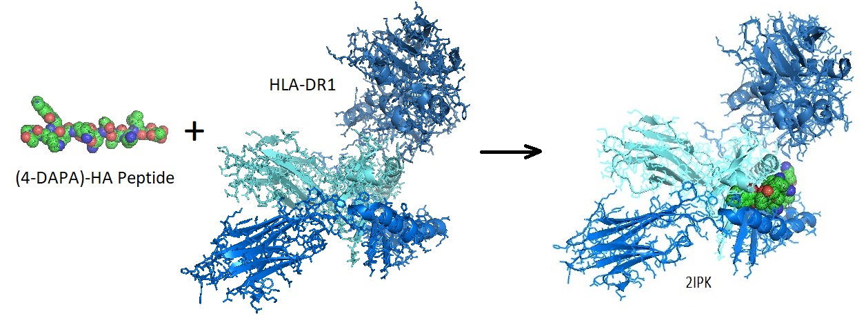
Figure 2: Binding of a fluorophore-peptide to HLA-DR1. The Crystal Structure Of The Mhc Class Ii Molecule Hla-dr1 In Complex With The Fluorogenic 14 mer Peptide, Acpkxvkqntlklat (x=3-[5-(dimethylamino)-1,3- Dioxo-1,3-dihydro-2h-isoindol-2-yl]-l-alanine) And The Superantigen, Sec3 Variant 3b2 [2IPK] [Sequence: XPKXVKQNTLKLAT].

(4-DAPA)-fluorophore peptides bind specifically to class II MHC proteins on antigen-presenting cells. The fluorescent peptides allow following peptide binding in vivo as well as tracking developmentally regulated cell-surface peptide-binding activity in primary human monocyte-derived dendritic cells. Peptides designed similarly enable the development of a myriad of fluorogenic peptide probes utilizing a wide range of peptide-based recognition events. Examples are peptide binding to antibodies, other binding proteins, protease-specific peptide sequences, and epitope-derived peptide sequences.

Reference

Venkatraman P, Nguyen TT, Sainlos M, Bilsel O, Chitta S, Imperiali B, Stern LJ. Fluorogenic probes for monitoring peptide binding to class II MHC proteins in living cells. Nat Chem Biol. 2007 Apr;3(4):222-8. doi: 10.1038/nchembio868. Epub 2007 Mar 11. PMID: 17351628; PMCID: PMC3444530. 
https://www.ncbi.nlm.nih.gov/pmc/articles/PMC3444530/?report=reader

 ---…---


Monitoring Peptide Binding with a Fluorogenic Probe

$
0
0

Fluorescent probes allow detection of protein location and activation, help identify protein complex formation and conformational changes as well as monitoring biological processes in vivo. Fluorescent probes are biosensor molecules. Biosensor molecules allow direct detection of analytes or target molecules.

A molecular biosensor contains a biological recognition element called a receptor covalently connected to a transducer molecule. The transducer is generally a fluorophore needed for signaling. The receptor molecule is selected or designed, such as to recognize the target molecule or analyte specifically. Upon binding to the target, the fluorophore responds to the local environment transforming the recognition event into a measurable signal. Fluorescent molecular biosensors allow detection and quantification of analytes. In general, target molecules are nucleic acids, DNA or RNA, or proteins, often enzymes or antibodies. Monitoring the peptide loading process onto class II MHC proteins is an example for the design of fluorogenic biosensor molecules.

Monitoring the peptide loading process onto class II MHC proteins

To visualize the peptide loading process onto class II MHC proteins, Venkatraman et al. designed a series of novel fluorogenic probes that incorporate the environment-sensitive amino acid analogs 6-N,N-dimethylamino-2-3-naphthalimidoalanine and 4-N,N-dimethylaminophthalimidoalanine. When these fluorophores bind to the protein, they experience substantial changes in emission spectra, quantum yield, and fluorescence lifetime.

Peptides containing these fluorophores bind specifically to class II MHC proteins on antigen-presenting cells allowing the monitoring of peptide binding in vivo. Venkatraman et al. used these probes to track developmentally regulated cell-surface peptide-binding activity in primary human monocyte-derived dendritic cells.

Figure 1: Structure of the Ac-PK(4-DAPA)VKQNTLKLAT peptide.


Figure 2: Binding of a fluorophore-peptide to HLA-DR1. The crystal structure of the Mhc Class II Molecule Hla-dr1 in complex with the fluorogenic 14 mer peptide, AcPKXVKQNTLKLAT (x=3-[5-(dimethylamino)-1,3- Dioxo-1,3-dihydro-2h-isoindol-2-yl]-l-alanine) And The Superantigen, Sec3 Variant 3b2 [2IPK].

(4-DAPA)-fluorophore peptides bind specifically to class II MHC proteins on antigen-presenting cells. The fluorescent peptides allow following peptide binding in vivo as well as tracking developmentally regulated cell-surface peptide-binding activity in primary human monocyte-derived dendritic cells. Peptides designed similarly enable the development of a myriad of fluorogenic peptide probes utilizing a wide range of peptide-based recognition events. Examples are peptide binding to antibodies, other binding proteins, protease-specific peptide sequences, and epitope-derived peptide sequences.

Reference

Venkatraman P, Nguyen TT, Sainlos M, Bilsel O, Chitta S, Imperiali B, Stern LJ. Fluorogenic probes for monitoring peptide binding to class II MHC proteins in living cells. Nat Chem Biol. 2007 Apr;3(4):222-8. doi: 10.1038/nchembio868. Epub 2007 Mar 11. PMID: 17351628; PMCID: PMC3444530.  https://www.ncbi.nlm.nih.gov/pmc/articles/PMC3444530/?report=reader

 ---…---


Selective 2’-Hydroxyl Acylation and Primer Extension or SHAPE

$
0
0

Selective 2’-Hydroxyl Acylation and Primer Extension or SHAPE

The majority of RNA molecules only appear to function when they exist in the correct secondary or tertiary structure by folding back onto themselves into difficult to predict three-dimensional structures. Many RNAs have specific regions with local structural flexibility or can transition from one conformation to another. Known RNA motif families are GNAA tetraloop, kink-turn, sarcin-ricin, and T-loop, but other potential motifs are also present in ribosomal RNA, sgRNA, SRP RNA, riboswitch and ribozyme.

To understand the function of RNAs, accurate, and comprehensive knowledge of the base-paired secondary structure and the identification of nucleotides involved in tertiary structure is needed. Known functional RNA components with three-dimensional (3D) conformation fall under the category of RNA motifs.


With that in mind, in years past, researchers have used a veriety of chemical and enzymatic reagents for mapping RNA secondary structures. However, many of these approaches suffer from shortcomings such as: 

  • Low discriminative power:  For many reagents, the magnitude of discrimination between single-stranded and base-paired regions can be small, and helix termini and some base pairs are often reactive.

  • Infrequent results:  The obtained information is often of limited value since a given reagent may react with only a subset of the four RNA nucleotides or backbone sites.

  • Low selectivity:  Therefore, multiple reagents are often employed for the interrogation of all positions in an RNA molecule

  • No unifying rule sets:  Historically, it has been challenging to develop unifying rule sets for classes of structures that react versus that do not. 

In 2005, Merino et al. reported an alternative approach for mapping RNA structures using the observation that the chemical reactivity of the 2’-ribose position is influenced by the adjacent 3’-phosphodiester anion.

Earlier in 1999, Chamberlin and Weeks started evaluating the chemical selectivity of 2′-amine acylation using a model oligonucleotide substrate containing either all ribose nucleotides or a single 2′-amino-cytidyl substitution. Chamberlin and Weeks investigated reagents allowing selective acylation of the 2'-ribose position for their use in mapping RNA structures. Also, the magnesium ion-dependent conformational changes in tRNAAsp transcripts containing single 2′-amine substitutions per transcript were studied as well. Chamberlin and Weeks found that acylation of synthetic 2’-amine-substituted nucleotides forming the 2’-amide product is strongly confined by the underlying local flexibility of the nucleotide. 


Figure 1: Selective Acylation of 2′-Amine Positions in RNA. The oligonucleotide substrate was treated with a succinimidyl ester. The incorporation of the biotinyl group was monitored as a slowly migrating band in a denaturing polyacrylamide gel. The all-ribose substrate reacted very slowly with the reagent (4% conversion in 60 min) as compared to the 2′-amine substituted oligonucleotide (97% conversion in 60 min). The slow modification of the all-ribose substrate is attributed to the reaction of base arylamines with the succinimidyl ester.

The researchers found that under denaturing conditions, all 2′-amine substituted RNA positions show similar reactivity. However, when tRNAAsp transcripts are refolded under native conditions (10 mM Mg2+, 100 mM NaCl), positions involved in base pairing and known tertiary interactions, including base triples and loop-loop interactions, are protected from modification. When no magnesium ions are present, the acceptor, T- and anticodon stems forms stable helices leading to a relatively low 2′-amine reactivity. These results suggested an interdependence between the formation of the D-stem helix and tertiary structure folding for yeast tRNAAsp transcripts. The researchers used this chemical approach for mapping local RNA flexibilities. Reported experimental results were consistent with prior biophysical and biochemical studies showing its utility for mapping local nucleotide environments on small amounts of RNA molecules.

Figure 2: Models of the 3D structure of yeast tRNAAsp from Asp and Phe transfer RNA crystals 2TA (Westhof et al. 1988).

Mortimer and Weeks set out to find a more selective fast-acting reagent needed for accurate analysis of RNA secondary and tertiary structures using SHAPE chemistry. The reagent selected needs to allow chemically assisted RNA structure analysis with the following criteria:

  • Enable a chemically straightforward approach,
  • Use of a single reagent reactive to all four nucleotides,
  • Is self-quenching,
  • Has a short reaction time, and
  • Yields accurate results with complex RNA molecules of known structure.

After testing the reagent N-methylisatoic anhydride (NMIA) for is use in SHAPE the scientists decided to develop a faster reacting reagent. The reagent 1-methyl-7-nitroisatoic anhydride (1M7) appears to fulfill these criteria.

Figure 3: Structures of reagents NMIA and 1M7.

In 1M7, the para nitro substituent is strongly electron-withdrawing leading to an increase in adduct formation as well as hydrolysis rates. Experimental verification allowed the researcher to conclude that 1M7 has the correct chemical characteristics for a fast-acting and self-quenching reagent for its use in SHAPE chemistry.

A SHAPE experiment performed on the RNase P domain under condition that stabilized the native tertiary fold allowed identifying 2’-O-adduct formation as stops of primer extension, using fluorescently labeled DNA primers, as resolved by capillary electrophoresis. Experiments were performed in the presence and absence of Mg2+ ions to test how magnesium ions influence the flexibility of the structure.  

To conclude, SHAPE chemistry performed with 1M7 allowed the researchers to generate an accurate report of the known structure of the RNase P specificity domain under native conditions.

Reference

1988: Westhof, Dumas, and Moras; Restrained refinement of two crystalline forms of yeast aspartic acid and phenylalanine transfer RNA crystals. Acta Crystallogr A 1988 Mar 1;44 (Pt 2): 112-123.

2000: Chamberlin and Weeks; Mapping local nucleotide flexibility by selective acylation of 2-amine substituted RNA. J. Am. Chem. Soc. 2000, 122, 216-224.


2005:
Edward J. Merino, Kevin A. Wilkinson, Jennifer L. Coughlan, Kevin M. Weeks; RNA Structure Analysis at Single Nucleotide Resolution by Selective 2‘-Hydroxyl Acylation and Primer Extension (SHAPE). J. Am. Chem. Soc. 2005, 127, 12, 4223-4231. https://doi.org/10.1021/ja043822v.

2007: Stefanie A. Mortimer and and Kevin M. Weeks; A Fast-Acting Reagent for Accurate Analysis of RNA Secondary and Tertiary Structure by SHAPE Chemistry. Journal of the American Chemical Society 2007 129 (14), 4144-4145. DOI: 10.1021/ja0704028.
https://pubs.acs.org/doi/full/10.1021/ja0704028.

2018: RNA motifs: Ping Ge, Shahidul Islam, Cuncong Zhong, Shaojie Zhang, De novo discovery of structural motifs in RNA 3D structures through clustering, Nucleic Acids Research, Volume 46, Issue 9, 18 May 2018, Pages 4783–4793, 
https://doi.org/10.1093/nar/gky139.


---…----

Primer and Probe Collection for 2019 Coronavirus Detection

$
0
0
 Primer and Probe Collection for 2019 Coronavirus Detection


The new coronavirus (2019-nCoV) was first detected in Wuhan City, Hubei Province China. Tens of thousands of infections with 2019-nCoV have been reported in China. The virus is spreading from person-to-person in parts of the world. Coronaviruses are a large family of viruses that are common in many animals, including camels, cattle, cats, and bats. However, some animal coronaviruses can infect people and spread between people, for example
MERS, SARS, and now with 2019-nCoV {CDC}. 

Corona virus outbreaks in January 2020. 


Source: Wikimedia Commons


The following is a list of primers and probes for the detection of the new 2019 coronavirus (2019-nCoV) in human clinical specimens. Please use indicated concentrations as a guide. Actual concentrations used should be experimentally verified. 

!! Primers and probes for the detection of the new 2019 coronavirus are available from BSI !! 


Genome: Wuhan seafood market pneumonia virus isolate 2019-nCoV/USA-AZ1/2020, complete genome  Sequence ID: MN997409.1 Length: 29882

Note: A, Adenine; C, Cytosine; G, Guanine; T, Thymine; U, Uracil; R, A or G; Y, C or T; S, G or C; W, A or T; K, G or T; M, A or C; B, C or G or T; D, A or G or T; V, A or C or G; N,  any base.

 CDC Primer and Probes

Name

Label 5’

Oligonucleotide Sequence (5’>3’)

Label 3’

Working Conc. [μM]


N Protein


2019-nCoV_N1-F


GACCCCAAAATCAGCGAAAT

28287------------28306


2019-nCoV_N1-R


TCTGGTTACTGCCAGTTGAATCTG

28358---------------28335


20

2019-nCoV_N1-P

FAM

ACCCCGCATTACGTTTGGTGGACC

28309---------------28332

BHQ1

5

2019-nCoV_N2-F


TTACAAACATTGGCCGCAAA

29164-------------29183


20

2019-nCoV_N2-R


GCGCGACATTCCGAAGAA

29230------------29213


20

2019-nCoV_N2-P

FAM

ACAATTTGCCCCCAGCGCTTCAG

29188---------------29210

BHQ1

5

2019-nCoV_N3-F


GGGAGCCTTGAATACACCAAAA

28681--------------28702


20

2019-nCoV_N3-R


TGTAGCACGATTGCAGCATTG

28752--------------28732


20

2019-nCoV_N3-P

FAM

AYCACATTGGCACCCGCAATCCTG

28706--------------28727 

BHQ1

5


Name

Label 5’

Oligonucleotide Sequence (5’>3’)

Label 3’

Working Conc. [μM]


RNAse Protein


RP-F


AGATTTGGACCTGCGAGCG


20

RP-R


GAGCGGCTGTCTCCACAAGT


RP-P RNAse P

FAM

TTCTGACCTGAAGGCTCTGCGCG

BHQ-1

5


Name

Label 5’

Oligonucleotide Sequence (5’>3’)

Label 3’

Working Conc. [μM]


ORF1ab


2019-nCoV-OFP


CCCTGTGGGTTTTACACTTAA

13342------------13362


0.6

2019-nCoV-ORP


ACGATTGTGCATCAGCTGA

13460------------13442


0.8

2019-nCoV-OP

FAM

CCGTCTGCGGTATGTGGAAAGGTTATGG

13377-------------------13404

BBQ1

0.1


Nucleoprotein- protein N


2019-nCoV-NFP


GGGGAACTTCTCCTGCTAGAAT

28881-------------28902


0.6

2019-nCoV-NRP


CAGACATTTTGCTCTCAAGCTG

28979-------------28958


0.8

2019-nCoV-NP

FAM

TTGCTGCTGCTTGACAGATT

28934-------------28953

TAMRA

0.1


ORF1b-nsp14


HKU-ORF1b-nsp14F


TGGGGYTTTACRGGTAACCT

18778-----------18797


0.6

HKU- ORF1b-nsp14R


AACRCGCTTAACAAAGCACTC

18909-------------18889


0.8

HKU-ORF1b-nsp141P

FAM

TAGTTGTGATGCWATCATGACTAG

18849---------------18872

TAMRA

0.1


Nucleoprotein – Protein N


HKU-NF


TAATCAGACAAGGAACTGATTA

29145--------------29166


0.6

HKU-NR


CGAAGGTGTGACTTCCATG

29254-----------29236


0.8

HKU-NP

FAM

GCAAATTGTGCAATTTGCGG

29196------------29177

TAMRA

0.1


Positive Control for insertion into plasmid

AGTTGACTTCGCAGTGGCTAACTAACATCTTTGGCACTGTTTATGAAAAACTCAAACCCGTCCTTGATTGGCTTG

AAGAGAAGTTTAAGGAAGGTGTAGAGACCCTGTGGGTTTTACACTTAAAAACACAGTCTGTACCGTCTGCGGTAT

GTGGAAAGGTTATGGCTGTAGTTGTGATCAACTCCGCGAACCCATGCTTCAGTCAGCTGATGCACAATCGTTTTT

ACTCCAGGCAGCAGTAGGGGAACTTCTCCTGCTAGAATGGCTGGCAATGGCGGTGATGCTGCT


Name

Label 5’

Oligonucleotide Sequence (5’>3’)

Label 3’

Working Conc. [μM]


RNA-dependent RNA polymerase


RdRP_SARSr-F2


GTGARATGGTCATGTGTGGCGG

15431-------------15452

2019-nCoV genome


0.6

RdRP_SARSr-R1


CARATGTTAAASACACTATTAGCATA

15460-----------------15435

Coronavirus BtRs-BetaCoV/YN2018D, complete genome. Sequence ID: MK211378.1 Length: 30213


0.8

RdRP_SARSr-P1

FAM

CCAGGTGGWACRTCATCMGGTGATGC

145---------------------170

Bat coronavirus isolate B18-171 RNA-dependent RNA polymerase (RdRp) gene, partial cds. Sequence ID: MK991947.1 Length: 387

BBQ1

0.1

RdRP_SARSr-P2

FAM

CAGGTGGAACCTCATCAGGAGATGC

15470----------------15494

Specific for 2019-nCoV, will not detect SARSCoV.

Wuhan seafood market pneumonia virus isolate 2019-nCoV/USA-AZ1/2020, complete genome Sequence ID: MN997409.1Length: 29882

BBQ1

0.1


Reference

https://www.who.int/docs/default-source/coronaviruse/peiris-protocol-16-1-20.pdf?sfvrsn=af1aac73_4 

Liu G, Li H, Zhao S, Lu R, Niu P, Tan W. Viral and Bacterial Etiology of Acute Febrile Respiratory Syndrome among Patients in Qinghai, China.Biomed Environ Sci. 2019 Jun;32(6):438-445. https://www.sciencedirect.com/science/article/pii/S0895398819301230

---...----


Towards development of quencher-free oligonucleotide probe with single base-discriminating fluorophore enhanced by bridged nucleic acid (BNA) for genotyping SNP (single nucleotide polymorphism) linked to cancer and other diseases

$
0
0
Towards development of quencher-free oligonucleotide probe with single base-discriminating fluorophore enhanced by bridged nucleic acid (BNA) for genotyping SNP (single nucleotide polymorphism) linked to cancer and other diseases


Single nucleotide polymorphisms (SNP) refers to nucleotide variations affecting a specific base position in the genome.  SNPs occurring in the coding region could either be ‘synonymous’ (does not alter amino acid sequence) or ‘nonsynonymous’ (changes polypeptide sequence) type.  SNPs located in the noncoding region can also affect gene expression by altering transcription, splicing or degradation of mRNA.  SNPs associated with the risk susceptibility for various disorders including Alzheimer’s disease, cystic fibrosis, sickle cell anemia or alcoholism have been identified.  For instance, a SNP in intron 32 of DMD gene deactivates splice donor site (Thi Tran et al., 2005).

The genetic susceptibility to cancer is associated with various SNPs linked to the genes regulating cell cycle, DNA repair, metabolism, immunity, etc (Deng et al., 2016).  SNPs in promoters may affect TATA box function (EDH17B2 gene), transcription factor binding (MDM2, MMP1, survivin  genes), epigenetic regulation (BRCA1 gene) or histone modification.  Nonsynonymous SNPs in exons may promote tumorigenesis (oncogenic mutation of p53) or decrease therapeutic efficacy by eliminating targets (EGF receptor) by altering their conformation.  Synonymous SNPs in exons can affect the stability, splicing or structure of RNA (stem-loop formation of COMT mRNA; MDR1 gene).  SNPs in introns were shown to affect ‘transcriptional silence element’ (elevates FGFR2 expression in breast cancer) or alter splicing (histocompatibility antigen in renal cell carcinoma).  While SNPs in 5’-UTR (untranslated region) may affect mRNA processing, translation, etc. (CDKN2A gene in melanoma), 3’-UTR SNPs may affect degradation, polyadenylation, etc. (affect micro-RNA regulation of estrogen receptor in breast cancer).   

The development of SNP diagnostics has been guided by the need for high-throughput data acquisition and simplicity, i.e. easy to perform, low cost, portable, accuracy.  To accommodate these concerns, current methods utilize high-throughput sequencing as well as PCR.  In the case of C/T detection, the SNP genotyping methods used in high-throughput assays include ‘flap endonuclease discrimination’ that involves cleavage of fluorescent probe from the triplex structure, ‘allele specific hybridization’ wherein quencher (for the probe) is removed during amplification, ‘primer extension’ which involves single base extension of fluorescent dideoxynucleotide at the polymorphic site.  Other SNP diagnostic methods are ‘allele specific digestion’ requiring cleavage of PCR extended product incorporating dUTP by uracil-DNA glycosylase, and ‘oligonucleotide ligation’ where ligation of two oligonucleotides occurs only if one is complementary to SNP (Jenkins et al., 2002).  


Several innovations are being introduced to further increase the sensitivity of SNP detection.  Molecular beacons have found great utility in detecting SNPs through PCR.  It consists of stem and loop structure with a quencher probe juxtaposed to a fluorophore to dampen its signal, which becomes restored through dissociation upon hybridization to target complementary strand.  Nevertheless, it requires dual labels that could contribute to the cost.  To improve, quencher-free probes that could distinguish the type of base in the opposite strand are being developed, i.e. probes whose fluorescence intensity changes upon hybridization to target strand (Hwang, 2018).  For instance, the quenched fluorescence of a fluorophore (ex. BODIPY, fluorescein) in a probe (when located adjacent to guanine residue) can be dequenched upon hybridizing to cytosine residue of target DNA.

One such advance involves incorporating pyrene labeled deoxyadenosine at the 5’-end of a hairpin (Panel A in Figure).  Its fluorescence was quenched when placed next to C, T or G base though was enhanced significantly upon interaction with target DNA (35 fold) or RNA (44 fold) (Seo et al., 2005).  Further increase in the ability to discriminate SNPs occurred when locked nucleic acid (LNA) was placed adjacent to pyrene-labeled uridine, i.e. emission increased with matched duplex than mismatched duplex (Kaura et al., 2015). 

Another approach involves the modification of nucleic acid analogues with higher affinity to complementary strands.  Briefly, the introduction of bridged nucleic acid has greatly increased the binding affinity of an oligonucleotide to target complementary strand.  The stability of the RNA:DNA or DNA:DNA duplex can be greatly augmented through incorporation of a small number of locked nucleic acids.  This has inspired the development of 2'-O,4'-aminoethylene bridged nucleic acid (2',4'-BNANC), the third-generation bridged nucleic acid containing a six-member bridged structure with an N-O linkage.  BNA provides greater binding affinity, better single-mismatch discrimination, enhanced RNA specificity, stronger/selective triplex-forming properties, and considerably higher nuclease resistance (Rahman et al., 2008; Miyashita et al., 2007).  To couple heightened binding affinity with greater fluorescence, novel (phenylethynyl)pyrene–BNA constructs were developed by the investigators at University of Southern Denmark (Panel B in Figure).  Using probes incorporating these fluorescent nucleoside analogues, the drug‐resistance‐causing mutation in HIV‐1 protease cDNA and RNA gene fragments was successfully detected (Astakhova et al., 2012).

Bio-synthesis, Inc. specializes in oligonucleotide modification and provides an extensive array of chemically modified nucleoside analogues (over ~200) including bridged nucleic acid (BNA).  A number of options are available to label oligonucleotides (DNA or RNA) with fluorophoreseither terminally or internally as well as conjugate to peptides. This includes pyrene-dU fluorescent baseoligonucleotide modification.  For oncogenic SNP genotyping, Bio-synthesis, Inc. provides a kit incorporating BNA for the rapid and convenient real-time PCR detection of the BRAF-V600E mutation with high sensitivity (<0.1% of mutant in wild-type background).  It recently acquired a license from BNA Inc. of Osaka, Japan, for the manufacturing and distribution of BNANC, a third generation of BNA oligonucleotides.  To meet the demands of therapeutic application, its oligonucleotide products are approaching GMP grade.  Bio-Synthesis, Inc. has recently entered into collaborative agreement with Bind Therapeutics, Inc. to synthesize miR-21 blocker using BNA for triple negative breast cancer.  The BNA technology that we offer provides superior, unequalled advantages in base stacking, binding affinity, aqueous solubility and nuclease resistance.  It also improves the formation of duplexes and triplexes by reducing the repulsion between the negatively charged phosphates of the oligonucleotide backbone.  Its single-mismatch discriminating power was especially useful for diagnosis (ex. FISH using DNA probe).  More importantly, BNA oligonucleotide exhibits lesser toxicity than other modified nucleotides for clinical application.

https://www.biosyn.com/oligonucleotideproduct/pyrene-du-fluorescent-base-oligonucleotide-modification.aspx#!

https://www.biosyn.com/oligonucleotide-modification-services.aspx

https://www.biosyn.com/braf-mutation-analysis-kit.aspx


References

Astakhova IK, Samokhina E, Babu BR, Wengel J.  Novel (phenylethynyl)pyrene–LNA constructs for fluorescence SNP sensing in polymorphic nucleic acid targets.  (2012)  ChemBiochem  13:1509-19.  PMID: 22761036 DOI: 10.1002/cbic.201200079

Deng N, Zhou H, Fan H, Yuan Y.  Single nucleotide polymorphisms and cancer susceptibility.  (2017)  Oncotarget  8:110635-110649.   PMID: 29299175  doi: 10.18632/oncotarget.22372. eCollection

Hwang GT. Single-Labeled Oligonucleotides Showing Fluorescence Changes Upon Hybridization with Target Nucleic Acids. 2018) Molecules. 23. pii: E124. PMID: 29316733  doi: 10.3390/molecules23010124. Review.

Jenkins S, Gibson N.  High-throughput SNP.  (2002) Comp Funct Genomics.  3:57-66.   PMID: 18628885   doi: 10.1002/cfg.130.

Kaura, M.; Hrdlicka, P.J.  Locked nucleic acid (LNA) induced effect on the hybridization and fluorescence properties of oligodeoxyribonucleotides modified with nucleobase-functionalized DNA monomers. (2015)  Org. Biomol. Chem. 13, 7236–7247.  PMID: 26055658  DOI: 10.1039/c5ob00860c

Miyashita, K., Rahman, S. M., Seki, S., Obika, S., and Imanishi, T. N-Methyl substituted 2′,4′- BNANC: a highly nuclease resistant nucleic acid analogue with high-affinity RNA selective hybridization. (2007) Chem. Commun. (Cambridge, U. K.), 3765−3767.

Rahman SM, Seki S, Obika S, Yoshikawa H, Miyashita K, Imanishi T. Design, synthesis, and properties of 2',4'-BNA(NC): a bridged nucleic acid analogue.  (2008)  J Am Chem Soc. 130:4886-96. PMID: 18341342  doi: 10.1021/ja710342q.

 Seo YJ, Ryu JH, Kim BH. Quencher-free, end-stacking oligonucleotides for probing single-base mismatches in DNA.  (2005)  Org Lett. 7:4931-3.  PMID: 16235925  DOI: 10.1021/ol0518582

Thi Tran HT, Takeshima Y, Surono A, Yagi M, Wada H, Matsuo M.  A G-to-A transition at the fifth position of intron-32 of the dystrophin gene inactivates a splice-donor site both in vivo and in vitro.  (2005)  Mol Genet Metab  85:213-9.  PMID: 15979033  DOI: 10.1016/j.ymgme.2005.03.006

Blocking RNA for Treating Triple Negative Breast Cancer

$
0
0

In 2019, the US National Cancer Institute awarded Bound Therapeutics LLC, together with Thomas Jefferson University and Bio-Synthesis Inc., a Small Business Technology Transfer grant starting 15 June for their collaborative research project to develop a "microRNA-21 based Blockade of Triple-Negative Breast Cancer." 

Triple-negative breast cancer does not have receptors for estrogen, progesterone, or HER-2/neu hormones. Hence triple-negative breast cancer patients do not benefit from receptor-targeted treatments, such as tamoxifen and Herceptin, that are sometimes effective for treating hormone-receptor-positive cancers. 

“Triple-negative breast cancer strikes younger women, tragically killing half the patients within 4 years," said Dr. Yuan-Yuan Jin, Chief Operating Officer of Bound Therapeutics LLC. "Surgery, chemotherapy, and radiation are the current standard of care for triple-negative breast cancer."

"To provide effective molecular therapy that will keep patients alive with a good quality of life, we have designed a cancer cell-targeted drug that will block a tiny strand of ribonucleic acid, called microRNA-21," explained Dr. Eric Wickstrom, Professor of Biochemistry and Molecular Biology at Thomas Jefferson University, a partner in the award. “MicroRNA-21 drives cell division, and occurs at high levels in most triple negative breast cancer cells.”

Dr. Miguel Castro, President and CEO of Bio-Synthesis Inc., another research partner, said that "The drug candidates containing BNAs (bridged nucleic acid analogs) and peptide analogs that we are making to treat triple-negative breast cancer cells are extraordinarily specific and safe in mammalian models." 

Our clinical collaborator, Dr. Edith Mitchell, Clinical Professor of Medicine and Medical Oncology at Thomas Jefferson University, commented that "Patients with triple-negative disease have limited treatment options compared to patients with more common forms of breast cancer. There is an urgent need for targeted treatments in this area." Dr. Mitchell serves as the Director of the Jefferson Center to Eliminate Cancer Disparities in diagnosis, treatment, and survival of patients with different ancestries. She is a past President of the National Medical Association. 

The funds will enable Bound Therapeutics and its partners Bio-Synthesis Inc. and Thomas Jefferson University to test their drug design in a triple-negative breast cancer mouse model with a healthy immune system. Early results suggest that microRNA-21 blockade decloaks triple-negative breast cancer cells to enable attack by immune cells. 

Historically, drug development involved looking for functional sites or binding pockets in malfunctioning proteins that cause the disease, in order to find drugs for a treatment. However, not all diseases have faulty proteins that can be targeted by a drug binding to functional sites located on a protein. However, synthetic complementary RNA analogs can block pathogenic RNA molecules or speed up their degradation. RNA analogs are short sequences that bind or hybridize to RNA. RNA molecules such as messenger RNA (mRNA), microRNA (miRNA), long noncoding RNA (lncRNA) or ribosomal RNA (rRNA) are potential targets. To lengthen the lifetime of RNA-based drugs or to make them more specific, artificial RNA analogs such as BNAs can be incorporated. 

Bridged nucleic acids (BNAs) display (i) equal or higher binding affinity against an RNA complement with excellent single-mismatch discriminating power, (ii) much better RNA selective binding, (iii) stronger and more sequence selective triplex-forming characters, and (iv) high nuclease resistance, than regular DNAs or RNAs, or phosphorothioate analogs.

---...---


Structure of Coronavirus nCoV 2019/2020

$
0
0

Coronaviruses (CoVs) are enveloped positive-sense RNA viruses. The club-like spikes projecting out from their surface gave them the name. Coronaviruses possess an unusual large RNA genome as well as a unique replication strategy. Coronaviruses cause a variety of diseases in animals ranging from cows, pigs to chicken, and other birds. In humans, coronaviruses can cause potentially lethal respiratory infections.

Coronaviruses belong to the largest group of viruses called the Nidovirales order. Members of this order include the Coronaviridae, Arteriviridae, and Roniviridae families. The Coronvirinae are one of two subfamilies in the Coronaviridae family. Coronavirinae are further subdivided into for groups, the alpha, beta, gamma, and delta coronaviruses. Nowadays, these viruses are divided using phylogenetic clustering. These virus families have animal and human hosts. The Middle Eastern Respiratory Syndrome Coronavirus (MERS-CoV) and Severe Acute Respiratory Coronavirus (SARS-CoV) are examples.

Nidoviruses contain an infectious, linear, positive-sense RNA genome that is capped and polyadenylated. Based on their genome size, nidoviruses are divided into two groups large and small nidoviruses.

All Nidovirales viruses are enveloped, non-segmented positive-sense RNA viruses containing very huge genomes.

Common features of coronaviruses include

(i) a highly conserved genomic organization with a large replicase gene preceding structural and accessory genes,

(ii) expression of many non-structural genes by ribosomal frameshifting,

(iii) several unique of unusual enzymatic activities encoded within the large replicase-transcriptase polyprotein, and

(iv) expression of downstream genes by synthesis of 3’-nested sub-genomic mRNAs.

The typical organization of the genome is

5’-leader-UTR-replicase-S(Spike)-E(Envelope)-M(Membrane)-N(Nucleocapsid)-3’-UTR-poly(A) tail. Accessory genes are interspersed within the structural genes at the 3’-end of the genome.

Accessory proteins are not needed for replication in tissue culture but appear to be important in viral pathogenesis. The synthesis of polypeptide 1ab (pp1ab) involves programmed ribosomal frame shifting during translation of open-reading frame 1a (orf1a). Frame shifting results in a new reading frame that produces a trans-frame protein product. In coronaviruses, a fixed portion of the ribosomes translating orf1a change reading frame at a specific location now decoding information contained in orf1b.

U_UUA_AAC is a universal frame-shifting site

Coronaviruses contain a frameshifting stimulation element as a conserved RNA sequence forming a stem-loop that promotes ribosomal frameshifting. Ribosomal frameshifting is a mechanism in which open-reading frame 1b (orf1b) is expressed. Replicase-transcriptase proteins are encoded in open-reading frame 1a and 1b (orf1a and orf1b) and are synthesized initially as two large polyproteins termed pp1a and pp1b. A comparative analysis performed by Baranov et al. in 2004 revealed the sequence U_UUA_AAC as a universal shift site. Frameshifting was characterized in SARS-CoV cultured in mammalian cells using a dual luciferse reporter system and mass spectrometry. Tandem tRNA slippage on the sequence U_UUA_AAC was confirmed by mutagenic analysis of the shift site. Mass spectrometry was used for the analysis of affinity tagged frameshift products. Further analysis of the frameshifting site showed that a proposed RNA secondary structure in loop II and two unpaired nucleotides at the stem I-stem II junction in SARS-CoV are important for frameshift stimulation.    


Model of Coronavirus COVID19 Transcription. A possible model of the coronavirus COVID19 transcription mechanism is shown here. The model is based on the genomic sequence and the model for the transcription of coronaviruses as proposed by Sawicki et al. in 2007. The organization and the expression of the Wuhan seafood market pneumonia virus isolate Wuhan-Hu-1 genome is depicted here. Structural relationships of the genome and subgenome mRNAs are shown. Orfs are defined by the published genome sequence. Possible autoproteolytic processing of orfs1a and orf1ab polypeptides into protein nsp1 to 16 are shown as well.      

Reference

Baranov PV, Henderson CM, Anderson CB, Gesteland RF, Atkins JF, Howard MT (February 2005). "Programmed ribosomal frameshifting in decoding the SARS-CoV genome". Virology. 332 (2): 498-510. [Pubmed]

Buchan, J.R.; Stansfield, I. (2007). "Halting a cellular production line: responses to ribosomal pausing during translation". Biol Cell. 99 (9): 475–487. [Source]

Fehr & Perlman; Coronaviruses: An overview of their replication and pathogenesis. Method Mol Biol. 2015; 1282:1-23. [
PMC

Sawicki SG, Sawicki DL, Siddell SG. A contemporary view of coronavirus transcription. J Virol. 2007 Jan;81(1):20-9. doi: 10.1128/JVI.01358-06. Epub 2006 Aug 23. PMID: 16928755; PMCID: PMC1797243. [PMC]

Yang H, Yang M, Ding Y, Liu Y, Lou Z, Zhou Z, Sun L, Mo L, Ye S, Pang H, Gao GF, Anand K, Bartlam M, Hilgenfeld R, Rao Z; The crystal structures of severe acute respiratory syndrome virus main protease and its complex with an inhibitor. Proc. Natl. Acad. Sci. U.S.A. (2003) 100 p.13190-5. [Pubmed]


Genomic structure of Wuhan seafood market pneumonia virus [Now COVID-19]

Isolate 2019-nCoV/USA-AZ1/2020  -  2019 Outbreak Info


Source: Wiki Commonns; CDC Commons 


#

Position

 1..29903 Wuhan seafood market pneumonia virus

1

1-265

 5’-UTR

2

266-21555

Orf1ab: Polyprotein. Ribosomal slippage, id "QHQ82463.1"

3

266-805

Orf1abnsp1Leader protein produced by both pp1a and pp1ab.

Protein id "YP_009725297.1" .


Promotes cellular mRNA degradation and blocks host cell translation.

The result is blocking of the innate immune response.

4

806-2719

Orf1abnsp2, produced by both pp1a and pp1ab. protein_id="YP_009725298.1".


Binds to prohibitin proteins. No known function as of 2020 (?).

5

2720-8554

Orf1abnsp3: Contains conserved domains: N-terminal acidic (Ac), predicted phosphoesterase, papain-like proteinase, Y-domain, transmembrane domain 1 (TM1), adenosine diphosphate-ribose 1''-phosphatase (ADRP); produced by both pp1a and pp1ab.

Protein id "
YP_009725299.1".

Large, multidomain transmembrane protein. Activities include:


a) ubiquitin-like 1 (Ubl1) and Ac domains interacting with N protein;

b) ADRP activity promoting cytokine expression;

c) Papain-like protease (PLPro)/Deubiquitinase domain.


Cleaves viral polyprotein and blocks host immune response.

Ubiquitin-like 2 (UBl2), nucleic acid binding (NAB), G2M,

SARS-unique domain (SUD), Y domains of unknown function.

Known structures: 
https://www.ncbi.nlm.nih.gov/structure/?term=SARS-CoV+PLpro

6

8555-10054

Orf1abnsp4, contains transmembrane domain 2 (TM2);
produced by both pp1a and pp1ab.

Protein id "YP_009725300.1".

Potential transmembrane scaffold protein, important for proper structure of double-membrane vesicles (DMVs).

7

10055-10972

Orf1ab: 3C-like proteinase; nsp5: Main proteinase (Mpro). Mediates cleavages downstream of nsp4. 


The 3D structure [
1UK3] for the severe acute respiratory syndrome (SARS) virus main protease has been determined (Yang et al., 2003); produced by both pp1a and pp1ab.

Protein_id "YP_009725301.1". Cleaves viral polyprotein.


>pdb|1UK3|A Chain A, Crystal Structure Of Sars Coronavirus Main Proteinase (3clpro) At Ph7.6

1--------10--------20--------30--------40--------50

SGFRKMAFPSGKVEGCMVQVTCGTTTLNGLWLDDTVYCPRHVICTAEDML

NPNYEDLLIRKSNHSFLVQAGNVQLRVIGHSMQNCLLRLKVDTSNPKTPK

YKFVRIQPGQTFSVLACYNGSPSGVYQCAMRPNHTIKGSFLNGSCGSVGF

NIDYDCVSFCYMHHMELPTGVHAGTDLEGKFYGPFVDRQTAQAAGTDTTI

TLNVLAWLYAAVINGDRWFLNRFTTTLNDFNLVAMKYNYEPLTQDHVDIL

GPLSAQTGIAVLDMCAALKELLQNGMNGRTILGSTILEDEFTPFDVVRQC

SGVTFQ

8

10973-11842

Orf1abnsp6; putative transmembrane domain; produced by both pp1a and pp1ab. Protein id "YP_009725302.1".

9

11843-12091

Orf1abnsp7; produced by both pp1a and pp1ab. Protein id "YP_009725303.1". 

Forms a hexadecameric complex with nsp8 and may act as a processivity clamp for RNA polymerase.

Structures for SARS-CoV nsp12-nsp7-nsp8 cofactors are known.

10

12092-12685

Orf1abnsp8; produced by both pp1a and pp1ab. Protein id "YP_009725304.1".  

Forms a hexadecameric complex with nsp7 and may act as processivity clamp for RNA polymerase and/or primase.

11

12686-13024

Orf1abnsp9ssRNA-binding protein; produced by both pp1a and pp1ab.

Protein id "YP_009725305.1".

12

13025-13441

Orf1abnsp10; formerly known as growth-factor-like protein (GFL). Produced by both pp1a and pp1ab.

Protein id  "YP_009725306.1". Cofactor for nsp16 and nsp14.

Forms a heterodimer with both and stimulates viral  exoribonuclease (ExoN) and 2-O-methyltransferase

(2-O-MT) activity.

13

13442-13468,

13468-16236

Orf1abRNA-dependent RNA polymerasensp12;

RNA-dependent RNA-polymerase (RdRp).

Produced by pp1ab only. Protein id "YP_009725307.1".

14

16237-18039

Orf1abhelicasensp13zinc-binding domain (ZD), NTPase/helicase domain (HEL), RNA 5'-triphosphatase; produced by pp1ab only.

Protein id "YP_009725308.1".

15

18040-19620

Orf1ab3'-to-5' exonucleasensp14; produced by pp1ab only.

Protein id "YP_009725309.1

N7 methyl-transferase (MTase) and 3‘-5‘-exoribonuclease (ExoN).

ExoN activity is important for proofreading of viral genome.

16

19621-20658

Orf1abendoRNAsensp15; produced by pp1ab only.

Protein id  "YP_009725310.1". 

Viral endoribonuclease (NendoU). A structure for the nsp15 (F307L) protein from the MHV coronavirus

was solved in 2006.

17

20659-21552

Orf1ab2'-O-ribose methyltransferasensp16; 2'-O-MT; produced by pp1ab only.

Protein id "YP_009725311.1".

18

266-13483

Orf1ab: pp1a; orf1a polyprotein.

Protein id "
YP_009725295.1". GeneID:"43740578". 

2’-O-MT shielding viral RNA from Melanoma differentiation associated protein 5 (mMDA5) recognition.

19

13442-13480

Orf1abnsp11; produced by pp1a only". Protein_id="YP_009725312.1".

20

21563-25384

S gene = Surface glycoprotein. "QHQ82464.1"
S: Structural protein; spike protein.

Protein id "YP_009724390.1"; GeneID: "43740568"

21

25393-26220

orf3a, “orf3a protein",”QHQ82465.1".

1--------10--------20--------30--------40--------50

MDLFMRIFTIGTVTLKQGEIKDATPSDFVRATATIPIQASLPFGWLIVGV

ALLAVFQSASKIITLKKRWQLALSKGVHFVCNLLLLFVTVYSHLLLVAAG

LEPFLYLYALVYFLQSINFVRIIMRLWLCWKCRSKNPLLYDANYFLCWHT

NCYDYCIPYNSVTSSIVITSGDGTTSPISEHDYQIGGYTEKWESGVKDCV

VLHSYFTSDYYQLYSTQLSTDTGVEHVTFFIYNKIVDEPEEHVQIHTIDG

SSGVVNPVMEPIYDEPTTTTSVPL

22

26245-26472

E gene = Envelope Protein"QHQ82466.1"

1--------10--------20--------30--------40--------50

MYSFVSEETGTLIVNSVLLFLAFVVFLLVTLAILTALRLCAYCCNIVNVSL

VKPSFYVYSRVKNLNSSRVPDLLV

23

26523-27191

M gene: ORF5; structural protein. start=1.

Membrane glycoprotein.

Protein_id  “YP_009724393.1”. GeneID “43740571"

1--------10--------20--------30--------40--------50

MADSNGTITVEELKKLLEQWNLVIGFLFLTWICLLQFAYANRNRFLYIIKL

IFLWLLWPVTLACFVLAAVYRINWITGGIAIAMACLVGLMWLSYFIASFRL

FARTRSMWSFNPETNILLNVPLHGTILTRPLLESELVIGAVILRGHLRIAG

HHLGRCDIKDLPKEITVATSRTLSYYKLGASQRVAGDSGFAAYSRYRIGNY

KLNTDHSSSSDNIALLVQ

24

27192-27201

?

25

27202-27387

Orf6; Protein id"QHQ82468.1.“.

1--------10--------20--------30--------40--------50

MFHLVDFQVTIAEILLIIMRTFKVSIWNLDYIINLIIKNLSKSLTENKYSQ

LDEEQPMEID


ORF6; protein id "YP_009724394.1“. GeneID “43740572"              

1--------10--------20--------30--------40--------50

MFHLVDFQVTIAEILLIIMRTFKVSIWNLDYIINLIIKNLSKSLTENKYSQ

LDEEQPMEID

26

27387-27393

?

27

27394-27759

ORF7a: GeneID  43740573. ORF7a protein. Protein_id id "YP_009724395.1.

1--------10--------20--------30--------40--------50

MKIILFLALITLATCELYHYQECVRGTTVLLKEPCSSGTYEGNSPFHPLA

DNKFALTCFSTQFAFACPDGVKHVYQLRARSVSPKLFIRQEEVQELYSPI

FLIVAAIVFITLCFTLKRKTE

Protein id="YP_009724395.1. GeneID:43740573

MKIILFLALITLATCELYHYQECVRGTTVLLKEPCSSGTYEGNSPFHPLA

DNKFALTCFSTQFAFACPDGVKHVYQLRARSVSPKLFIRQEEVQELYSPI

FLIVAAIVFITLCFTLKRKTE

28

27894-28259


ORF8: ORF8 protein. Protein id "YP_009724396.1GeneID:”43740577".

MKFLVFLGIITTVAAFHQECSLQSCTQHQPYVVDDPCPIHFYSKWYIRVG

ARKSAPLIELCVDEAGSKSPIQYIDIGNYTVSCLPFTINCQEPKLGSLVV

RCSFYEDFLEYHDVRVVLDFI

29

28274-29533

N Protein: ORF9; structural protein. Nucleocapsid phosphoprotein.

Protein id "YP_009724397.2. GeneID: ”43740575.

1--------10--------20--------30--------40--------50

MSDNGPQNQRNAPRITFGGPSDSTGSNQNGERSGARSKQRRPQGLPNNTA

SWFTALTQHGKEDLKFPRGQGVPINTNSSPDDQIGYYRRATRRIRGGDGK

MKDLSPRWYFYYLGTGPEAGLPYGANKDGIIWVATEGALNTPKDHIGTRN

PANNAAIVLQLPQGTTLPKGFYAEGSRGGSQASSRSSSRSRNSSRNSTPG

SSRGTSPARMAGNGGDAALALLLLDRLNQLESKMSGKGQQQQGQTVTKKS

AAEASKKPRQKRTATKAYNVTQAFGRRGPEQTQGNFGDQELIRQGTDYKH

WPQIAQFAPSASAFFGMSRIGMEVTPSGTWLTYTGAIKLDDKDPNFKDQV

ILLNKHIDAYKTFPPTEPKKDKKKKADETQALPQRQKKQQTVTLLPAADL

DDFSKQLQQSMSSADSTQA

30

29558-29674

ORF10: ORF10 protein. Protein_id "YP_009725255.1"

GeneID "43740576"

MGYINVFAFPFTIYSLLLCRMNSRNYIAQVDVVNFNLT

31

29675-29903

3‘-UTR


----...---


Iron Responsive Element RNA or IRE

$
0
0

In humans, iron is a vital trace element. Iron is essential in oxygen transport, oxidative metabolism, cellular proliferation, as well as for many catalytic reactions. Iron metabolism appears to be a complicated process and needs to be tightly controlled and maintained within the ideal range for optimal iron homeostasis. Since no active mechanism for iron excretion exists, the absorbed amount of iron by the intestine is tightly controlled (Yiannikourides, Latunde-Dada; 2019).

Genes critical for the uptake, sequestering, and utilization of iron are post-transcriptionally regulated. The mRNAs of these genes contain conserved stem-loop structures called iron-responsive elements (IREs). Iron-responsive elements are found in untranslated regions of mRNAs of proteins involved in iron metabolism. IREs are nucleic-acid binding sites for a cytosolic RNA-binding protein called the IRE-binding protein (IRE-BP). The iron status of a cell reversibly regulates the binding of IRE-BP to IREs. The iron-responsive element (IRE) is found in the 5’-untranslated region (5’-UTR) of ferritin messenger RNA (mRNA), delta aminolevulinic acid synthase mRNAs, and in the 3'-UTR of transferrin receptor mRNA.

Figure 1: (A and B) Structural models of the Iron Responsive Element RNA (IRE), 1AQO.  Sequence: GGA GTG CTT CAA CAG TGC TTG GAC GCT CC. The IRE is an approximately 30 nucleotide RNA hairpin located in the 5’-untranslated (5’-UTR) region of all ferritin mRNAs and in 3’-UTR region of all transferrin receptor mRNAs. IREs are bound by two related IRE-binding proteins (IRPs) which help control intracellular levels of iron by regulating the expression of both ferritin and transferrin receptor genes (Address et al. 1997). (C) Structural model of Iron Regulatory Protein 1 in complex with ferritin IRE-RNA, 3SNP. Iron regulatory protein 1 (IRP1) binds iron-responsive elements (IREs) in messenger RNAs (mRNAs), to repress translation or degradation, or binds an iron-sulfur cluster, to become a cytosolic aconitase enzyme (Walden et al, 2006). Images were created using PyMOL and PDB files.

A part of the 3' untranslated region of the mRNA regulates transferrin receptor mRNA levels post-transcriptional in response to the presence or absence of iron. In-vitro assays showed that IREs from the ferritin and transferrin receptor mRNAs compete for interaction with a cytoplasmic protein, indicating that the activity of the IRE-binding protein dependents on the cells iron status. At low iron concentrations or if the iron is absent, IRE-BPs bind the IRE motif in the ferritin mRNA, causing reduced translation rates. The presence of iron releases the mRNA via interaction with the IRE. Furthermore, the transferrin receptor mRNA is more stable when IRE-BPs are bound to the IREs 3'-UTR of transferrin receptor mRNA.

RNA sequences with stem-loop structures, as found in iron-responsive elements look like naturally occurring aptamers. Regulatory RNA sequence motifs are similar to aptamers made of single-stranded 
oligonucleotides with sequences that precisely fold into a well-defined three-dimensional (3D) structure binding to selected molecules.

Purification of IREs-binding Proteins

Biotinylation of regulatory sequences such as the IREs allows the purification of proteins binding to the regulatory elements. First, prepare biotinylated oligonucleotides that contain the IRE sequence motifs. Select the cells or tissue to be studied. Next, incubate the unfractionated cytosol with the biotinylated oligonucleotides with a solution of avidin. Capture specific RNA-protein complexes with the help of biotin-agarose beads. Release the proteins from the RNA in the presence of high salt. The second round of purification will yield relative homogeneous protein solutions.

Rouault et al. used this approach for the purification and identification of the protein in the human liver that binds to the iron-responsive RNA sequences. Furthermore, gel-mobility shift assays or cross-linking induced by UV irradiation allow the study of RNA-protein interaction. 

Biotinylated-oligonucleotides containing regulatory elements stabilized at selected nucleotide positions with bridged-nucleic acids (BNAs) may allow enhanced purification of regulatory RNA-binding proteins. 

Reference

Addess KJ, Basilion JP, Klausner RD, Rouault TA, Pardi A; Structure and dynamics of the iron responsive element RNA: implications for binding of the RNA by iron regulatory binding proteins. J Mol Biol. 1997 Nov 21;274(1):72-83. [Pubmed]

John L. Beard, Iron Biology in Immune Function, Muscle Metabolism and Neuronal Functioning, The Journal of Nutrition, Volume 131, Issue 2, February 2001, Pages 568S–580S. [Pubmed]


Casey JL, Koeller DM, Ramin VC, Klausner RD, Harford JB. Iron regulation of transferrin receptor mRNA levels requires iron-responsive elements and a rapid turnover determinant in the 3' untranslated region of the mRNA. EMBO J. 1989 Dec 1;8(12):3693-9. PMID: 2583116; PMCID: PMC402052. 
[Pubmed]

Harrell CM, McKenzie AR, Patino MM, Walden WE, Theil EC. Ferritin mRNA: interactions of iron regulatory element with translational regulator protein P-90 and the effect on base-paired flanking regions. Proc Natl Acad Sci U S A. 1991 May 15;88(10):4166-70. doi: 10.1073/pnas.88.10.4166. PMID: 1903535; PMCID: PMC51619. 
[Pubmed

Rouault TA, Hentze MW, Haile DJ, Harford JB, Klausner RD. The iron-responsive element binding protein: a method for the affinity purification of a regulatory RNA-binding protein. Proc Natl Acad Sci U S A. 1989 Aug;86(15):5768-72. doi: 10.1073/pnas.86.15.5768. PMID: 2474819; PMCID: PMC297711. [Pubmed]

Yiannikourides, A.; Latunde-Dada, G.O. A Short Review of Iron Metabolism and Pathophysiology of Iron Disorders. Medicines 2019, 6, 85. [PMC]

---...---

RNA cap epitranscriptome analysis reveals novel caps

$
0
0

Almost all naturally occurring types of RNA are post-transcriptionally modified. In general, modifications occur on the nucleobases or ribose, including on the 5'-terminal caps, on messenger RNA (mRNA), as well as on other RNAs. Newly formed eukaryotic RNA transcripts are post-transcriptionally modified. The precursor 5'-guanosine-triphosphate (5'-Gppp) undergoes several modifications before transferred into the cytoplasm. Initially, Eukaryotic mRNAs are transcribed by RNA polymerase II as direct copies of a gene that include several non-coding sequences such as introns. Nuclear RNA experiences three main events during its life: (i) addition of a 5' cap, (ii) addition of a 3' cap, (iii) removal of unwanted intronic sequences by a process called RNA splicing. The regulation of the intracellular deoxynucleotide triphosphate (dNTP) pool is essential for genomic stability.

The chemical nature of the 5′ end of RNA appears to determine the stability of RNA during RNA processing, and localization, as well as the translation efficiency. This type of modification provides an additional layer of gene regulation called "epitranscriptomic" gene regulation. A model of a synthetic 7N-Methyl-8-Hydroguanosine-5’-triphosphate capped RNA hexamer is shown in figure 1.


Figure 1: Molecular model of a synthetic 7N-Methyl-8-Hydroguanosine-5'-triphosphate capped RNA hexamer. Images were generated using the structure of vaccinia methyltransferase VP39 complexed with M7G capped RNA hexamer and S-adenosylhomocysteine, PDB 1AV6, and PyMol. The left image shows the cap as van-der-Waals spheres and the nucleotides in the RNA hexamer as sticks. The image to the right illustrates the hexamer as the surface structure.

Recently, Wang et al. developed a system-level method for the quantitative analysis of the cap epitranscriptome called CapQuant. Twenty-one (21) different caps were analyzed. Wang et al. discovered new cap structures in human and mouse cells as well as cell- and tissue-specific variations in cap methylation and high proportions of caps lacking 2'-O-methylation. Also, Wang et al. reported that CapQuant accurately captured the preference for purine nucleotides at eukaryotic transcription start sites as well as the correlation between metabolite levels and metabolite caps
.  

CapQuant Method

The method for transcriptome-wide quantification of RNA-caps combines off-line HPLC enrichment of cap nucleotides with isotope-dilution, and chromatography-coupled triple-quadrupole mass spectrometry (LC-MS/MS using a TSQ mass spectrometer).

Figure 2: Outline of workflow for CapQuant method for mRNA capture. Legend: SIL, stable isotope label; NP1, Nuclease P1; TSS, Transcription Start Site.

The CapQuant method utilizes nuclease P1 (NP1) for the hydrolysis of RNA to nucleoside monophosphates (NMPs). Di- and tri-phosphate linkages in NpppN and NpppN caps are not hydrolyzed. After NP1 removal, cap structures and 5’-NMPs in the limited digest are analyzed by reversed-phase ion-pairing HPLC and cap-containing fractions are isolated for subsequent quantification using LC–MS/MS.

Reference

Helm M, Alfonzo JD. Posttranscriptional RNA Modifications: playing metabolic games in a cell's chemical Legoland. Chem Biol. 2014 Feb 20;21(2):174-85. doi: 10.1016/j.chembiol.2013.10.015. Epub 2013 Dec 5. PMID: 24315934; PMCID: PMC3944000. [PMC

Hodel AE, Gershon PD, Quiocho FA.; Structural basis for sequence-nonspecific recognition of 5'-capped mRNA by a cap-modifying enzyme. Mol Cell. 1998 Feb;1(3):443-7. [PMC

Wang J, Alvin Chew BL, Lai Y, Dong H, Xu L, Balamkundu S, Cai WM, Cui L, Liu CF, Fu XY, Lin Z, Shi PY, Lu TK, Luo D, Jaffrey SR, Dedon PC. Quantifying the RNA cap epitranscriptome reveals novel caps in cellular and viral RNA. Nucleic Acids Res. 2019 Nov 18;47(20):e130. doi: 10.1093/nar/gkz751. PMID: 31504804; PMCID: PMC6847653. [PMC

BNA gapmers allow selective downregulation of PCSK9 mRNA.

$
0
0

A gapmer oligonucleotide modified with bridged nucleic acids (BNAs) used as an antisense oligonucleotide (ASO) for the reduction of circulating LDL-cholesterol levels lowers the level of hepatic PCSK9. 

In Hypercholesterolemia, high levels of cholesterol are present in the blood. In humans with this condition, during a blood test, elevated levels of cholesterol and lipoproteins are detected. The presence of high plasma cholesterol levels, together with normal plasma triglyceride levels, causes the rise of cholesterol and apolipoprotein B (apoB)-rich lipoproteins, called low-density lipoprotein (LDL) that characterize Hypercholesterolemia.

Cholesterol, a fatty molecule, occurs naturally in the human body since it is essential in cell walls. Cholesterol is also the primary molecule for the biosynthesis of hormones. The intestine absorbs eaten fat and cholesterol transports them to the liver through the blood. Two main types of cholesterol exist in blood: low-density lipoprotein (LDL) cholesterol (the “bad” cholesterol) and high-density lipoprotein (HDL) cholesterol (the “good” cholesterol).

Figure 1: Chemical structure and model of cholesterol.

The low-density lipoprotein (LDL) receptor binds and internalizes cholesterol-containing particles.

Since cholesterol is insoluble in water and in body fluids, it must be transported by a water-soluble carrier. The LDL particle is a sphere 20 to 25 nm in diameter with an outer surface that is a monolayer membrane made up of phospholipids and cholesterol in which one molecule of the large protein called apo-B is embedded. In the nonpolar core, cholesterol is esterified through the single hydroxyl group to a long-chain fatty acid. After transport into the cell by a process called endocytosis, LDL particles are transported to lysosomes. In lysosomes, the apo-B protein is degraded to amino acids and the cholesterol esters are hydrolyzed to cholesterol and fatty acids. The cell incorporates cholesterol directly into the cell membranes or re-esterifies it and stores it as lipid droplets.

Familial Hypercholesterolemia is characterized by very high levels of cholesterol in the blood. This condition is inherited. The body of people that have familial Hypercholesterolemia is unable to excrete extra cholesterol, and therefore it builds up in the blood. Humans with Familial Hypercholesterolemia are at a high risk of developing a form of heart disease called coronary artery disease at a young age.

The proprotein convertase subtilisin/kexin type 9 (PCSK9) protein controls the number of low-density lipoprotein receptors. PCSK9 is a regulatory factor in Familial Hypercholesterolemia (FH). Hence it is an attractive therapeutic target for the reduction of low-density lipoprotein (LDL) cholesterol. PCSK9 is responsible for the degradation of hepatic low-density lipoprotein receptor (LDLR). Low levels of hepatic PCSK9 activities are associated with reduced levels of circulating LDL-cholesterol.

Figure 2:  Structure of Proprotein Convertase Subtilisin/kexin Type 9 protein in complex with 2 peptides. [PDB 5VLK]. 

In 2020, Gupta et al. reported the use of a gapmer oligonucleotide modified with bridged nucleic acids (BNAs) as an antisense oligonucleotide (ASO) for the reduction of circulating LDL-cholesterol levels by lowering the level of hepatic PCSK9. Gupta et al. designed an LNA antisense oligonucleotide (LNA ASO) complementary to the human and mouse PCSK9 mRNA. A 13-nucleotide long gapmer (Sequence: GTctgtggaaGCG; uppercase BNA/LNA, lowercase DNA) with phosphorothioate internucleoside linkages was used. Gupta et al. used Human hepatocytes derived cell lines HepG2 and HuH7 and a pancreatic mouse β-TC3 cell line known to express high endogenous levels of PCSK9 for the study. The study showed that the ASO efficiently reduced the mRNA and protein levels of PCSK9. Also, after transfection, levels of LDL-receptor (LDLR) increased. In vivo efficacy of the ASO was further investigated in mice by tail vein intravenous administration of BNA ASO in saline solution. The level of PCSK9 mRNA was reduced by ∼60 %, lasting more than 16 days. Hepatic LDLR protein levels were significantly up-regulated by 2.5 to 3 folds for at least eight days and ∼two-fold for 16 days. Analysis of liver alanine aminotransferase (ALT) levels showed that the cells tolorated long term LNA ASO treatment. Also, the treatment did not cause hepatotoxicity.

A second study investigated two bridged nucleic acids (BNA/LNA) antisense oligonucleotides targeting PCSK9 for a lowering of low-density lipoprotein cholesterol levels in nonhuman primates (Lindholm et al. 2012). The study showed that antisense bridged nucleic acid gapmers reduced PCSK9 messenger RNA and serum PCSK9 protein by 85%, resulting in a 50% reduction in circulating low-density lipoprotein cholesterol. Also, the treatment reduced serum total cholesterol levels to the same extent as circulating low-density lipoprotein cholesterol with no change in high-density lipoprotein levels.

Gapmers used for the study were complementary to human (accession #NM174936) and Macaca fascicularis (cynomolgus monkey) PCSK9 mRNA with the sequences: TGCtacaaaacCCA and GTctgtggaaGCG.


Figure 3: Gapmer Design Example. A gapmer is a chimeric antisense oligonucleotide that contains a central block of deoxynucleotide monomers sufficiently long to induce RNase H cleavage. The central block of a gapmer is flanked by blocks of 2’-O modified ribonucleotides or other artificially modified ribonucleotide monomers such as bridged nucleic acids (BNAs). In a gapmer these modified nucleic acids protect the internal block from nuclease degradation. Natural unmodified DNA, as well as modified DNA analogs such as phosphorothiote DNA analogs can be used to stabilize RNA molecules useful as gapmers for therapeutic approaches. 


Reference

Gupta N, Fisker N, Asselin MC, Lindholm M, Rosenbohm C, Ørum H, Elmén J, Seidah NG, Straarup EM (2010). Deb S (ed.). "A locked nucleic acid antisense oligonucleotide (LNA) silences PCSK9 and enhances LDLR expression in vitro and in vivo". PLoS ONE. 5 (5): e10682. Bibcode:2010PLoSO...510682G. doi:10.1371/journal.pone.0010682. [
PMC]

Lindholm MW, Elmén J, Fisker N, Hansen HF, Persson R, Møller MR, Rosenbohm C, Ørum H, Straarup EM, Koch T. PCSK9 LNA antisense oligonucleotides induce sustained reduction of LDL cholesterol in nonhuman primates. Mol Ther. 2012 Feb;20(2):376-81. doi: 10.1038/mt.2011.260. Epub 2011 Nov 22. PMID: 22108858; PMCID: PMC3277239. [PMC]

Lodish, Baltimore, Berk, Darnel, Matsudaira, Zipursky; Molecular Cell Biology. Scientific American Books. 1995.

Zhang Y, Ultsch M, Skelton NJ, Burdick DJ, Beresini MH, Li W, Kong-Beltran M, Peterson A, Quinn J, Chiu C, Wu Y, Shia S, Moran P, Di Lello P, Eigenbrot C, Kirchhofer D.; Discovery of a cryptic peptide-binding site on PCSK9 and design of antagonists. Nat Struct Mol Biol. 2017 Oct;24(10):848-856. doi: 10.1038/nsmb.3453. Epub 2017 Aug 21. [
PDB]

---...---

The Search for Antitumor Peptides 1 - Target MDM2

$
0
0

The transcription factor p53 regulates many cellular processes, including cell-cycle progression, apoptosis, senescence, DNA repair, and metabolism. Also, p53 is a potent tumor suppressor. Mice that do not have p53 develop normally but develop a variety of tumors. The gene encoding p53, TP53, is deleted or mutated in ~50% of human cancers. When p53 is mutated or erased, the protein no longer functions as a tumor suppressor. Since p53 is essential for the regulation of many cellular processes, p53 levels and activity need to be tightly controlled.

The murine double minute 2 (MDM2) oncogene is a primary regulator and inhibitor of p53. MDM2 inhibits the function of p53 via multiple mechanisms by direct protein-protein interaction through an autoregulatory feedback loop. MDM2 functions as an effective p53 antagonist or inhibitor in cells through direct interaction. Therefore, molecules that block the MDM2–p53 protein-protein interaction can lead to an increase of p53 protein and transcriptional activation of p53. Activating the tumor suppressor function of p53 is thought to have therapeutic potential for the treatment of human cancers retaining wild-type p53. Biochemical studies and the availability of a high-resolution cocrystal structure of MDM2 in complex with a p53 peptide enabled the design of molecular inhibitors blocking the MDM2-p53 interaction.

Figure 1: Structural models for the MDM2 oncoprotein in complex with a p53 tumor suppressor peptide. The image on the left shows the surface model, and the image on the right shows the ribbon model of the MDM2 oncoprotein bound to the p53 tumor suppressor transactivation domain, PDB 1YCR.

The crystal structure illustrates the oncogenic p53/MDM2 interaction, in which an N-terminal α-helix of the tumor suppressor p53 binds a hotspot on MDM2. MDM2 is a E3 ubiquitin ligase that downregulates p53 that is overexpressed in some cancers. Numerous peptide therapeutics have already been developed to target this interaction.


Miyachi et al. in 2009 suggested since p53 and MDM2 play important roles in tumor development and growth, inhibition of the MDM2-p53 pathway to consider as a promising therapeutic target for malignancies with wild-type p53. Inhibition of MDM2 expression using antisense oligonucleotides was shown to activate p53 and to suppress tumor growth in mouse xenograft models. However, it is still challenging to apply antisense therapy in clinical settings. Hence, the development of molecular inhibitors is also needed.


The small-molecule inhibitor nutlin-3 restored the p53 pathway in rhabdomyosarcoma (RMS) cell lines with wild-type p53. Miyachi, therefore, suggested that p53 restoration therapy is a potential therapeutic strategy for refractory RMS with wild-type p53.

An altogether different approach is the use of stabled peptides for the inhibition of protein-protein interactions. When cyclized peptides mimic native binding motifs, these peptides can inhibit protein-protein interactions and therefore the function of a protein. Macrocyclization, as used in stapled peptides, is a strategy for the synthesis of peptides with stable secondary structures. Cyclization can stabilize native bioactive conformations in peptides derived from proteins. Peptide mimicking native binding motifs can inhibit protein-protein interactions. 

Recently, Lau et al. reported the design of stabled peptides acting as macrocyclic alpha-helical inhibitors of protein-protein interactions. The research group used a stabling technique based on a double strain-promoting azide-alkyne reaction. In their proof of concept experiment, MDM2-binding peptides were stapled in parallel, directly in the cell culture medium, and evaluated using a p53 reporter assay. For the double strain-promoting azide-alkyne reaction, Lau et al. prepared a strained diyne and tested the reaction using Fmoc-azido-homoalanine which resulted in the expected product. See figure 2.


Figure 2: Strain-promoted azide-alkyne cycloaddtion (SPAAC).

Next, the stapling of a p53-derived diazidopeptide with the diyene linker in 1:1 H2O/tBuOH allowed the synthesis of the stapled peptide. However, minor byproducts of the same mass indicated that a modified peptide with a different conformation was also observed.

Figure 3: Double-SPAAC stabling reaction for the synthesis of a p53-derived diazidopeptide accroding to Lau et al. 2015.


Figure 4: Structural models of the stabled peptide (A dn B) and the model when bound to MDM2.


Reference

Lau YH, Wu Y, Rossmann M, Tan BX, de Andrade P, Tan YS, Verma C, McKenzie GJ, Venkitaraman AR, Hyvönen M, Spring DR. Double Strain-Promoted Macrocyclization for the Rapid Selection of Cell-Active Stapled Peptides. Angew Chem Int Ed Engl. 2015 Dec 14;54(51):15410-3. doi: 10.1002/anie.201508416. Epub 2015 Nov 2. PMID: 26768531; [PMCID: PMC5868729].

Mitsuru Miyachi, Naoki Kakazu, Shigeki Yagyu, Yoshiki Katsumi, Satoko Tsubai-Shimizu, Ken Kikuchi, Kunihiko Tsuchiya, Tomoko Iehara and Hajime Hosoi; Restoration of p53 Pathway by Nutlin-3 Induces Cell Cycle Arrest and Apoptosis in Human Rhabdomyosarcoma Cells. Cancer Therapy: Preclinical. DOI: 10.1158/1078-0432.CCR-08-2955 Published June 2009. [Pubmed]

Wang S, Zhao Y, Aguilar A, Bernard D, Yang CY. Targeting the MDM2-p53 Protein-Protein Interaction for New Cancer Therapy: Progress and Challenges. Cold Spring Harb Perspect Med. 2017 May 1;7(5):a026245. doi: 10.1101/cshperspect.a026245. PMID: 28270530; [PMCID: PMC5411684].


---...---

8- BROMO-2’-DEOXYGUANOSINE and familial amyotrophic lateral sclerosis

$
0
0

8-Bromo-2'-deoxyguanosine, 8-Br-dG, C10H12BrN5O4 , CAS 13389-03-2, 2-Amino-8-bromo-9-((2R,4S,5R)-4-hydroxy-5-(hydroxymethyl)tetrahydrofuran-2-yl)-1H-purin-6(9H)-one, 8-bromo-2'-dG, 8-Br-2'-dG. Mw: 346.14 g/mol.

The organobromine compound 8-bromo-2’-deoxyguanosine is a modified
2'-deoxyguanosine having a bromo substituent at position 8 of the guanine ring system and a member of guanosines. A dG to 8Br-dG substitution is useful approach for the study of DNA and RNA structures, for example, the study of DNA and RNA G-quadruplexes.

Purine nucleosides with bulky substituents at the 8-position favor the syn conformation at the N-glycoside bond. The substitution of Guanosine by 8-bromoguanine on an alternating CG decamer stabilizes the Z-form in such a way that the B-form is not observed. Also, measurements of melting temperatures indicated that duplexes in which 8-bromo-2'-deoxyguanosine paired with natural bases are less stable. The incorporation of 8-bromo-2'-deoxyguanosine into oligonucleotides allows probing syn-anti conformational preferences in G-quartet structures. The bromination of poly (d(GC)) stabilizes the Z-DNA form. For example, a modification of 38% 8-bromoguanine and 18% 5-bromocytosine is enough to favor a stable Z-DNA helix in physiological conditions.

An extended expansion of the GGGGCC repeat within the non-coding region of the C9orf72 gene has been identified as the most common cause of familial amyotrophic lateral sclerosis (ALS) and frontotemporal dementia (FTD). Both are devastating neurodegenerative disorders. Therefore, the formation of an unusual secondary structures within expanded GGGGCC repeat, including DNA and RNA G-quadruplexes and R-loops was proposed as a driver of ALS and FTD pathogenesis. NMR structural investigation of DNA oligonucleotides with four repeat units with the ability to form an unimolecular G-quadruplex indicated their folding into multiple G-quadruplex structures in the presence of K(+) ions.

A single dG to 8Br-dG substitution at position 21 in oligonucleotide d[(G4C2)3G4] and optimization of folding conditions enabled the formation of mostly a single G-quadruplex species.
The G-quadruplex structure of d[(G4C2)3GG(Br)GG] is composed of four G-quartets, connected by three edgewise C-C loops. All four strands adopt antiparallel orientation to one another and have alternating syn-anti progression of glycosidic conformation of guanine residues. One of the cytosines in every loop is stacked upon the G-quartet, contributing to a very compact and stable structure (Brčić and Plavec 2015).

Molecular models of 8-Br-dG, dG, and the GGGGCC repeat derived from the solution structure of a DNA quadruplex containing ALS and FTD related GGGGCC repeat stabilized by 8-bromodeoxyguanosine substitution [PDB 2N2D].


Reference

Brčić J, Plavec J. Solution structure of a DNA quadruplex containing ALS and FTD related GGGGCC repeat stabilized by 8-bromodeoxyguanosine substitution. Nucleic Acids Res. 2015 Sep 30;43(17):8590-600. doi: 10.1093/nar/gkv815. Epub 2015 Aug 7. PMID: 26253741; [PMCID: PMC4787828].

Fábrega, C., Macías, M.J., Eritja, R.; Synthesis and properties of oligonucleotides containing 8-bromo-2’-deoxyguanosine. Nucleosides Nucleotides & Nucleic Acids, 20(3), 251- 260 (2001). PMID: 11393401, doi: 10.1081/NCN-100002085. [ACS]

DeJesus-Hernandez M, Mackenzie IR, Boeve BF, Boxer AL, Baker M, Rutherford NJ, Nicholson AM, Finch NA, Flynn H, Adamson J, Kouri N, Wojtas A, Sengdy P, Hsiung GY, Karydas A, Seeley WW, Josephs KA, Coppola G, Geschwind DH, Wszolek ZK, Feldman H, Knopman DS, Petersen RC, Miller BL, Dickson DW, Boylan KB, Graff-Radford NR, Rademakers R. Expanded GGGGCC hexanucleotide repeat in noncoding region of C9ORF72 causes chromosome 9p-linked FTD and ALS. Neuron. 2011 Oct 20;72(2):245-56. doi: 10.1016/j.neuron.2011.09.011. Epub 2011 Sep 21. PMID: 21944778; [PMCID: PMC3202986].

Syn conformation

---...---

Viewing all 591 articles
Browse latest View live