Quantcast
Channel: Latest Articles of Bio-Synthesis Inc.
Viewing all 591 articles
Browse latest View live

Splice switching oligonucleotides

$
0
0

What are splice-switching oligonucleotides (SSOs)?

Splice-switching oligonucleotides are short, synthetic, oligonucleotides that act as antisense oligonucleotides. Oligonucleotides containing modified nucleic acids, for example bridged nucleic acids (BNAs), base-pair with pre-mRNA with high affinity and disrupt normal splicing of transcripts by blocking the RNA–RNA base-pairing or protein–RNA binding interactions that occur between components of the splicing machinery and pre-mRNAs. Splice-switching antisense oligonucleotides containing a backbone modified with 2’-O-methyl-phosphorothioate groups and approximately 60% BNAs, as well as two BNA-modified nucleotides at the 3’-end and one BNA-modified nucleotide at the 5’-end appear to work well for controlling and modulating the expression of specific exons hence they can act as antisense oligonucleotides enabling modulation and regulation of splicing events.

Exon skipping oligonucleotides can be designed rationally. Design approaches involve the selection of an antisense sequence targeting the exon that is supposed to be skipped.  Design parameters to be taken into account include the activity of the splice-switching oligonucleotide, the melting temperature, the guanine-cytosine content, the length of the oligonucleotide, as well as the secondary structure or sequence motif corresponding to a splicing signal of the target RNA that influence the activity of the splice-switching oligonucleotide. Since the synthesis of modified oligonucleotides is now routinely done in an automated fashion, splice-switching oligonucleotides, containing modified or unmodified nucleic acid, are now commercially available.

What is splicing? 

According to the NCI Dictionary of Genetics Terms “splicing” is defined as the “process by which introns, the noncoding regions of genes, are excised out of the primary messenger RNA transcript, and the exons (i.e., coding regions) are joined together to generate mature messenger RNA. Mature mRNA then serves as the template for synthesis of a specific protein.” Proper expression of most protein-coding genes requires the splicing of pre-mRNA. In other words, splicing refers to the editing of newly transcribed pre-messenger RNA (pre-mRNA), the removal of introns and the joining or ligation of exons. Genes encoded in the nucleus are spliced within the nucleus either during or after transcription.

Illustration explaining the process of “Alternative, or Differential Splicing.” As a result of splicing a single gene can code for multiple proteins. During alternative splicing a specific exons of a gene may be included within or excluded from the final messenger RNA (mRNA) produced from that gene.

Reference

Alternative splicing

Havens MA, Hastings ML.
Splice-switching antisense oligonucleotides as therapeutic drugs. Nucleic Acids Res. 2016;44(14):6549-63.

Ryszard Kole, Tiffany Williams and Lisa Cohen;
RNA modulation, repair and remodeling by splice switching oligonucleotides  Acta Biochemica Polonica. Vol. 51 No. 2/2004 373–378.

NCI Dictionary of Genetics Terms splicing

Pires VB, Simões R, Mamchaoui K, Carvalho C, Carmo-Fonseca M. Short (16-mer) locked nucleic acid splice-switching oligonucleotides restore dystrophin production in Duchenne Muscular Dystrophy myotubes. PLoS One. 2017;12(7):e0181065. Published 2017 Jul 24. doi:10.1371/journal.pone.0181065.


---...---


Inosine and the RNA World Theory

$
0
0

Kim et al. recently demonstrated that inosine serves as a guanosine substitute. Szostak’s research group tested if 8-oxo-purines could have acted as substrates in primordial RNA to test a new hypothesis of the RNA world theory. However, their results indicated that during their investigation inosine worked almost as well as guanosine. Therefore the researchers reported that inosine enabled RNA to replicate with high speed and few errors. Finally the researchers concluded that inosine could have served as a surrogate for guanosine in the early emergence of life and that the earliest forms of life (using A, U, C, and I) may have arisen from a different set of nucleobases than those found in modern life (now using A, U, C, and G).

Many biologists now also agree that bacterial cells cannot form from nonliving chemicals in one step. For life to arise from nonliving chemical in one step there must have been intermediate molecules, or at least "pre-cellular life forms." The leading contender of
various theories for the pre-cellular wolrd now is the RNA World Theory.

Figure 1: Itp complex of human inosine triphosphatase and inosine triphoshate.The crystal structure of human ITPA is shown in complex with its prime substrate ITP. These structures also revealed the site of the substrate and Mg2+ coordination. According to PubChem “Inosine is a purine nucleoside that has hypoxanthine linked by the N9 nitrogen to the C1 carbon of ribose. It is an intermediate in the degradation of purines and purine nucleosides to uric acid and pathways of purine salvage. It also occurs in the anticodon of certain transfer RNA molecules. Inosine is found associated with purine nucleoside phosphorylase deficiency and xanthinuria type I, which are inborn errors of metabolism."Inosine triphosphate pyrophosphohydrolase (ITPase; EC 3.6.1.19) catalyzes the pyrophosphohydrolysis of inosine triphosphate (ITP) to inosine monophosphate (IMP).

The RNA world theory assumes that life on Earth originated from a mixture of self-replicating molecules that can store or code for information. Self-catalyzing molecules are known to undergo natural selection, and chemical experiments indicate that the RNA pyrimidine nucleotides, uridine, and cytosine, could have formed under primordial conditions. However, the formation of the purine nucleotides adenosine and guanosine under these conditions has cast the theory in doubt. Now Kim et al. suggest that RNA could have started with a different set of nucleotide bases. Instead of guanine, RNA could have relied on inosine.

As we know modern life needs three major components to function:  Proteins, DNA, and RNA. But unlike DNA molecules, RNA can fold in different conformations or folds permitting RNAs to carry out multiple specific functions in a cell. DNA needs protein for replication and proteins are coded for by DNA yet RNA can act as a code and as replication machinery.

Aptamers are an excellent example of code recognition molecules and ribozymes and self-splicing group I introns are an example for catalytic RNA molecules. Furthermore, DNA can be viewed as a modified RNA. Hence RNA is perceived as a precursor molecule to DNA. Also, self-catalyzing molecules can undergo natural selection. For example, ribozymes are RNA molecules that can catalyze or accelerate chemical reactions similar to protein-based enzymes.


Compared to DNA sequences RNA sequences are aligned or compared differently since sequence variations in RNA maintain base-pairing pattern. Therefore alignment of RNA sequences will exhibit covariation at interacting base pairs. Also, RNA specifying genes will have conserved regions reflecting a common ancestry.

Inosine is present in tRNAs in three different positions, at position 34 in both eukaryotes and prokaryotes, and position 34 is the first nucleotide position of the anticodon loop.

Adenosine-to-inosine (A-to-I) RNA editing is a prevalent mode of transcription modification in higher eukaryotes. Adenosine deaminases acting on RNA (ADARs) proteins catalyze the reaction. Also, A-to-I editing adds another layer of gene regulation in RNA metabolisms, including RNA folding, processing, localization, and degradation. Furthermore, A-to-I editing events in exonic regions contribute to proteome diversity since the translational machinery decodes inosine as guanosine. However, the precise regulatory mechanisms for this critical cellular process are not yet fully understood. In addition, it is also known that primers with an inosine chain at the 5′-terminus improve the reliability of single nucleotide polymorphism (SNP) analysis when using the PCR-amplified product length polymorphism method.

Reference

Seohyun Chris Kim, Derek K. O’Flaherty, Lijun Zhou, Victor S. Lelyveld, Jack W. Szostak; Inosine, but none of the 8-oxo-purines, is a plausible component of a primordial version of RNA. Proceedings of the National Academy of Sciences Dec 2018, 115 (52) 13318-13323;  DOI: 10.1073/pnas.1814367115.  https://www.pnas.org/content/115/52/13318https://www.ncbi.nlm.nih.gov/pmc/articles/PMC4371269/

Inosine:

https://phys.org/news/2018-12-inosine-potential-route-rna-life.html#jCp, https://pubchem.ncbi.nlm.nih.gov/compound/inosine,
https://news.harvard.edu/gazette/story/2018/12/inosine-could-be-a-potential-route-to-the-first-rna-harvard-study-says/
, https://www.sciencedirect.com/topics/neuroscience/inosine

Molecular Biology of the Cell:

https://www.ncbi.nlm.nih.gov/books/NBK26876/

Ribozymes:
http://exploringorigins.org/ribozymes.html

RNA world:
https://en.wikipedia.org/wiki/RNA_world,
https://www.ncbi.nlm.nih.gov/pubmed/7523187
,
https://www.panspermia.org/rnaworld.htm

Primers:
https://journals.plos.org/plosone/article?id=10.1371/journal.pone.0136995

---...---


Crook siRNA

$
0
0

Crook siRNA is a modified siRNA-DNA construct that can function as a PCR primer. Jiang et al. in 2005 reported that the "Crook" siRNA-DNA construct functions as a primer for PCR and when transfected into mammalian cells induces selective mRNA knock-down equivalent to its unmodified siRNA counterpart.

Furthermore, the research group suggested that his bifunctional siRNA construct is useful for future in vivo studies on the uptake, distribution, and pharmacokinetics of siRNA.  The application of this type of siRNA-DNA construct could turn out to become quite important for the development of siRNA-based therapeutics and potentially as well as for PCR-based detection of siRNA used for RNAi reverse genetics.


Selective gene silencing by interference RNA or RNAi promised to be a powerful technique for neutralizing or inhibiting gene expression by neutralizing targeted mRNA molecules, however, harnessing this method turned out to be more challenging than anticipated.  https://en.wikipedia.org/wiki/RNA_interference.

In RNA interference (RNAi) double-stranded small interfering RNA (ds siRNA) composed of single-stranded (ss) guide and passenger RNAs are recognized and processed by Ago2 and C3PO, endonucleases of the RNA-induced silencing complex (RISC). When RISC cleaves passenger RNA, the guide RNA is available for base-pairing with its homologous mRNA target. As a result, the degradation of complementary messenger RNA reduces or completely stops gene expression.

Researchers discovered that the 3′ end of passenger RNA could accommodate a DNA extension of 19-nucleotides without the loss of RNAi function. This construct called passenger-3′-DNA/ds siRNA includes a 3′-nuclease-resistant mini-hairpin structure.

Allison and Milner compared different constructs:  Construct (I) contained guide-3′-DNA and ds siRNA, construct (II) included passenger-3′-DNA and ds siRNA, construct (III) contained guide-3′-DNA and ss siRNA, and construct (IV) included passenger-3′-DNA and ss siRNA. The RNAi target selected was SIRT1, a cancer-specific survival factor.

Constructs I–III induced selective knock-down of SIRT1 mRNA and protein in both non-cancer and cancer cells. Apoptotic cell death in the cancer cells was the result. However, construct IV lacking the SIRT1 guide strand was not inducing cell death. Also, adding the 3′-DNA mini-hairpin to the construct made the double-stranded RNA structure resistant to nucleases. In comparison, a similar length DNA extension that did not form a hairpin structure offered little protection to nucleases. Therefore, Allison and Milner suggested that this DNA extension with its mini-hairpin structure, when added to any double-stranded siRNA, protects from degradation by serum nucleases.


Reference

Allison SJ, Milner J.; RNA Interference by Single- and Double-stranded siRNA With a DNA Extension Containing a 3' Nuclease-resistant Mini-hairpin Structure. Mol Ther Nucleic Acids. 2014;2(1):e141. Published 2014 Jan 7. doi:10.1038/mtna.2013.68.

Jiang M, Arzumanov AA, Gait MJ, Milner J.; A bi-functional siRNA construct induces RNA interference and also primes PCR amplification for its own quantification. Nucleic Acids Res. 2005;33(18):e151. Published 2005 Oct 7. doi:10.1093/nar/gni144.


Humans take their microbes into space

$
0
0

A recent study sampled the microbe diversity that can survive in the International Space Station (ISS). The study revealed that humans take their microbes with them into space. Molecular and culture-based methods allowed the assessment of microbial communities on ISS surfaces. Bacterial and fungal DNA was detected using amplicon sequencing. The study revealed a diverse population of bacteria and fungi on ISS environmental surfaces and the dominant organisms were associated with the human microbiome.


ISS

Table 1 lists primer pairs used by the study useful for the detection of pathogens present in the human microbiome.

Table 1: Primers for PCR amplification and sequencing

Primers

Sequences


16S rRNA gene


Forward

5′-AGA GTT TGA TCC TGG CTC AG-3′

27F

Reverse

5′-GGT TAC CTT GTT ACG ACT T-3′

1492R


Partial 16S rRNA gene


Forward

5′-CGG TGA ATA CGT TCY CGG-3′

1369F

Reverse

5′-GGW TAC CTT GTT ACG ACT T-3′

1492R


ITS region


Forward

5′-TTG GTC ATT TAG AGG AAG TAA-3′

ITS1F

Reverse

5′-GGT CCG TGT TTC AAG ACG-3′

Tw13

Forward

5′-GTC CCT GCC CTT TGT ACA CAC-3′

NS91

Reverse

5′-ACC TTG TTA CGA CTT TTA CTT CCT C-3′

ITS51


Bacterial diversity can be assessed by analyzing the V4 hypervariable region of the 16S rRNA gene coding sequence.


Analysis of Bacterial Diversity


The V4 hypervariable region of the 16S rRNA gene coding sequence can be used for this.


Forward

5′-CAG CMG CCG CGG TAA-3′

A519F (S-D-Arch-0519-a-S-15)

Reverse

5′-TAC NVG GGT ATC TAA TCC-3′

802R (S-D-Bact-0785-b-A-18)


Analysis of Fungal Diversity


The ITS1 region between 18S and 5.8S rRNA coding sequences can be used.


Forward

5′-TAG AGG AAG TAA AAG TCG TAA-3′

ITS1-F_KYO2

Reverse

5′-TTY RCT RCG TTC TTC ATC-3′

ITS2_KYO2


Expected amplicon length distribution is 271 ± 90 bp for Ascomycota, 284 ± 42 bp for Basidiomycota, and 216 ± 94 bp for non-Dikarya species.


Reference

Checinska Sielaff, Aleksandra, Urbaniak, Camilla, Mohan, Ganesh Babu Malli, Stepanov, Victor G., Tran, Quyen, Wood, Jason M., Minich, Jeremiah, McDonald, Daniel, Mayer, Teresa, Knight, Rob, Karouia, Fathi, Fox, George E., Venkateswaran, Kasthuri, (2019); Characterization of the total and viable bacterial and fungal communities associated with the International Space Station surfaces. Microbiome 2019 7:50.


---...---

L-DNA, the mirror-image form of d-DNA

$
0
0

L-DNA is a mirror-image form of the naturally occurring d-conformation of DNA. L-DNA forms a left-helical double-helix but has similar physical characteristics in regards to solubility, duplex stability, and selectivity as D-DNA. However, L-DNA cannot form stable duplexes with d-DNA.

L-DNA is useful for the design of functional nucleic acid probes, for example in biological applications. Advancements made in solid-phase and liquid-phase synthesis of DNA now allows incorporation of L-DNA into regular DNA as well as into nucleic acid-based conjugates or other nucleic acid-based constructs.

In 1980, Arnott et al. observed a ladder-like DNA (L-DNA) structure in the X-ray diffraction pattern obtained from calf thymus DNA saturated with intercalating [(bipy)2Pt(en)]2+. The analysis of the x-ray pattern revealed that when saturation with the intercalator, the DNA double helix is unwound into a linear, duplex arrangement similar to a ladder. Therefore this DNA was named L-DNA. The intercalator molecule is inserted in the structure into every second, alternate site. In each Watson-Crick base-pair of L-DNA, one base is in syn, but the partner in anti orientation. The syn base is associated with the C3’-endo sugar pucker, and the anti with a c2’-endo sugar pucker.

In 2003, Cherrak et al. reported the structure of the complex of d(C1m8G2C3G4C5LG6LC7G8C9G10)2 containing L-nucleotides and 8-methylguanine. Solving the structure of the complex revealed that the nucleotides act cooperatively to promote a left-handed helix under physiological salt conditions.

Structure of the d(C1m8G2C3G4C5LG6LC7G8C9G10)2 containing L-nucleotides and 8-methylguanine.

In 2006, Hauser et al. reported that L-DNA allows analyzing different marker types on a single universal microarray platform.

And in 2007, Kim et al. showed that incorporating L-DNA into the stem region of a molecular beacon reduces intra- and intermolecular stem invasions. Furthermore, this incorporation increased the melting temperature, and the selectivity to its target is also improved, leading to enhanced bio-stability.

More recently, in 2011, Corradini et al. reviewed how chiral, or mirror image based molecules, influence DNA binding, and interactions, as well as the major structural factors underlying the formation of stable heterochiral complexes. Complexes studied were obtained by incorporation of modified nucleotides into natural duplexes, or by hybridization between homochiral opposite strands.

In summary, the incorporation of modified DNA such as L-DNA or BNAs into regular DNA allows the generation of a large variety of DNA helices at low salt concentrations by manipulating internal factors such as sugar configuration, duplex length, nucleotide composition, and base methylation.


Reference

Cherrak I, Mauffret O, Santamaria F, Hocquet A, Ghomi M, Rayner B, Fermandjian S; L-nucleotides and 8-methylguanine of d(C1m8G2C3G4C5LG6LC7G8C9G10)2 act cooperatively to promote a left-handed helix under physiological salt conditions. Nucleic Acids Res. (2003) 31 p.6986-95.

Corradini R, Sforza S, Tedeschi T, Marchelli R.; Chirality as a tool in nucleic acid recognition: principles and relevance in biotechnology and in medicinal chemistry. Chirality. 2007 May 5;19(4):269-94.

D'Alonzo D, Guaragna A, Palumbo G.; Exploring the role of chirality in nucleic acid recognition. Chem Biodivers. 2011 Mar;8(3):373-413. doi: 10.1002/cbdv.201000303.

Hauser NC, Martinez R, Jacob A, Rupp S, Hoheisel JD, Matysiak S. Utilising the left-helical conformation of L-DNA for analysing different marker types on a single universal microarray platform. Nucleic Acids Res. 2006 Oct;34(18):5101-11. doi: 10.1093/nar/gkl671. Epub 2006 Sep 20. PubMed PMID: 16990248; PubMed Central PMCID: PMC1636439. [Pubmed]

Kim Y, Yang CJ, Tan W. Superior structure stability and selectivity of hairpin nucleic acid probes with an L-DNA stem. Nucleic Acids Res. 2007 Dec;35(21):7279-87. doi: 10.1093/nar/gkm771. Epub 2007 Oct 24. PubMed PMID: 17959649; PubMed Central PMCID: PMC2175343.


Sanger, W.; 1984. Principles of Nucleic Acid Structure. Springer-Verlag. New York, Berlin, Heidelberg, Tokyo.


---...---


Genotypes, Phenotypes and Mutagenesis

$
0
0

In cells, DNA damage leads to heritable mutations that can cause and accelerate disease progression and drug resistance in pathogenic bacteria as well as in cancers. However, all cells possess DNA-repair enzymes that can minimize the number of naturally occurring mutations. New imaging tools now allow monitoring real-time dynamics of mutagenesis to reveal the repair of DNA  and the establishment of damage tolerance in cells.

The term “Mutagenesis” refers to a change in the genetic information of an organism resulting in a mutation. Genetic mutations can occur spontaneously or as a result of exposure to mutagens, agents that cause mutations. When mutations occur, the nucleotide sequence of a small genomic region is changed. The majority of mutations appear to be point mutations in which one nucleotide gets replaced with another. However, some mutations involve the insertion or a deletion of one or a few nucleotides. Also, errors in DNA replication or damaging effects of mutagens that change the structure of individual nucleotides can cause mutations. Chemicals such as bromouracil, or 5-bromouracil (5-Bru), induce DNA mutations that can be used as experimental mutagens for example for the study of repair mechanisms. 5-BrU causes point mutations via base substitution resulting in a base pair change from an A-T to a G-C or from a G-C to an A-T after for er of replication cycles. However, the formation of the exact base-pair depends on whether 5-BrU is positioned within the DNA molecule or is an incoming base when it is enolized or ionized.

Figure 1:  STRUCTURE OF A BROMOURACIL-GUANINE BASE PAIR MISMATCH IN A Z-DNA FRAGMENT. 5-BrU is a brominated derivative of uracil that acts as an antimetabolite or base analog. 5-BrU can be substituted for thymine in DNA where it can induce DNA mutation similarly as 2-aminopurine. It is often used as an experimental mutagen. https://www.rcsb.org/structure/1DA1


Mutagenesis can lead to cancer and various heritable diseases. However, some individuals with disease-causing mutations fail to show or express the disease phenotype. This phenomenon is known as ‘reduced or incomplete penetrance.’  Therefore not all ‘disease-causing’ mutations cause the specific disease. Therefore, reduced penetrance can be a function of the particular mutation(s) involved or of allele dosage. The newly emerged technology called ‘next-generation sequencing’ may shed more light on how genetic variants influence clinical phenotypes soon.

Genomes have a maintenance system that repairs, regulates and maintains DNA in response to DNA damage or drug treatments potentially causing lethal mutations. For decades, intensive research has been done to enhance our understanding of the molecular mechanisms of mutagenesis during healthy cell growth and in response to DNA damage.


In the last decades, many genes affecting mutation rates and regulatory mechanisms controlling their expression were identified. Despite this, we still lack a clear understanding of how replication and repair mechanisms as a whole define mutation rates. Furthermore, it also is not yet clear what determines what mutagenic DNA lesion is accurately getting repaired or converted into a mutation? Therefore it is still unclear which molecular machinery controls mutation rates, and despite extensive characterization of individual DNA repair and damage tolerance pathways, it is still unclear how they influence mutation rates.

Many chemicals naturally present or synthetically introduced to the environment can cause mutations. These mutagens can cause mutations in different ways. For example, base analogs are mistakenly used as substrates during the synthesis of new DNA in cells. Mutagens that react directly with the nucleotides in DNA cause structural changes. Other mutagens may not react directly with DNA but rather cause the cell to synthesize cell-damaging chemicals such as peroxides. However, the range of mutagens is so extensive that it is difficult to classify them all. Besides, radiation can also be a cause of mutations.

Modified nucleic acids, when incorporated into DNA or RNA, are useful tools for the study of mutagenesis as well as repair mechanisms and it machinery.

Reference

Cooper DN, Krawczak M, Polychronakos C, Tyler-Smith C, Kehrer-Sawatzki H. Where genotype is not predictive of phenotype: towards an understanding of the molecular basis of reduced penetrance in human inherited disease. Hum Genet. 2013;132(10):1077-130. doi: 10.1007/s00439-013-1331-2. Epub 2013 Jul 3. PubMed PMID: 23820649; PubMed Central PMCID: PMC3778950.

https://www.ncbi.nlm.nih.gov/pmc/articles/PMC3778950/

https://www.ncbi.nlm.nih.gov/books/NBK21114/

https://www.ncbi.nlm.nih.gov/pmc/articles/PMC339574/

Stovall GM, Bedenbaugh RS, Singh S, Meyer AJ, Hatala PJ, Ellington AD, Hall B. In vitro selection using modified or unnatural nucleotides. Curr Protoc Nucleic Acid Chem. 2014 Mar 26;56:9.6.1-33. doi: 10.1002/0471142700.nc0906s56. PubMed PMID: 25606981; PubMed Central PMCID: PMC4068349.https://www.ncbi.nlm.nih.gov/pmc/articles/PMC4068349/

Uphoff S. Real-time dynamics of mutagenesis reveal the chronology of DNA repair and damage tolerance responses in single cells. Proc Natl Acad Sci U S A. 2018 Jul 10;115(28):E6516-E6525. doi: 10.1073/pnas.1801101115. Epub 2018 Jun 25. PubMed PMID: 29941584; PubMed Central PMCID: PMC6048535. https://www.ncbi.nlm.nih.gov/pmc/articles/PMC6048535/

---…---


A PEPTIDE BASED ZINC PROBE

$
0
0

Fluorescent probes used as biological indicators can provide useful information about physiological changes in cells. Rationally designed probes containing dyes that respond linearly to the target molecule or ion allow quantitation of the target molecule or ion independent of the dye, the instrumentation as well as the thickness of the sample. Many molecular indicators based on intermolecular Fluorescence Resonance Energy Transfer (FRET), as well as Bioluminescence Resonance Energy Transfer (BRET), were developed in the last decades. Among them are chemically and genetically encoded FRET-based sensor proteins or peptides that have significantly enhanced our understanding of the intracellular functions of Zn2+.

Zinc is an essential structural component of many DNA binding proteins that plays a significant role in cellular signaling including in mitosis and the suppression of apoptosis in cells. Furthermore, zinc is essential for the function of the immune system. Both, the innate and adaptive immune system involve regulating intracellular signaling pathways based on zinc ions. The expression and action of zinc “importers” (ZIP 1–14), zinc “exporters” (ZnT 1–10), and zinc-binding proteins control the role of zinc in cells.

Hence, zinc is an essential micronutrient, and the leading cause of zinc deficiency is malnutrition, for example, cell-mediated immune dysfunctions.

In 1996, Godwin and Berg showed that a zinc finger consensus peptide, CP, can be used for the design of a cellular zinc probe. Since zinc finger peptides bind zinc tightly and selectively, they provide an ideal framework for selective zinc probes

Figure 1 illustrates the consensus peptide sequence and its location within the protein fold of a zinc finger protein used for the design of the probe.


Figure 1: The crystal structure of a complex between a protein with three consensus-sequence-based zinc finger domains and an oligonucleotide corresponding to a favorable DNA binding site (1MEY) is illustrated here. The structure of this sequence-specific DNA binding protein based on tandem arrays of Cys2His2 zinc finger domains revealed the modular interactions and structural adaptations that compensate for differences in contact residue side-chain lengths.

Peptide-based FRET-probes are synthesized using Fmoc-chemistry based solid phase peptide synthesis. For this particular probe, the dyes fluorescein (495, 521 nm) and lissamine (578, 596 nm) are covalently conjugated to the peptide. The fluorophore fluorescein (F2) is the donor and lissamine (F1) is the acceptor molecule of the peptide probe. In the absence of zinc (II) the peptide is unfolded and the dyes are relatively far apart. Therefore, the amount of intermolecular energy transfer between the chromophores is small. Upon binding of the zinc ion, the peptide folds, and the fluorophores are brought closer together which now increases the amount of intermolecular interaction and energy transfer. The peptide probe takes advantage of the highly efficient, specific, and tunable zinc binding characteristics of this peptide-based FRET-probe.

Reference

Aper SJ, Dierickx P, Merkx M. Dual Readout BRET/FRET Sensors for Measuring Intracellular Zinc. ACS Chem Biol. 2016 Oct 21;11(10):2854-2864. doi: 10.1021/acschembio.6b00453. Epub 2016 Aug 22. PubMed PMID: 27547982; PubMed Central PMCID: PMC5080634.

Gammoh NZ, Rink L. Zinc in Infection and Inflammation. Nutrients. 2017 Jun 17;9(6):624. doi: 10.3390/nu9060624. PubMed PMID: 28629136; PubMed Central PMCID: PMC5490603.

Hilary Arnold Godwin, and Jeremy M. Berg; A Fluorescent Zinc Probe Based on Metal-Induced Peptide Folding. J. Am. Chem. Soc., 1996, 118 (27), pp 6514–6515. DOI: 10.1021/ja961184d.

Kim CA, Berg JM.; A 2.2 A resolution crystal structure of a designed zinc finger protein bound to DNA. Nat Struct Biol. 1996 Nov;3(11):940-5.

Wessels I, Maywald M, Rink L. Zinc as a Gatekeeper of Immune Function. Nutrients. 2017 Nov 25;9(12):1286. doi: 10.3390/nu9121286. PubMed PMID: 29186856; PubMed Central PMCID: PMC5748737.
https://www.ncbi.nlm.nih.gov/pmc/articles/PMC5748737/

---…---


A Molecular Beacon for Cancer Point-of-Care

$
0
0

A Molecular Beacon for Cancer Point-of-Care Diagnostics

“Point-of-Care” can positively impact health care delivery as well as address challenges of health disparities.

What is “Point-of-Care”?

In the early days of modern medicine, medical doctors would visit patients at their home resulting in a first form of “Point-of-Care”. However, as modern medicine developed, the overall care of patients shifted to specialized hospitals aiming to cure diseases and doctors now no longer consider it essential to visited their patients at home, rather patients now, especially new patients, have to wait in line for a visit at their family doctor or any other specialized doctor. Luckily, in recent decades, sensor technologies were developed, enabling the rapid analysis of blood samples for a multitude of critical care assays, including blood chemistry, electrolytes, blood gases, and hematology. Also, modern biosensors now allow toxicology and drug screening, the measurement of blood cells and blood coagulation, bedside diagnosis of heart disease through detection of cardiac markers in the blood, as well as glucose self-testing. Current developments in point-of-care testing utilize nanotechnology in medicinal chemistry to develop “Point-of-Care” tests for the diagnosis and treatment of cancer, stroke, and cardiac arrest.

New approaches in “Point-of-Care” can positively impact health care delivery as well as address challenges of health disparities. Furthermore, portable diagnostic and monitoring devices for point-of-care testing could shift modern medicine from curative medicine to predictive and personalized medicine.

Utilizing nanotechnology allows the design of improved functionalized biosensors. For example, employing nanophotonic technology enables the design of high-performance biosensors that can provide high sensitivity, compactness, and a shorter time to result, as well as label-free detection, and reduced sample volumes.

Figure 1:  Image of a molecular beacon construct.


Nanophotonics or nano-optics refers to the use of light at the nanometer scale often involving metallic components that can transport and focus light via surface plasmon polaritons, infrared or visible-frequency electromagnetic waves traveling along a metal-dielectric or metal-air interface. Gonçalves et al. recently reported the design of a molecular beacon conjugated to a light-activated cysteine peptide for label-free detection of the miRNA-21 by using integrated photonic sensing structures. A photonic technique called Light Assisted Molecular Immobilization (LAMI) enables detection of the miRNA with the help of the molecular beacon. The construct contains a sequence complementary to the cancer biomarker miRNA-21 allowing the detection of the microRNA via hybridization. Also, the construct consists of a Cy3 labeled molecular beacon covalently attached to a light-switchable peptide. However, crucial to the success of the molecular beacon construct is the use of the light-activated peptide KAMHAWGCGGGC-NH2 conjugated to the molecular beacon. This peptide contains a sequence motif with tryptophan in close spatial proximity to a disulfide bridge. The peptide allows the light-activated immobilization to thiol-reactive surfaces. The light-activation via UV excitation at 275 to 295 nm of the aromatic residue side chain induces electron ejection that is thought to react with the nearby disulfide bridge forming a disulfide electron adduct. The resulting short-lived adduct dissociates to free thiol groups that covalently binds the peptide to the thiol-reactive surface.

MicroRNA miR-21 as a cancer biomarker is frequently upregulated in solid tumors, as well as in B cell lymphomas. miR-21 inhibits the expression of phosphatases, thereby limiting the activity of signaling pathways AKT and MAPK. miR-21 is associated with a wide variety of cancers which occurs in breasts, ovaries, cervix, colon, lung, liver, brain, esophagus, prostate, pancreas, and thyroid.


Reference

Gonçalves, Odete,Wheeler, Guy, Dalmay, Tamas, Dai, Houquan, Castro, Miguel,Castro, Patrick, Garcci­a-Rupѐrez, Jaime, Ruiz-Tórtola, Ãngela, Griol, Amadeu, Hurtado, Juan, Bellieres, Laurent, Baňuls, Marria, González, Daniel, Lopez-Guerrero, Jose, Neves-Petersen, Maria; (2019). Detection of miRNA cancer biomarkers using light activated Molecular Beacons. RSC Advances. 10.1039/c9ra00081j.

---...---


Crosslinking Ligation And Sequencing of Hybrids or CLASH

$
0
0

A technique called CLASH as described in 2014 by Helwak and Tollervey allows high-throughput identification of RNA-RNA interaction sites without bioinformatic predictions. Next-generation deep sequencing has revealed that most of the eukaryotic genome is transcribed. However, the biological functions of the majority of transcripts are not known yet. MicroRNAs (miRNAs) play a key role in gene regulation. Therefore Tollervey’s group developed a technique for ligation and sequencing of miRNA-target RNA duplexes associated with the human Argonaut protein AGO1. The goal for the development of this method was to provide an unbiased view of human miRNA targets since the reliable bioinformatic or experimental identification of their targets is still difficult.


Figure 1: Crystal structure of Arabidopsis Ago1 Mid domain.

MiRNAs play key roles in the posttranscriptional regulation of gene expression by guiding the association between the RNA-induced silencing complex (RISC) and target RNAs. More than 1,000 miRNAs are expressed in human cells. Each miRNA can potentially bind to hundreds of messenger RNAs (mRNAs) however, only a small fraction of these interactions has been validated experimentally.

CLASH allows studying the human miRNA interactome as well as other RNA interactomes or RNA-RNA interactions in cells. The method relies on UV based cross-linking of a tagged bait protein in vivo to stabilize RNA interaction that can be purified under denaturing conditions.

The following is a brief overview of the Experimental and Bioinformatic Procedure as described by Helwak et al. in 2013.

(A) Growing cells are UV irradiated, and the Argonoute-TEV cleavage site-His6 tripartite tag (PTH-AGO1) is purified. RNA fragmentation, ligation, cDNA synthesis, and sequencing of AGO1-associated RNAs allows the identification of sites of AGO1 binding as well as RNA-RNA interactions at AGO1-binding sites.

(B) Sequencing reads are mapped to a database of human transcripts using BLAST. Sequences reliably mapped to two different sites are folded in silico using UNAFold to identify the interaction site of the RNA molecules that gave rise to the chimeric cDNA.

During CLASH, RNAs associated with the bait protein are partially truncated, and the ends of RNA-duplexes are ligated together. Next linkers are added, cDNA libraries are prepared and high-throughput sequencing is performed. The ligated duplexes give rise to chimeric cDNAs and unambiguously identify RNA-RNA interaction sites independent of bioinformatic predictions.

CLASH has various significant features:

1.    UV irradiation specifically stabilizes direct protein-RNA interactions. When applied to live cells the recovered complexes should represent a snap-shot of physiological interactions.

2.    Protein-RNA complexes are purified stringently under denaturing conditions.

3.    Several samples can be prepared in parallel.

4.    According to the authors, the CLASH method is flexible and adaptable to the analysis of other RNA-RNA interactions.

Reference

Helwak A, Tollervey D. Mapping the miRNA interactome by cross-linking ligation and sequencing of hybrids (CLASH). Nat Protoc. 2014 Mar;9(3):711-28. doi: 10.1038/nprot.2014.043. Epub 2014 Feb 27. PubMed PMID: 24577361; PubMed Central PMCID: PMC4033841.https://www.ncbi.nlm.nih.gov/pmc/articles/PMC4033841/

Helwak A, Kudla G, Dudnakova T, Tollervey D. Mapping the Human miRNA Interactome by CLASH Reveals Frequent Noncanonical Binding. Cell. 2013;153:654–65.[PMC free article] [PubMed]

https://www.ncbi.nlm.nih.gov/Structure/pdb/3VNB

https://www.ncbi.nlm.nih.gov/pubmed/29447113   Niaz S.; The AGO proteins: an overview.Biol Chem. 2018 May 24;399(6):525-547. doi: 10.1515/hsz-2017-0329.

---…---

mRNA Capping and Decapping Enzymes

$
0
0

Capping and decapping of mRNA are cellular events during gene expression. The chemical nature of the 5′ end of RNA appears to determine the stability of RNA during RNA processing, and localization, as well as the translation efficiency. Therefore it has been proposed that this type of modification provides an additional layer of gene regulation called “epitranscriptomic” gene regulation.

Enzymes that are known to decap mRNA belong to two different protein families, the Nudix pyrophosphohydrolases and the HIT family of pyrophosphatases.

The nucleoside diphosphates linked to moiety-X (NUDIX) hydrolases belong to a superfamily of enzymes conserved throughout all species. Originally this protein family was called the MutT protein family. The MutT protein family hydrolyzes a nucleoside diphosphate linked to some other moiety, X. Therefore researchers proposed to call this protein family the “nudix” family instead of the “MutT protein family.”

Nudix decapping enzymes hydrolyze the phosphodiester bond between the beta and the alpha phosphates in a metal-dependent manner resulting in a 5’-monophosphate RNA (pRNA) and a 7-methyl-guanosine diphosphate (m7-GDP).

However, the HIT decapping enzyme family hydrolyzes the phosphodiester bond between the gamma and beta phosphates. The result is a 5′-diphosphate-RNA (ppRNA) and a 7-methyl-guanosine monophosphate (m7GMP).

In general, the HIT protein family based hydrolyzes is independent of divalent metal ions. The so-called scavenger decapping enzyme DcpS is a member of the HIT family of pyrophosphatases. The DcpS scavenger decapping enzyme clears the cell of residual cap structures that would otherwise accumulate. This reaction is the final step of the 3’ to 5’ end mRNA decay pathway. Therefore it is suggested that DcpS influences the pool of available cap-binding proteins and impacts their downstream functions. Dcps appears to be a regulatory protein. However, more research is needed to clarify this.

Recent findings indicate that bacterial as well as eukaryotic transcripts are capped with cellular cofactors. Specific RNA polymerases add the cofactors during transcription initiation. Mitochondrial RNA binding proteins RNAP) also cap transcripts with ADP-containing cofactors. However, the role of universal RNAP-catalyzed capping is not yet clear.

Nudix hydrolases occur in all organism and hydrolyze a wide range of organic pyrophosphates. Nudiz enzymes hydrolyze nucleoside di- and triphosphates, dinucleoside and diphosphoinositol polyphosphates, nucleotide sugars and RNA caps with varying degrees of substrate specificity. Some of these enzymes degrade potentially mutagenic, oxidized nucleotides. Other enzymes of these protein family control the levels of metabolic intermediates and signaling compounds.

In 2017, Bird et al. showed that NAD+, NADH, and dpCoA function as initiating nucleotides (NCINs) for de novo transcription initiation. The research group reported the crystal structures of transcription initiation complexes containing NCIN-capped RNAs and the mechanism and structural basis of NCIN capping, suggesting that NCIN-mediated “ab initio capping” may occur in all organisms, also reported as the capping of cellular RNAs.

Figure 1: Solution Structure of the MutT Pyrophosphohydrolase Complexed with Mg(2+) and 8-oxo-dGMP. PDB 1PUS.

Figure 2: Structure Of Human Dcps Bound To M7GDP. PDB 1XMM.

Metabolically labile nucleoside phosphoramidates can be incorporated into nucleotide analogs either chemically or enzymatically. These types of nucleotide analogs have found applications as prodrugs and are called “ProTides”. ProTides can be cleaved by the human histidine triad nucleotide binding protein 1 (hHint1) to expose the nucleotide monophosphate which evate the highly selective nucleoside kinases that limit the use of nucleosides as prodrugs.

A combination of enzyme kinetic methods with X-ray crystallography and isothermal titration calorimetry allows exploration of the energetics of substrate binding and studying the structural basis for catalytic efficiency of ProTides.

Reference

Abeygunawardana C., Weber D. J., Gittis A., Frick D. N., Lin J., Miller A.-F., Bessman M. J., Mildvan A. S.; Solution structure of the MutT enzyme, a nucleoside triphosphate pyrophosphohydrolase. (1995) Biochemistry 34, 14997–15005.

Bird JG, Zhang Y, Tian Y, Panova N, Barvík I, Greene L, Liu M, Buckley B, Krásný L, Lee JK, Kaplan CD, Ebright RH, Nickels BE. The mechanism of RNA 5? capping with NAD+, NADH and desphospho-CoA. Nature. 2016 Jul 21;535(7612):444-7. doi: 10.1038/nature18622. PubMed PMID: 27383794; [
PubMed]

Maize KM, Shah R, Strom A, Kumarapperuma S, Zhou A, Wagner CR, and Finzel BC; A Crystal Structure Based Guide to the Design of Human Histidine Triad Nucleotide Binding Protein 1 (hHint1) Activated ProTides. Mol Pharm. 2017 Nov 6;14(11):3987-3997. doi: 10.1021/acs.molpharmaceut.7b00664. Epub 2017 Oct 26.

Massiah MA, Saraswat V, Azurmendi HF, Mildvan AS; Solution structure and NH exchange studies of the MutT pyrophosphohydrolase complexed with Mg(2+) and 8-oxo-dGMP, a tightly bound product. Biochemistry (2003) 42 p.10140-54

Maurice J. Bessman, David N. Frick and Suzanne F. O'Handley;  The MutT Proteins or “Nudix” Hydrolases, a Family of Versatile, Widely Distributed, “Housecleaning” Enzymes. The Journal of Biological Chemistry 271, 25059-25062.

McLennan AG;  The Nudix hydrolase superfamily. Cell Mol Life Sci. 2006 Jan;63(2):123-43. [Pubmed]

Julius C, Riaz-Bradley A, Yuzenkova Y. RNA capping by mitochondrial and multi-subunit RNA polymerases. Transcription. 9(5):292-297. doi: 10.1080/21541264.2018.1456258. Epub 2018 Apr 25. PubMed PMID: 29624107; PubMed Central PMCID: PMC6150613. [Pubmed

Carreras-Puigvert J, Zitnik M, Jemth AS, Carter M, Unterlass JE, Hallström B, Loseva O, Karem Z, Calderón-Montaño JM, Lindskog C, Edqvist PH, Matuszewski DJ, Ait Blal H, Berntsson RPA, Häggblad M, Martens U, Studham M, Lundgren B, Wählby C, Sonnhammer ELL, Lundberg E, Stenmark P, Zupan B, Helleday T. A comprehensive structural, biochemical and biological profiling of the human NUDIX hydrolase family. Nat Commun. 2017 Nov 16;8(1):1541. doi: 10.1038/s41467-017-01642-w. PubMed PMID: 29142246; [PubMed]

Wulf MG, Buswell J, Chan SH, Dai N, Marks K, Martin ER, Tzertzinis G, Whipple JM, Corrêa IR Jr, Schildkraut I. The yeast scavenger decapping enzyme DcpS and its application for in vitro RNA recapping. Sci Rep. 2019 Jun 13;9(1):8594. doi: 10.1038/s41598-019-45083-5. PubMed PMID: 31197197; [PubMed]

---...---

Efficient intracellular delivery of prodrugs with ProTides

$
0
0

ProTides are prodrugs allowing the efficient intracellular delivery of nucleoside analog monophosphates and monophosphonates. In ProTides the hydroxyls of the monophosphate or monophosphonate groups are masked by aromatic groups and amino acid ester moieties. In the cell, the amino acid ester groups are enzymatically cleaved-off to release the free nucleoside monophosphate and monophosphonate species.

Figure 1: Schematic structure of Protides.

Historically several nucleoside analogs were used for the treatment of various diseases including cancer and viral infections, and more than twenty different nucleoside analogs are now available for the treatment of viral infections and cancer. Inside the cell, nucleoside analogs nucleoside and nucleotide kinases activate the analogs. Kinases phosphorylate the nucleoside analogs in a stepwise manner to form physiologically active mono-, di-, and triphosphorylated metabolites.

For example, phosphinate and phosphate triester derivatives of the nucleoside analog arabinosyl adenosine (araA) inhibit DNA synthesis in mammalian cells. Their mode of action involves the release of the free nucleoside araA and the nucleotide araAMP.

As therapeutics, activated phosphorylated antiviral nucleoside analogs target and inhibit intracellular enzymes such as virus-encoded DNA or RNA polymerases. Additionally, they are incorporated into viral nucleic acid chains leading to the termination of the elongation process.

Different strategies are available for the synthesis of ProTides. Protide synthesis is possible by

1)  coupling of a nucleoside with a diarylphosphite followed by subsequent oxidative amination,

2)  coupling of the nucleoside with a phosphorochloridate reagent, or

3)  coupling of an amino acid to a nucleoside aryl phosphate.

Once inside the cell, ProTides utilize the metabolism of the cells to release the nucleoside monophosphate.

Reference

McGuigan C, Shackleton JM, Tollerfield SM, Riley PA. Synthesis and evaluation of some novel phosphate and phosphinate derivatives of araA. Studies on the mechanism of action of phosphate triesters. Nucleic Acids Res. 1989 Dec 25;17(24):10171-7. doi: 10.1093/nar/17.24.10171. PubMed PMID: 2602149; PubMed Central PMCID: PMC335291. [PMC]

Sarr A, Bré J, Um IH, Chan TH, Mullen P, Harrison DJ, Reynolds PA. Genome-scale CRISPR/Cas9 screen determines factors modulating sensitivity to ProTide NUC-1031. Sci Rep. 2019 May 21;9(1):7643. doi: 10.1038/s41598-019-44089-3. PubMed PMID: 31113993; PubMed Central PMCID: PMC6529431. [PMC]

Wiki book  DNA and its nucleotides.

---…---


Bio-Synthesis, Inc. to supply peptide-BNA for the treatment of triple negative breast cancer

$
0
0
 Bio-Synthesis, Inc. to supply peptide-BNA for the treatment of triple negative breast cancer


Cancer continues to pose a problem as approximately ~1500 are expected to die daily from cancer in the U.S. alone.  Breast cancer is the leading type of cancer for women and is more common with higher survival rates in developed world.  In addition to conventional therapies, treatments include hormone blocking therapy and targeted therapy, ex. trastuzumab inhibiting human epidermal growth factor receptor 2 (Her2) receptor. (Alvarez et al., 2010). 

For therapy, breast cancers are often classified according to their ‘triple status’.  Triple status refers to the expression level of human epidermal growth factor receptor 2 receptor (Her2), estrogen receptor (ER), and progesterone receptor (PR).  Triple negative breast cancer lacks the expression of these receptors, undermining the efficacy of drugs targeting these receptors.  Given their poor survivability, an urgent need exists to develop novel therapies.


Oncogenic signaling pathways driven by protein kinase B (AKT) or mitogen activated protein kinase (MAPK) play a critical role in regulating the growth of various human cancers including breast cancer (Samadi et al. 2018).  The activity of these kinases is negatively regulated by phosphatases.  


MicroRNAs (miRNAs) are small noncoding RNAs that regulate gene expression by targeting mRNAs for degradation or suppressing mRNA translation.   The microRNA miR-21 was shown to be upregulated in human breast cancer cells (Iorio et al., 2005).  As the targets of miR-21 include tumor suppressors phosphatase and tensin homolog (PTEN) (Meng et al., 2007) and human MutS protein homolog 2 (hMSH2) (Valeri et al, 2010), suppressing the activity of miR-21 represents an attractive approach for treating triple negative breast cancer.


A promising therapy is to devise an oligonucleotide-based blocking agent that hybridizes to miR-21 in the RNA-induced silencing complex (RISC) to free target mRNAs from degradation.  The potency of the miR-21 blocker could be greatly improved by using aminomethyl bridged nucleic acid (BNA) as it allows better base-pair stacking and a high stability, elevates Tm, and lowers toxicity (Rahman et al 2007).  Thus, the therapeutic efficacy of the miR-21 blocker could be increased through the BNA technology.   


Bio-Synthesis, Inc. is uniquely poised to provide BNA as it has acquired a license from BNA Inc. of Osaka, Japan for the manufacturing and distribution of BNA-NC, a third generation of BNA oligonucleotides.  Recently, Bio-Synthesis, Inc. has entered into collaborative agreement with Bind Therapeutics, Inc. to synthesize miR-21 blocker using BNA.  To provide tumor selectivity, the miR-21 blocker will be conjugated to a peptide that allows selective uptake by breast cancer cells.


https://www.biospace.com/article/rna-blockade-to-treat-triple-negative-breast-cancer/


References


Alvarez RH, Valero V, Hortobagyi GN. Emerging targeted therapies for breast cancer.   (2010). J Clin Oncol. 28:3366-79.  PMID: 20530283  DOI: 10.1200/JCO.2009.25.4011


Iorio MV, Ferracin M, Liu CG, Veronese A, Spizzo R, Sabbioni S, Magri E, Pedriali M, Fabbri M, Campiglio M, Ménard S, Palazzo JP, Rosenberg A, Musiani P, Volinia S, Nenci I, Calin GA, Querzoli P, Negrini M, Croce CM. MicroRNA gene expression deregulation in human breast cancer.   (2005) Cancer Res. 65:7065-70. PMID: 16103053   DOI: 10.1158/0008-5472.CAN-05-1783


Li S, Shen Y, Wang M, Yang J, Lv M, Li P, Chen Z, Yang J.  Loss of PTEN expression in breast cancer: association with clinicopathological characteristics and prognosis.  (2017)  Oncotarget. 8:32043-32054. PMID: 28410191 PMCID: PMC5458267 DOI: 10.18632/oncotarget.16761


Meng F, Henson R, Wehbe-Janek H, Ghoshal K, Jacob ST, Patel T.  MicroRNA-21 regulates expression of the PTEN tumor suppressor gene in human hepatocellular cancer.   (2007) Gastroenterology. 133: 647–58. PMC 4285346. PMID 17681183  doi:10.1053/j.gastro.2007.05.022. 


Rahman, S.M.; Seki, S.; Utsuki, K.; Obika, S.; Miyashita, K.; Imanishi, T. 2',4'-BNA(NC): a novel bridged nucleic acid analogue with excellent hybridizing and nuclease resistance profiles. (2007) Nucleosides Nucleotides Nucleic Acids 26, 1625-1628.  PMID: 18066840  DOI: 10.1080/15257770701548980


Samadi P, Saki S, Dermani FK, Pourjafar M, Saidijam M.  Emerging ways to treat breast cancer: will promises be met?  (2018)  Cell Oncol (Dordr).  41:605-621.  PMID: 30259416  doi: 10.1007/s13402-018-0409-1. 


Valeri N, Gasparini P, Braconi C, Paone A, Lovat F, Fabbri M, Sumani KM, Alder H, Amadori D, Patel T, Nuovo GJ, Fishel R, Croce CM. MicroRNA-21 induces resistance to 5-fluorouracil by down-regulating human DNA MutS homolog 2 (hMSH2).  (2010). Proc of Natl Acad of Sci. USA. 107: 21098–103.  PMID: 21078976 PMCID: PMC3000294  DOI: 10.1073/pnas.1015541107


A clinical study demonstrates the feasibility of RNA interference therapy targeting ribonucleotide reductase in melanoma patients

$
0
0
 A clinical study demonstrates the feasibility of RNA interference therapy targeting ribonucleotide reductase in melanoma patients


Melanoma continues to be the focus of an intensive investigation therapeutically. While most melanomas occur in the skin, it may also occur in other tissues like eye or intestine.  Though melanoma accounts for a minority (less than 1%) of skin cancers, it is responsible for a large fraction of skin cancer-associated mortality.  Presently, over 1,190,000 individuals are living in the U. S. alone with melanoma and the rate for new cases continues to rise.  In addition to the conventional therapies, other innovative treatments such as targeted therapy and immunotherapy have been increasingly used.


Antimetabolite drugs represent a significant arsenal against cancer (Tiwari, 2019).  Numerous commonly used chemotherapeutics fall under this category, which include gemcitabine, capecitabine, 5-fluorouracil, cytosine arabinoside (ara-C), and methotrexate.  Antimetabolites have been used extensively to treat cancer as they interfere with the biosynthesis of deoxyribonucleotides, the building blocks of DNA necessary for replication.  As cancer cells tend to divide more frequently than the normal cells, these drugs have been used extensively to treat leukemia and various solid human tumors.


One of the current targets of antimetabolite chemotherapy is ribonucleotide reductase (RR), which is highly conserved as it represents the sole enzyme catalyzing the reduction of ribonucleotides to their corresponding deoxyribonucleotides.  Its catalysis requires the generation of tyrosine free radicals by its iron center.  Among the RR inhibitors are gemcitabine and hydroxyurea, with the latter quenching tyrosyl free radicals in the active site to inactivate the enzyme (Shao et al, 2013).


RNA interference by small interfering RNA (siRNA) refers to a post-transcriptional mechanism through which gene expression could be silenced through degrading the target mRNA molecules.  It involves cleaving long double stranded RNA (dsRNA) into short dsRNA fragments, which is then unwound into single strand RNA to hybridize to the complementary target mRNA for degradation by the RNA-induced silencing complex (RISC) (Wilson et al, 2013). Its discovery has generated an intense pharmaceutical interest as it provided a unique opportunity to suppress gene expression without permanently inactivating the gene.


In a landmark phase I clinical trial conducted at the City of Hope National Medical Center, the plausibility of RNA interference therapy targeting ribonucleotide reductase was assessed in melanoma patients (Davis et al, 2010).  The siRNA was delivered using a nanoparticle self-assembled using cyclodextrin-based polymers displaying human transferrin designed at the California Institute of Technology.  After a systemic administration, the siRNA reduced the expression of the endogenous RR gene by degrading its mRNA in melanoma cells.   The study demonstrated the feasibility of triggering RNA interference using systemically delivered siRNA in humans, establishing siRNA as a novel therapeutic.


Bio-Synthesis, Inc. is the major supplier of the RNA interference technology as it has extensive experience in the synthesis and modification of siRNA or shRNA.  The constructs consist of highly purified double-stranded RNA molecules, made of up to 30 RNA nucleotides with two nucleotides 3' overhangs.  They have been used extensively to suppress the expression of a specific gene of interest for identifying its function, elucidating pathways, and screening for potential new drug targets.  Highly modified RNA oligomers of various lengths (100mers or longer) are available as well as bioconjugates to tether the RNAs (single or double stranded) to a number of other moieties. 


https://www.biosyn.com/sirna-synthesis.aspx


References


Tiwari M.  Antimetabolites: established cancer therapy. (2012). J Cancer Res Ther. 8:510-9. PMID: 23361267  doi: 10.4103/0973-1482.106526.


Davis ME, Zuckerman JE, Choi CH, Seligson D, Tolcher A, Alabi CA, Yen Y, Heidel JD, Ribas A.  Evidence of RNAi in humans from systemically administered siRNA via targeted nanoparticles. (2010). Nature. 464:1067-70. PMID: 20305636   doi: 10.1038/nature08956.


Shao J, Liu X, Zhu L, Yen Y.  Targeting ribonucleotide reductase for cancer therapy.  (2013).  Expert Opin Ther Targets. 17:1423-37.  PMID: 24083455   doi: 10.1517/14728222.2013.840293.


Wilson RC, Doudna JA.  Molecular mechanisms of RNA interference.  (2013).  Annu Rev Biophys. 42:217-39. PMID: 23654304 PMCID: PMC5895182   doi: 10.1146/annurev-biophys-083012-130404.  


BNA miRNA Probes for the Study of Osteoarthritis

$
0
0

BNA enhanced microRNA (miRNA) probes are powerful tools for the development of highly specific in situ hybridization (ISH) assays allowing studying miRNA expression levels in cellular tissues. In recent decades, miRNA have emerged as essential regulators in skeletal physiology. However, their exact roles in mesenchymal progenitor cells-derived skeletal tissue cells still need to be clarified. miRNAs regulate cellular growth and survival post-transcriptionally. Therefore, misregulated miRNA pathways can lead to cellular growth defects, as well as the development of chronic diseases and possibly chemotherapeutic resistance.

Huang et al. recently showed that two homologous microRNAs, miR-204 and miR-211, maintain joint homeostasis to suppress osteoarthritic pathogenesis. During osteoarthritis, the joint cartilage and the underlying bone degenerate leading to pain and stiffness, mainly occurring in the hip, knee, and thumb joints. More than 10% of the adult population is affected by this disease.

In their recent study, Huang et al. utilized genetically modified mice with a miR-204/-211 deficiency in mesenchymal progenitor cells. The study revealed that the miR-204/-211- Runt-related transcription factor 2 (Runx2) axis is crucial for maintaining joint tissue homeostasis. Runx2 regulates osteoblastic differentiation and skeletal morphogenesis.

Joint tissue homeostasis is essential for the maturation of osteoblasts, intra-membranous and endochondral bone formation, as well as bone remodeling known as ossification.

BNA enhanced miRNA probes are well suited for the study of complex molecular mechanisms of pathogenic osteoarthritis and to help elucidate pathways involved in osteoarthritis progression.

Reference

Huang J, Zhao L, Fan Y, Liao L, Ma PX, Xiao G, Chen D. The microRNAs miR-204 and miR-211 maintain joint homeostasis and protect against osteoarthritis progression. Nat Commun. 2019 Jun 28;10(1):2876.[Pubmed].

---...---


Chemical synthesis of artificial nucleotides and their incorporation by modified enzymes: BNA-oligonucleotides for therapy

$
0
0
 Chemical synthesis of artificial nucleotides and their incorporation by modified enzymes: BNA-oligonucleotides for therapy

The naturally occurring DNA or RNA possesses multiple vulnerabilities as therapeutic agents.  These polymeric biomolecules can be easily degraded by nucleases such as endonuclease or exonuclease targeting DNA or RNA.   Nucleases function in diverse intracellular processes including DNA repair, replication and recombination as well as anti-viral defensive system.  Double stranded RNA or mRNA could also be degraded through the RNA interference mechanism (Bernstein et al, 2001).    

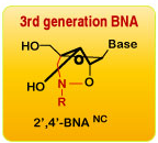
To improve, xeno nucleic acids (XNAs) representing artificially modified nucleic acid analogues have been developed.  Whereas modification of the base moiety and the phosphodiester backbone may render greater complementary base pairing ability and nuclease resistance, respectively, modification of the sugar group may affect both properties.  One type of XNAs include 2’- modified analogues, which include 2’-deoxy-2’-fluoroarabino nucleic acid (FANA) and 2’-O-methoxyethyl-RNA (MOE).  Bridged nucleic acid (BNA) represents another category comprised of conformation-locked analogues such as locked nucleic acid (LNA) with a bridge linking 2’-O and 4’-C of the ribose moiety.   2'-O,4'-aminoethylene bridged nucleic acid (2',4'-BNANC) is the third -generation BNA containing a six-member bridged structure with an N-O linkage.  An increased conformational inflexibility of ribose renders greater binding affinity to complementary single-stranded RNA or double-stranded DNA.  XNAs with a 6-membered ring sugar group have also been developed, ex. 3’-fluoro hexitol nucleic acid (FHNA) (Morihiro et al., 2017). 


 The development of XNAs have ushered in the opportunity to utilize oligonucleotides for therapy that may not be as limited as their unmodified precursors.  With greater resistance to nucleases, higher specificity, and increased base-pairing potential, multiple clinical applications are being considered.  These include antisense LNA oligodeoxynucleotides that bind to complementary H-Ras mRNA or miR-17-5p microRNA (miRNA) to inhibit cancer progression (Fluiter et al., 2005; Jin et al, 2015).  A gapmer incorporating BNANC efficiently degraded CUG expanded repeat RNA, which causes myotonic dystrophy (Manning et al., 2017).  For Duchenne muscular dystrophy, splice-switching oligonucleotides with 2′-O-MOE was effective in modulating the pre-messenger RNA splicing of dystrophin (Yang et al, 2013).  For RNA interference, the LNA based siRNA therapeutics exhibited greater functionality/stability (Elmén et al., 2005). 

Aptamers consist of single-stranded RNA or DNA oligonucleotides whose 3-dimensional structure has been exploited to isolate species that bind specifically to therapeutic targets.  The RNA aptamer Pegaptanib targeting vascular endothelial growth factor (VEGF) was the first to be approved by FDA for age-related macular degeneration (lee et al., 2015).  To identify, a library consisting of randomly sequenced DNA or RNA oligonucleotides is screened for binding to a target ligand.  The bound oligonucleotides are recovered and amplified by PCR before repeating the selection cycle to enrich for the specifically bound sequences.  To identify a RNA aptamer, the isolated oligonucleotides need to be reverse transcribed before amplifying.

The stability of aptamers could be greatly improved by incorporating XNAs like BNA to render nuclease resistance.  However, most modified nucleosides are poor substrates for polymerases including reverse transcriptase.  One solution is to introduce XNAs after the aptamers have been identified.   The other approach is to generate polymerase mutants capable of recognizing XNA-based aptamers.  Using Tgo DNA polymerase derived from Thermococcus gorgonarius, PolC7, a variant was generated capable of reverse transcribing LNA-based oligonucleotides (Pinheiro et al, 2012), which was used to isolate a HNA-based aptamer binding to HIV’s trans-activating response RNA (TAR).   Another group reported that LNA-based oligonucleotides could be reversed transcribed using SuperScript III Reverse Transcriptase (Crouzier et al., 2012).  A polymerase mutant capable of amplifying 2’-OMe containing oligonucleotide was also generated (Chen et al 2016). 

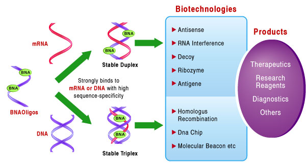
Bio-Synthesis, Inc. has been at the forefront of the oligonucleotide technologies for 35 years.  Ever since the introduction of solid phase nucleotide synthesis, it has kept pace with the latest breakthroughs in the nucleotide modification chemistry.  Its recent acquisition of a license from BNA Inc. of Osaka, Japan, for the manufacturing and distribution of BNANC, a third generation of BNA oligonucleotides, is in keeping with this commitment.  In recent days, the company has expanded its operation to align closely with the developments in therapy.  To meet the demands of therapeutic application, its oligonucleotide products are approaching GMP grade.  Bio-Synthesis, Inc. has recently entered into collaborative agreement with Bind Therapeutics, Inc. to synthesize miR-21 blocker using BNA.  The BNA technology that we offer provides superior, unequalled advantages in base stacking, binding affinity, aqueous solubility and nuclease resistance.  It also improves the formation of duplexes and triplexes by reducing the repulsion between the negatively charged phosphates of the oligonucleotide backbone.  Its single-mismatch discriminating power was especially useful for diagnosis (ex. FISH using DNA probe).  More importantly, BNA oligonucleotide exhibits lesser toxicity than other modified nucleotides for clinical application.

 https://www.biosyn.com/bna-synthesis-bridged-nucleic-acid.aspx


References 

Bernstein E, Caudy AA, Hammond SM, Hannon GJ.  Role for a bidentate ribonuclease in the initiation step of RNA interference.  (2001). Nature. 409, 363–366. PMID 1120174  . doi:10.1038/35053110.

Chen T, Hongdilokkul N, Liu Z, Adhikary R, Tsuen SS, Romesberg FE.  Evolution of thermophilic DNA polymerases for the recognition and amplification of C2'-modified DNA.  (2016)  Nat Chem.  8:556-62  PMID: 27219699 PMCID: PMC4880425 DOI: 10.1038/nchem.2493

Crouzier L, Dubois C, Edwards SL, Lauridsen LH, Wengel J, Veedu RN.  Efficient Reverse Transcription Using Locked Nucleic Acid Nucleotides towards the Evolution of Nuclease Resistant RNA Aptamers. (2012).  PLoS One. 2012; 7: e35990.  PMCID: PMC3338489  PMID: 22558297  doi: 10.1371/journal.pone.0035990

Elmén J, Thonberg H, Ljungberg K, Frieden M, Westergaard M, Xu Y, Wahren B, Liang Z, Ørum H, Koch T, Wahlestedt C.  Locked nucleic acid (LNA) mediated improvements in siRNA stability and functionality. (2005).  Nucleic Acids Res. 33:439-47.  PMID: 15653644

Fluiter K1, Frieden M, Vreijling J, Rosenbohm C, De Wissel MB, Christensen SM, Koch T, Ørum H, Baas F.  On the in vitro and in vivo properties of four locked nucleic acid nucleotides incorporated into an anti-H-Ras antisense oligonucleotide. (2005). Chembiochem. 6:1104-9. PMID: 24244378 PMCID: PMC3824000 DOI: 10.1371/journal.pone.0078863

Manning KS, Rao AN, Castro M, Cooper TA.   BNANC Gapmers Revert Splicing and Reduce RNA Foci with Low Toxicity in Myotonic Dystrophy Cells. (2017). ACS Chem Biol. 12:2503-2509.  PMID: 28853853 PMCID: PMC5694563  doi: 10.1021/acschembio.7b00416.

Morihiro K, Kasahara Y, Obika S.  Biological applications of xeno nucleic acids.  (2017). Mol Biosyst. 13:235-245.  PMID: 27827481   doi: 10.1039/c6mb00538a.  

Lee JH, Canny MD, De Erkenez A, Krilleke D, Ng YS, Shima DT, Pardi A, Jucker F.  A therapeutic aptamer inhibits angiogenesis by specifically targeting the heparin binding domain of VEGF165.  (2005).  Proc Natl Acad Sci U S A.  102:18902-7.

Pinheiro VB1, Taylor AI, Cozens C, Abramov M, Renders M, Zhang S, Chaput JC, Wengel J, Peak-Chew SY, McLaughlin SH, Herdewijn P, Holliger P.  Synthetic genetic polymers capable of heredity and evolution.  (2012) Science. 36: 341–344.  PMCID: PMC3362463   PMID: 22517858   doi: 10.1126/science.1217622  

Yang L, Niu H,   Gao X, Wang Q, Han G, Cao L, Cai C, Weiler J, Yin H.  Effective exon skipping and dystrophin restoration by 2′-O-Methoxyethyl antisense oligonucleotide in dystrophin-deficient mice.  (2013). PLoS One. 8: e61584. PMCID: PMC3637291 PMID: 23658612 doi: 10.1371/journal.pone.0061584 


The utility of 6-thio-2’-deoxyguanosine for novel telomerase-dependent anti-cancer therapy

$
0
0
 The utility of 6-thio-2’-deoxyguanosine for novel telomerase-dependent anti-cancer therapy


Unlike prokaryotic genomes consisting of circular DNA, the eukaryotic genomes consist of linear DNA, which is organized into distinct chromosomes.   The DNA winds around the basic protein histone to form nucleosomes, which undergo compaction to form a highly condensed chromatin.  Chromosomes are organized into distinct structural components, ex. centromere that mediates chromosome segregation during mitosis (Alberts, 2015).  Telomeres at the ends of chromosomes are comprised of numerous repeats of simple DNA sequence such as TTAGGG in the case of humans.


The main function of telomere is to protect chromosome ends from degradation and avoid genetic instability arising from the fusion with other chromosomes at the termini.  To prevent DNA repair/recombination from occurring at the chromosome ends, the DNA sequence repeats and a single stranded 3’-overhang of telomere fold back on itself to form a loop (T loop) with the single strand invading the duplex to anneal with one of the two strands (D loop) (Xu et al., 2016).  This ‘capping’ function of telomere is mediated by Shelterin proteins, which incudes TRF1, TRF2, POT1, RAP1 and others.


In most organisms, the length of telomere decreases after each round of DNA replication in normal cells.  The loss of telomere occurs as the terminal DNA sequence occupied by the RNA primer towards the end of the lagging strand (Richter et al., 2007) cannot be replicated by DNA polymerase (though other factors such as oxidative stress may also contribute).  A high copy number of DNA sequence repeats may ensure that shortening affects telomeric DNA primarily and avoid deleting important genetic information.  Further, it has been suggested that the progressive loss of terminal DNA may continue till it reaches a specific length that activates cell senescence.


The progressive shortening of telomeres can be reversed through telomerase, which regenerates repeat sequences.  Telomerase is comprised of telomerase RNA (TERC), telomerase reverse transcriptase (TERT) and several associating proteins (Venteicher et al., 2008; Xu et al., 2016).  To extend shorter telomeres, TERT uses TERC’s template to add sequence repeats at the 3' end.  Whereas most normal somatic cells express little telomerase, TERT activity is up-regulated in continuously dividing stem cells and ectopically expressing hTERT immortalizes non-stem cells, indicating that telomeres may affect cells’ lifespan.  Consistently, an elevated telomerase activity was observed in mostf cancer specimens (Kim et al., 1994).


The antimetabolite drug 6-thioguanine has been used to treat acute myelogenous leukemia and chronic myelogenous leukemia.  Its anticancer property requires activation via conversion to thioguanosine diphosphate (TGDP) or thioguanosine triphosphate (TGTP) catalyzed by hypoxanthine-guanine phosphoribosyltransferase and kinases, and the conversion to deoxyribosyl analogues by ribonucleotide reductase.  Its cytotoxicity is associated with the incorporation of 6-thioguanine nucleotides into DNA during S phase to interfere with replication or into RNA to impair protein synthesis. 


The impact of disrupting telomere on the growth of hTERT-expressing human cancer cells was examined.  Treatment with 6-thio-2’-deoxyguanosine led to its incorporation into telomeres, rendering them dysfunctional.  It resulted in the selective death of lung cancer cells expressing telomerase (Mender et al., 2015), therapy-resistant melanoma (Zhang et al., 2018) and pediatric brain tumor (Sengupta et al., 2018).  The results point to the potential utility of 6-thio-2’-deoxyguanosine as a novel telomerase-dependent anti-cancer therapeutic.

                                                              
Bio-Synthesis, Inc. provides extensive options for the application of various modified nucleosides for research or therapy purposes.  For instance, with 6-thio-2'-deoxyguanosine (6-thio dG), we offer oligonucleotide modification to selectively conjugate to DNA binding proteins to study their interaction.  For bridged nucleic acid (BNA), we have acquired license from BNA Inc. of Osaka, Japan, to supply BNA-oligonucleotide to Bound Therapeutics LLC. to develop a novel peptide-miroRNA therapeutic for triple negative breast cancer.


https://www.biosyn.com/oligonucleotideproduct/6-thio-2%27-deoxyguanosine,6-thio-dg-oligonucleotide-modification.aspx


References

Alberts B.  Molecular Biology of the Cell.  Sixth edition.  (2015).  Garland Science, Taylor and Francis Group, New York.

Kim NW, Piatyszek MA, Prowse KR, Harley CB, West MD, Ho PL, Coviello GM, Wright WE, Weinrich SL, Shay JW.  Specific association of human telomerase activity with immortal cells and cancer.  (1994). Science  266:2011-5.    PMID: 7605428 DOI: 10.1126/science.7605428

Mender I, Gryaznov S, Dikmen ZG, Wright WE, Shay JW.  Induction of telomere dysfunction mediated by the telomerase substrate precursor 6-thio-2'-deoxyguanosine.  (2015).  Cancer Discov  5:82-95.   PMID: 25516420  PMCID: PMC4293221  DOI: 10.1158/2159-8290.CD-14-0609

Richter T, von Zglinicki T.  A continuous correlation between oxidative stress and telomere shortening in fibroblasts.  (2007)  Exp Gerontol. 42:1039-42. PMID: 17869047 DOI: 10.1016/j.exger.2007.08.005

Sengupta S, Sobo SM, Lee K, Kumar SS, White AR, Mender I, Fuller C, Chow LML, Fouladi M, Shay JW, Drissi R.  Induced telomere damage to treat telomerase expressing therapy-resistant pediatric brain tumors.  (2018)  Mol Cancer Ther 17:1504-1514.  PMID: 29654065  DOI: 10.1158/1535-7163.MCT-17-0792

Venteicher A.S., Meng Z., Mason P.J., Veenstra T.D., Artandi S.E. Identification of APTases pontin and reptin as telomerase components essential for holoenzyme assembly. (2008).  Cell 132:945–957.   PMID: 18358808 PMCID: PMC2291539 DOI: 10.1016/j.cell.2008.01.019

Xu Y,  Goldkorn A.  Telomere and telomerase therapeutics in cancer. (2016).  Genes (Basel) 7: 22. PMCID: PMC4929421  PMID: 27240403

Zhang G, Wu LW, Mender I, Barzily-Rokni M, Hammond MR, Ope O, et al.  Induction of telomere Ddsfunction prolongs disease control of therapy-resistant melanoma.  (2018).  Clin Cancer Res 24:4771-4784.   PMID: 29563139  PMCID: PMC6150856   DOI: 10.1158/1078-0432.CCR-17-2773

Genome editing: modified CRISPR/Cas9 corrects genetic mutation causing Alzheimer’s disease or cancer without cleaving DNA

$
0
0

Genome editing: modified CRISPR/Cas9 corrects genetic mutation causing Alzheimer’s disease or cancer without cleaving DNA

Genetic mutations account for a significant fraction of human disorders.  Genetic analyses have shown that inherited mutations may predispose individuals to developing various illnesses.  These include the mutant alleles of RB (retinoblastoma), p53 (Li-Fraumeni syndrome), APC (familial adenomatous polyposis), BRCA1/BRCA2 (breast cancer), VHL (von Hippel–Lindau disease) and other tumor suppressor genes (Knudson, 2002; Lee et al., 1994).  Mutations leading to other types of disorders (ex. Bloom syndrome, xeroderma pigmentosum, hemophilia, cystic fibrosis and Duchenne muscular dystrophy) have also been identified.  Increasingly, mutations affecting neural pathways, resulting in mental disorders affecting memory (ex. APP, APOEe4 for Alzheimer’s disease), behavior (ex. MAOA for aggression, autism), etc. are being uncovered.

Genetic mutations can also interfere with therapy.  For the anticancer chemotherapeutic Taxol, specific mutations in the b-tubulin subunit gene confer resistance (Giannakakou et al., 1997).  For tyrosine kinase inhibitors, the point mutation T790M in epidermal growth factor (EGF) receptor represents a common mechanism of resistance (Yun et al., 2008).  In chronic myeloid leukemia, resistance to the targeted drug Gleevec could occur through specific point mutations in BCR/ABL tyrosine kinase.  Consequently, researchers have strived to develop a means through which the predisposing mutation could be corrected to preempt the disorder or to restore therapeutic efficacy.

Initially, genome editing was attempted using zinc-finger nuclease (ZFN).  ZFN is comprised of a DNA binding domain formed by zinc fingers (from transcription factors) and the DNA cleavage domain of bacterial restriction endonuclease Fok-1 (Gaj et al. 2013).  Recognition of specific sequence by its DNA binding domain enables site-specific double strand cleavage.  This property could be exploited to abolish a dominantly acting mutant allele if subsequently repaired by error-prone nonhomologous end joining (NHEJ) pathway.  Alternatively, it may allow gene editing if repaired via homologous recombination (HR) by supplying a template DNA.  A similar principle underlies genome editing by TALEN (transcription activator-like effector nucleases), which represents the DNA recognition motif of TAL effector (secreted bacterial protein) fused to Fok-1’s cleavage domain.  The main caveat is that both require custom protein engineering for target sequence.

Clustered regularly interspaced short palindromic repeats (CRISPR) represent a region in bacterial genome where the double stranded DNA fragments of previously infected virus’ genome are captured and inserted at the ‘spacer’ loci by Cas1 and Cas2.  The RNA transcribed from the spacer is processed into crRNA, which serves as guide RNA to direct Cas9 endonuclease to target DNA.  Upon identifying a foreign DNA complementary to the spacer, Cas9 (also requires trans-activating tracrRNA) would cleave it to compromise the invading agent.  To harness this bacterial immunity function for genome editing, tracrRNA and crRNA have been fused into a single guide RNA (sgRNA) molecule (Deltcheva et al, 2011).  The CRISPR gene editing system can be directed to cleave cellular genome at any location by modifying guide RNA.


Nevertheless, CRISPR genome editing poses problems associated with mutagenesis at off-target sites as it can be inherited in the case of germ line.  To resolve, investigators (Harvard University) modified Cas9 to edit genome without cleaving DNA.  Catalytically inactive Cas9 was tethered to rAPOBEC1 cytidine deaminase, which was termed ‘based editor’ (Komor et al., 2016).  It deaminated cytidine (C) to uridine, which converted to thymidine (T) following replication.  The base editor (3rd generation) was able to correct mutation in apolipoprotein variant APOE4 (associated with Alzheimer’s disease) or p53 (linked to cancer) by causing C to T conversion.  Using a similar strategy, another base editor containing TadA (E. coli tRNA adenine deaminase; converts adenine to inosine in tRNAArg) was used to deaminate adenine, driving A to G conversion in target DNA (Gaudelli et al., 2017).  Subsequent analyses, however, revealed that cytosine base editor alters single nucleotides at a number of off-target sites in the genome (Zuo et al., 2019) whereas both cytosine and adenine base editors mutate numerous coding or non-coding RNAs in the transcriptome (Grunwald et al., 2019), suggesting that further improvements are necessary. 

As an alternative approach, recent works have shown that the genome editing efficacy of CRISPR-Cas system can be improved by modifying sgRNA, i.e. by incorporating chemically modified nucleosides in the synthesis of sgRNA (Hendel et al. in 2015). Bio-Synthesis, Inc. specializes in oligonucleotide modification and provides an extensive array of chemically modified nucleoside analogues (over ~200) including bridged nucleic acid (BNA). It recently acquired a license from BNA Inc. of Osaka, Japan, for the manufacturing and distribution of BNANC, a third generation of BNA oligonucleotides. To meet the demands of therapeutic application, its oligonucleotide products are approaching GMP grade. Bio-Synthesis, Inc. has recently entered into collaborative agreement with Bind Therapeutics, Inc. to synthesize miR-21 blocker using BNA.  The BNA technology that we offer provides superior, unequalled advantages in base stacking, binding affinity, aqueous solubility and nuclease resistance. It also improves the formation of duplexes and triplexes by reducing the repulsion between the negatively charged phosphates of the oligonucleotide backbone. Its single-mismatch discriminating power was especially useful for diagnosis (ex. FISH using DNA probe). More importantly, BNA oligonucleotide exhibits lesser toxicity than other modified nucleotides for clinical application.

https://www.biosyn.com/tew/Chemically-Modified-Nucleic-Acids-for-CRISPR-Cas.aspx

References

Deltcheva E, Chylinski K, Sharma CM, Gonzales K, Chao Y, Pirzada ZA, Eckert MR, Vogel J, Charpentier E.  CRISPR RNA maturation by trans-encoded small RNA and host factor RNase III.  (2011)  Nature. 471: 602–607. PMC 3070239  PMID 21455174  doi:10.1038/nature09886.

Gaj T, Gersbach CA, Barbas CF 3rd. ZFN, TALEN, and CRISPR/Cas-based methods for genome engineering. (2013) Trends Biotechnol. 31:397-405. PMID: 23664777 doi: 10.1016/j.tibtech.2013.04.004.  

Gaudelli NM, Komor AC, Rees HA, Packer MS, Badran AH, Bryson DI, Liu DR.  Programmable base editing of A•T to G•C in genomic DNA without DNA cleavage.  (2017)  Nature 551:464-471. PMID: 29160308 PMCID: PMC5726555 doi: 10.1038/nature24644.  (Erratum: Nature. 2018 PMID: 29720650)

Giannakakou P, Sackett DL, Kang YK, Zhan Z, Buters JT, Fojo T, Poruchynsky MS. Paclitaxel-resistant human ovarian cancer cells have mutant beta-tubulins that exhibit impaired paclitaxel-driven polymerization. (1997)  J Biol Chem. 272:17118-25.  PMID:  9202030  

Grünewald J, Zhou R, Garcia SP, Iyer S, Lareau CA, Aryee MJ, Joung JK.   Transcriptome-wide off-target RNA editing induced by CRISPR-guided DNA base editors.  (2019) Nature  569:433-437. PMID: 30995674   doi: 10.1038/s41586-019-1161-z

Hendel A, Bak RO, Clark JT, Kennedy AB, Ryan DE, Roy S, Steinfeld I, Lunstad BD, Kaiser RJ, Wilkens AB, Bacchetta R, Tsalenko A, Dellinger D, Bruhn L, Porteus MH.  Chemically modified guide RNAs enhance CRISPR-Cas genome editing in human primary cells.  (2015)  Nat Biotechnol. 33:985-989. PMID: 26121415  doi: 10.1038/nbt.3290

Knudson AG.  Cancer genetics.  (2002)  Am J Med Genet. 111:96-102. PMID: 12124744 DOI: 10.1002/ajmg.10320

Komor AC, Kim YB, Packer MS, Zuris JA, Liu DR.  Programmable editing of a target base in genomic DNA without double-stranded DNA cleavage.  (2016)  Nature 533:420-4.  PMID: 27096365  doi: 10.1038/nature17946

Lee WH, Xu Y, Hong F, Durfee T, Mancini MA, Ueng YC, Chen PL, Riley D.   The corral hypothesis: a novel regulatory mode for retinoblastoma protein function.  (1994)  Cold Spring Harb Symp Quant Biol. 59:97-107.  PMID: 7587136

Yun CH, Kristen Mengwasser E, Toms AV, Woo MS, Greulich H, Wong KK, Meyerson M, Eck MI.  The T790M mutation in EGFR kinase causes drug resistance by increasing the affinity for ATP.  (2008).  PNAS 105: 2070-2075.  PMID: 18227510 PMCID: PMC2538882 DOI: 10.1073/pnas.0709662105

Zuo E, Sun Y, Wei W, Yuan T, Ying W, Sun H, Yuan L, Steinmetz LM, Li Y, Yang H.  Cytosine base editor generates substantial off-target single-nucleotide variants in mouse embryos.  (2019)  Science  364:289-292.    PMID: 30819928  doi: 10.1126/science.aav9973 


Phase II clinical trial of Hedgehog pathway inhibitor with low-dose cytarabine (Ara-C) extends the survival of acute myeloid leukemia (AML) patients

$
0
0
Phase II clinical trial of Hedgehog pathway inhibitor with low-dose cytarabine (Ara-C) extends the survival of acute myeloid leukemia (AML) patients

Acute myeloid leukemia (AML) accounts for >140,000 deaths globally per year with nearly ~10,600 deaths in the United States.  AML is the 2nd most common adult leukemia with the 5-year overall survival rate of ~27% though it’s higher (60-70%) for children.  AML is caused by myeloid cells forming leukemic blasts instead of undergoing normal differentiation.  The rapid accumulation of abnormal cells in bone marrow with the resultant deficiency in red blood cells, platelets or certain white blood cells negatively affects immune and other physiological functions.  The current efforts to manage AML include chemotherapy, hematopoietic stem cell transplantation and tyrosine kinase inhibitors.

The malignancy in AML is attributed to the acquiring of genetic mutations that arrest cell differentiation and cause uncontrolled proliferation.  The block in differentiation, which may occur at distinct stages, arrests hematopoietic stem cells as myeloid stem cells or myeloid blasts instead of developing into monocytes or granulocytes.  A number of chromosomal rearrangements (e.g. translocation, inversion) have been documented in AML and the resultant gene product may affect tumor progression.  In the case of acute promyelocytic leukemia (a subtype of AML), translocation t(15;17) encodes the fusion protein PML-RARα, which transcriptionally regulates the genes inhibiting myeloid differentiation (Melnick et al., 1999).

For older AML patients, fewer treatment options are available as they are ineligible for intensive chemotherapy, opting for less aggressive treatment, i.e. low-dose cytarabine [also known as aracytidine or cytosine arabinoside (Ara-C)] or hypomethylating agents.  Administering the hypomethylating agent decitabine (5-aza-2'-deoxycytidine), however, only slightly extended the median overall survival (Kantarjian et al., 2012).  Thus, new therapies are needed to improve their survival.


The Hedgehog signaling pathway plays a key role in embryonic development.   Hedgehog (HH) gene was originally discovered while studying the genetic mutants affecting the anterior-posterior body axis of Drosophila by Nüsslein-Volhard and colleagues (Nobel prize, 1995).  The HH protein defines the antero-posterior orientation of the Drosophila embryo and its mutants display abnormally short and stubby larvae (Lee et al., 1992).  In vertebrates, the HH homologue, Secreted Sonic Hedgehog (SHH) protein, binds to Patched-1 (PTCH1) receptor of the target cells and blocks it from inhibiting the downstream protein Smoothened (SMO).  This leads to the activation of GLI transcription factors to regulate the transcription of Hedgehog-target genes.

Intriguingly, the hedgehog signaling pathway is activated in basal cell carcinoma as well as the cancers of the breast, brain and prostate (Sari et al., 2018).  The uncontrolled activation of the signaling pathway may contribute to the persistence of cancer stem cells exhibiting drug resistance.  In myeloid leukemic cells, inhibiting the HH signaling reduced P-glycoprotein associated with multi-drug resistance, suggesting that activation of the HH pathway may confer resistance to chemotherapy (Queiroz et al, 2010).

To pharmacologically abrogate the Hedgehog signaling pathway, Glasdegib was developed by Pfizer Inc., which inhibits SMO protein (Munchhof et al., 2011).  Pre-clinical studies demonstrated that Glasdegib suppresses the growth of AML cells and sensitizes chemotherapy-resistant AML cells to cytarabine (Fukushima et al, 2016).  These results inspired several clinical trials.  Recently, a Phase II clinical study was conducted at the M. D. Anderson Cancer Center and other medical centers to examine the therapeutic efficacy of Glasdegib and low-dose cytarabine in newly diagnosed AML patients (Cortes et al., 2019).  It showed that the combined regimen extends overall survival (~4 months), which led to its approval by FDA for the treatment of newly diagnosed older AML patients, who cannot receive intensive chemotherapy.   Sonidegib is another inhibitor of SMO developed by Novartis, Inc.

Bio-Synthesis, Inc. provides extensive options for the application of various modified nucleosides for research or therapy purposes.  It specializes in oligonucleotide modification and provides an extensive array of chemically modified nucleoside analogues (over ~200).  For instance, with Ara-C, we offer oligonucleotide modification to selectively conjugate to DNA binding proteins to study their interaction.   For bridged nucleic acid (BNA), it has recently acquired a license from BNA Inc. of Osaka, Japan, for the manufacturing and distribution of BNANC, a third generation of BNA oligonucleotides.  Bio-Synthesis, Inc. has recently entered into collaborative agreement with Bind Therapeutics, Inc. to synthesize miR-21 blocker using BNA.  The BNA technology that we offer provides superior, unequalled advantages in base stacking, binding affinity, aqueous solubility and nuclease resistance.  More importantly, BNA oligonucleotide exhibits lesser toxicity than other modified nucleotides for clinical application.

https://www.biosyn.com/oligonucleotideproduct/aracytidine-ara-c-oligonucleotide-modification.aspx#!


References

Cortes JE, et al.  Randomized comparison of low dose cytarabine with or without glasdegib in patients with newly diagnosed acute myeloid leukemia or high-risk myelodysplastic syndrome. (2019)  Leukemia 33:379-389.  PMID: 30555165  doi: 10.1038/s41375-018-0312-9. Epub 2018 Dec 16. 

Fukushima N, et al.  Small-molecule Hedgehog inhibitor attenuates the leukemia-initiation potential of acute myeloid leukemia cells. (2016) Cancer Sci  107:1422-1429.  PMID: 27461445  doi: 10.1111/cas.13019

Kantarjian HM, et al.  Multicenter, randomized, open-label, phase III trial of decitabine versus patient choice, with physician advice, of either supportive care or low-dose cytarabine for the treatment of older patients with newly diagnosed acute myeloid leukemia. (2012)  J Clin Oncol 30:2670-7.  PMID: 22689805

Lee JJ, von Kessler DP, Parks S, Beachy PA.  Secretion and localized transcription suggest a role in positional signaling for products of the segmentation gene hedgehog. (1992) Cell 71:33-50. PMID: 1394430 DOI: 10.1016/0092-8674(92)90264-d

Melnick A, Licht JD. Deconstructing a disease: RARalpha, its fusion partners, and their roles in the pathogenesis of acute promyelocytic leukemia. (1999)  Blood  93:3167-215. PMID: 10233871

Munchhof MJ, et al.  Discovery of PF-04449913, a Potent and Orally Bioavailable Inhibitor of Smoothened.  (2011)  ACS Med Chem Lett 3:106-11.  PMID: 24900436  doi: 10.1021/ml2002423. eCollection

Queiroz KC, et al. Hedgehog signaling maintains chemoresistance in myeloid leukemic cells. (2010)  Oncogene  29:6314-22.   PMID: 20802532   DOI: 10.1038/onc.2010.375

Sari IN, Phi LTH, Jun N, Wijaya YT, Lee S, Kwon HY.  Hedgehog Signaling in Cancer: A Prospective Therapeutic Target for Eradicating Cancer Stem Cells.  (2018)  Cells 7(11). pii: E208.   PMID: 30423843   doi: 10.3390/cells7110208 


Bio-Synthesis, Inc. supplied modified oligonucleotide identified the strand tracked by CMG complexes to unwind double helix to form bi-directional forks for DNA replication

$
0
0

 Bio-Synthesis, Inc. supplied modified oligonucleotide identified the strand tracked by CMG complexes to unwind double helixto form bi-directional forks for DNA replication

DNA replication, which is fundamental to the propagation of all living organisms, is a highly conserved and regulated process both temporally and spatially.  Understanding the underlying mechanism is significant as its deregulation leads to uncontrolled cell proliferation, resulting in cancer and other diseases. The process is divided into three stages: initiation, elongation and termination.  In prokaryotes, the initiation phase involves unwinding of the DNA sequence specifying the origin of replication (oriC) and loading of the replication complex.  The oriC region encodes multiple binding sites for the initiator protein, which become occupied by DnaA with the resultant unwinding of the DNA.  After loading DnaC, which recruits DnaB helicase, the primase DnaG synthesizes RNA primer to initiate replication by DNA polymerase III (Kaguni et al., 2011).

In eukaryotes, during G1 phase, the ‘pre-replication complex’ is formed at the origin of replication comprised of origin recognition complex (ORC), Cdc6, Cdt1 (chromatin licensing and DNA replication factor) and minichromosome maintenance proteins Mcm2-7.  In S phase, phosphorylation by cyclin dependent kinase 2 (CDK2) and DDK activates the complex to transit to the ‘initiation complex’.  Mcm2-7 forms double hexamer rings on DNA that serves as scaffold for the assembly of ‘CMG’ (cdc45-MCM-GINS) helicase and the replisome complex (Parker et al., 2016).  Intriguingly, Mcm7 has been shown to associate with the tumor suppressor Rb linked to retinoblastoma/osteosarcoma and mutated in ~30% of human cancers including non-small cell lung cancer, prostate cancer, glioma and breast cancer (Sterner et al.., 1998; Bookstein et al., 1990).  Cdc45 loads DNA polymerase, PCNA, RPA and others.  GINS and Cdc45 augment the helicase activity.

The exact mechanism through which bi-directional replication forks are generated is not clearly understood.  Previous works have shown that the two head-to-head CMG complexes assembled on dsDNA undergo separation, and, upon binding to Mcm10, both complexes engage opposite single strands of the unwound DNA and go past one another to form bi-directional forks.  However, whether CMG and Mcm10 can untwist double helix sufficiently to allow CMG to translocate to ssDNA is not known.

To determine if the ring-shaped CMG motor could force open duplex of sufficient length, researchers (Rockefeller University) designed a T-shaped dsDNA probe, which allows CMG to load and slide but will encounter a block at the T-junction.  Here, the ability of CMG to melt the DNA would allow a labeled oligonucleotide annealed at the T-junction to dissociate for monitoring.  By varying the length of DNA at the T-junction, they determined that CMG (plus Mcm10) could unwind 60-bp dsDNA (Fig 3 inLangston et al., 2019).

Next, to determine how it opens dsDNA, either strand of the probe (where CMG slides before the T-junction) was partly replaced with 10 neutral methylphosphonate linkages acquired from Bio-Synthesis, Inc.  The duplex unwinding was significantly reduced when the methylphosphonate linkage was incorporated on the 3’-5’ strand in the direction of unwinding, suggesting that CMG tracks along this strand primarily while encircling dsDNA (Langston et al., 2019).  Subsequent experiments corroborated the above finding, suggesting that the head-to-head CMG complexes plus Mcm10 generate sufficient force to open several turns of the duplex.


Modified oligonucleotides (DNA or RNA) have been useful in investigating the mechanistic/stereo-chemical aspects of various biochemical reactions/processes.  Bio-Synthesis, Inc. provides a full spectrum of high-quality custom services by direct solid-phase chemical synthesis or enzyme-assisted approach to prepare oligonucleotides with artificially modified backbone, base, sugar or inter-nucleotidic linkage.  Its integrated Flex Oligo Synthesis platform results in considerable cost-saving by increasing chemical-coupling efficiency and allowing for significant scale-up production in a short time period. We specialize in complex oligonucleotide modifications using phosphodiester backbone, purine/pyrimidine heterocyclic bases or sugar-modified nucleotides such as our patented 3rd generation bridged nucleic acids.  They have been used as structural or mechanistic probes in diverse diagnostic or therapeutic applications.

For this, Bio-Synthesis, Inc. provides an extensive array of chemically modified nucleoside analogues (over ~200) including bridged nucleic acid (BNA).  It recently acquired a license from BNA Inc. of Osaka, Japan, for the manufacturing and distribution of BNANC, a third generation of BNA oligonucleotides.  To meet the demands of therapeutic application, its oligonucleotide products are approaching GMP grade.  Bio-Synthesis, Inc. has recently entered into collaborative agreement with Bind Therapeutics, Inc. to synthesize miR-21 blocker using BNA.  The BNA technology that we offer provides superior, unequalled advantages in base stacking, binding affinity, aqueous solubility and nuclease resistance.  It also improves the formation of duplexes and triplexes by reducing the repulsion between the negatively charged phosphates of the oligonucleotide backbone.  Its single-mismatch discriminating power was especially useful for diagnosis (ex. FISH using DNA probe).  More importantly, BNA oligonucleotide exhibits lesser toxicity than other modified nucleotides for clinical application.

https://www.biosyn.com/oligonucleotide-modification-services.aspx


References

Bookstein R, Rio P, Madreperla SA, Hong F, Allred C, Grizzle WE, Lee WH.  Promoter deletion and loss of retinoblastoma gene expression in human prostate carcinoma.  (1990)  Proc Natl Acad Sci USA  87:7762-6.  PMID: 2217208 PMCID: PMC54828  DOI: 10.1073/pnas.87.19.7762

Kaguni JM.  Replication initiation at the Escherichia coli chromosomal origin.  (2011) Curr Opin Chem Biol 15: 606–13.  PMC 3189269  PMID 21856207   doi:10.1016/j.cbpa.2011.07.016

Langston LD, O'Donnell ME.  An explanation for origin unwinding in eukaryotes.  (2019)  Elife.  Jul 8;8. pii: e46515.   PMID: 31282859  doi: 10.7554/eLife.46515.

Parker MW, Botchan MR, Berger JM.  Mechanisms and regulation of DNA replication initiation in eukaryotes. (2017) Crit Rev Biochem Mol Biol 52:107-144. PMID: 28094588 doi: 10.1080/10409238.2016.1274717

Sterner JM, Dew-Knight S, Musahl C, Kornbluth S, Horowitz JM.  Negative regulation of DNA replication by the retinoblastoma protein is mediated by its association with MCM7.  (1998)  Mol Cell Biol  18:2748-57.  PMID: 9566894  PMCID: PMC110654  DOI: 10.1128/mcb.18.5.2748


Spermine-modified cationic ZNA oligonucleotide for in situ hybridization, qPCR, SNP detection, antisense/antigene strategies, and self-delivery into various types of cells

$
0
0
For hybridization, the rate of renaturation is thought to be proportional to the square root of the length of the complementary single strands (Niranjani et al., 2016).  In practice, shorter oligonucleotides are preferred as probes to achieve efficient delivery into cells or permeate across dense chromosomes to find target. To optimize, various parameters affecting hybridization such as probe length or concentration, temperature, and pH have been modulated.  Another contributing factor is the electrostatic repulsion between the phosphate moieties of the two single strands of the duplex.  Modifying this property affects hybridization as increasing the monovalent cation concentration (to neutralize the anionic phosphate groups) enhances the stability of the duplex.  Likewise, lowering the salt concentration (to increase stringency) affects hybridization.


To attenuate the electrostatic repulsion, Zip Nucleic Acids (ZNA) was developed, consisting of oligonucleotides conjugated to positively charged spermine derivatives (Noir et al., 2008).  It facilitates target recognition by allowing the oligonucleotide to scan the single stranded DNA till it finds the complementary sequence. Upon hybridization, the interaction of the positively charged moiety with complementary strand augments the binding affinity and stabilizes the duplex without compromising specificity, resulting in an increased Tm value.  The ‘zipper’-like property enables ZNA primers to perform (more efficiently than LNAs) at lower concentrations of primer/Mg2+ and higher annealing temperatures, improving PCR amplification of AT-rich regions, RNA-to-cDNA conversion, and single-nucleotide polymorphism (SNP) discrimination (Moreau et al., 2009).  The spermine modification renders oligonucleotide conjugates more resistant to nuclease degradation.  ZNA-modified oligonucleotides were successfully used to detect miRNA expression via in situ hybridization (ISH) in maize seedlings (Trevisan et al., 2012).   


The potential of the positively charged moiety to mediate the transfer of ZNA oligonucleotides into cells was investigated.  For antisense strategy involving RNA interference, oligospermine-oligoribonucleotide siRNA conjugates, which were resistant to nucleases in serum, entered HeLa cells without carrier to silence gene expression (Paris et al., 2012).  Similarly, carrier-free ZNA-modified siRNA oligonucleotides (albeit non-tumor specific) entered A549Luc human lung carcinoma cells to suppress gene expression (Nothisen et al., 2009).  For antigene strategy requiring triple helix formation to inhibit transcription, spermine conjugated LNA-modified oligonucleotides alone could enter fibroblasts (derived from Huntington's disease patients) to reduce huntingtin (HTT) expression by targeting its promoter (Gagnon et al., 2011).   


Increasing the number of cationic units will alter the ‘overall charge’ of the ZNA oligonucleotides, with the resultant application of negatively charged conjugates for molecular biological/diagnostic applications and positively charged conjugates for enhanced target recognition (via potentially increasing ZNA’s attraction to nucleic acids) and the self-delivery of oligonucleotides into cells.  To design ZNA oligonucleotides, its ‘net charge’ can be calculated using the equation “3n-(m-1)”, with m and n values representing the length of the oligonucleotide and the number of spermine units, respectively.  To empirically determine the Tm value, the hybridization of dual complementary oligonucleotides with varying number of spermine units was examined (Noir et al., 2008).  The results showed that, for 9 to 12-mer oligonucleotides, the modification raises Tm by 4 to 7 °C per spermine independent of the base sequence or conjugation site (5’ or 3′).  The approximate Tm value of ZNA oligonucleotide can be calculated using the formula “Tm (ZNA™) = Tm (DNA) + 36z/(N-3.2)“, where z and N represent the number of cationic units and the number of nucleotides, respectively.  Increasing the positive charge may reduce solubility, however, which could be resolved by raising pH or dissolving in concentrated PBS stock.


Bio-Synthesis, Inc. provides extensive options for the application of various modified nucleosides for research or therapy purposes.  It specializes in oligonucleotide modification and provides an extensive array of chemically modified nucleoside analogues (over ~200) including ZNA.  For bridged nucleic acid (BNA), it has recently acquired a license from BNA Inc. of Osaka, Japan, for the manufacturing and distribution of BNANC, a third generation of BNA oligonucleotides.  Bio-Synthesis, Inc. has recently entered into collaborative agreement with Bind Therapeutics, Inc. to synthesize miR-21 blocker using BNA.  The BNA technology that we offer provides superior, unequalled advantages in base stacking, binding affinity, aqueous solubility and nuclease resistance.  More importantly, BNA oligonucleotide exhibits lesser toxicity than other modified nucleotides for clinical application. 


References


Gagnon KT, Watts JK, Pendergraff HM, Montaillier C, Thai D, Potier P, Corey DR.  Antisense and antigene inhibition of gene expression by cell-permeable oligonucleotide-oligospermine conjugates.  (2011)  J Am Chem Soc.  133(22):8404-7.   doi: 10.1021/ja200312y.   PMID: 21539318


Moreau V, Voirin E, Paris C, Kotera M, Nothisen M, Rémy JS, Behr JP,et al.  Zip Nucleic Acids: new high affinity oligonucleotides as potent primers for PCR and reverse transcription. (2009)  Nucleic Acids Res.  37(19):e130. doi: 10.1093/nar/gkp661.   PMID: 19696078


Niranjani G, Murugan R.  Theory on the Mechanism of DNA Renaturation: Stochastic Nucleation and Zipping.  (2016)  PLoS One 11(4):e0153172. doi: 10.1371/journal.pone.0153172. eCollection 2016.  PMID: 27074030


Noir R, Kotera M, Pons B, Remy JS, Behr JP.  Oligonucleotide-oligospermine conjugates (Zip Nucleic Acids): a convenient means of finely tuning hybridization temperatures.  (2008)  J Am Chem Soc  130:13500-5.  doi: 10.1021/ja804727a.  PMID: 18781743


Nothisen M, Kotera M, Voirin E, Remy JS, Behr JP.  Cationic siRNAs provide carrier-free gene silencing in animal cells.  (2009)  J Am Chem Soc  131:17730-1. doi: 10.1021/ja908017e.  PMID: 19928854


Paris C, Moreau V, Deglane G, Karim L, Couturier B, Bonnet ME, et al.  Conjugating phosphospermines to siRNAs for improved stability in serum, intracellular delivery and RNAi-mediated gene silencing.  (2012)  Mol Pharm  9:3464-75. doi: 10.1021/mp300278b.  PMID: 23148419


Trevisan S, Nonis A, Begheldo M, Manoli A, Palme K, Caporale G, et al. Expression and tissue-specific localization of nitrate-responsive miRNAs in roots of maize seedlings. (2012)  Plant Cell Environ  35:1137-55. doi: 10.1111/j.1365-3040.2011.02478.x.   PMID: 22211437

Viewing all 591 articles
Browse latest View live