Quantcast
Channel: Latest Articles of Bio-Synthesis Inc.
Viewing all 591 articles
Browse latest View live

N6-methyladenine is a newly discovered epigenetic mark in eukaryotes..

$
0
0

N6-methyladenosine is a modification found in eukaryotic mRNAs and long non-coding RNAs.  Recent research indicates that N6-methyladenosine is part of a controlling mechanism that regulates cellular functions such as the circadian rhythm, meiosis and stem cell development. In prokaryotic DNA, N6-methyladenosine primarily functions in the host defense system. However, until recently the significance of this modification in eukaryotes had been unclear.

DNA methylation is fundamental to epigenetic regulation. The most common DNA modification observed in eukaryotes is 5-methylcytosine (5mC, or m5A), whereas N6-methyladenosine (6mA) is most prevalent in prokaryotes. Because of the widespread distribution of 5mC in mammals and plants, earlier sequencing studies have focused on this methylated nucleoside. The presence of 5mC in eukaryotes has been unclear.  However, new research now indicates that adenine bases are also methylated in eukaryotes, including in mammals and humans. 


Figure 1: Structural models of 5-methylcytosine (5mC) and N6-methyladenine (6mA).


Figure 2: Structural models of Ythdf1 YTH domain in complex with 5mer 6mA RNA. PDB 4RCJ. The structure provides the molecular basis for the recognition of 6mA by the YTH domain of YTHDF2.

N6-methyladenosine and the regulation of  messenger RNA stability

According to Wang et al. (2014) N6-methyladenosine (6mA) is a prevalent internal modification in eukaryotic messenger RNAs. The recent discovery of 6mA demethylases in mammalian cells indicates that this modification may have some important function in cells. Also, its misregulation maybe a cause for diseases as well. Wang et al. were able to show that 6mA is selectively recognized by the human YTH domain family 2 protein (YTHDF2) to regulate mRNA degradation. The YTHDF2 protein family specifically recognizes and binds 6mA-containing RNAs. This protein family is known to regulate the stability of mRNAs and plays a role in the efficiency of mRNA splicing, processing, and stability. The binding of 6mA-containing RNAs results in localization to mRNA decay sites including the processing of P-bodies. Some 6mA methylation marks at the 5’-untranslated region (5’-UTR) promote cap-independent mRNA translation. 

Combined recent discoveries of 6mA modifications suggest that this modification is a prevalent internal modification in eukaryotic RNA as part of an essential RNA regulatory mechanism.
 

Writers, Readers, and Erasers

Similarly to the histone code this methylation mark is written, read, and erased by specific RNA binding proteins. The modification is post-transcriptionally installed, written or added by 6mA methyltransferases (METTL3-METTL14-WTAP complex. “Writer” protein), recognized or “Read” by the YTH domain of YTHDF2 proteins, and erased by 6mA demethylases (FTO and ALKBH5. “Eraser” proteins). Li et al. in 2014 solved the structure of the YTH domain of human YTHDF2 in complex with 6mA mononucleotide. They were also able to show that a 6mA-containing RNA probe sequence derived from mRNA binds to  YTH-YTHDF2. The RNA probe used was AUGG(6mA)CUCC.

N6-methyladenosine in eukaryotic DNA

The latest development of highly sensitive methods allowed for the detection of this N6-methyladenosine in eukaryotic DNA such as Chlamydomonas, Tetrahymena, C. elegans, Drosophila, and green algae. 

Luo et al. in 2015 discuss recent publications documenting the presence of m6A in Chlamydomonas reinhardtii, Drosophila melanogaster, and Caenorhabditis elegans. This paper considers possible roles for this DNA modification. Reported results imply that m6A takes part in regulating transcription, the activity of transposable elements and transgenerational epigenetic inheritance. Furthermore, Luo et al. propose 6mA as a new epigenetic mark in eukaryotes. However, the prevalence and significance of this modification in eukaryotes had been unclear until recently. 

Modern mass spectrometry now allows accurate quantification amounts of methylated DNA and next-generation sequencing (NGS) provides a powerful additional tool for the genome-wide study of DNA modifications. The combination of next-generation sequencing methods with enzymatic methods now allows for the characterization methylated nucleic acid bases in DNA.

Luo et al. in 2015 applied a sensitive restriction enzyme-assisted sequencing method for the study of m6A sites in DNA at single-base resolution. The research group used an approach that uses the restriction enzyme DpnI for the cleavage of methylated adenine sites in duplex DNA together with NGS. Luo et al. found that DpnI recognizes CATC, GATG and GATC sides and cleaves G(m6A)TC sites as well as G9m6A)TG sites.

The resulting sequencing data suggested that the m6A non-GATC sites are highly enriched at promoter regions and form a periodic pattern around transcription start sites.

To validate their results, the researchers used liquid chromatography in tandem with mass spectrometry (LC-MS/MS) to quantify m6A abundance in four candidate organisms. Modern mass spectrometry can detect the very low abundance of nucleotide modifications but can only provide the overall ratio of the modification in total DNA. On the other hand, high-throughput sequencing coupled with bioinformatic analysis offers a powerful tool for the study of m6A pattern and other modifications in eukaryotic DNA.  


Reference

http://www.uniprot.org/uniprot/Q9Y5A9

"N-methyladenosine-dependent regulation of messenger RNA stability." Wang X., Lu Z., Gomez A., Hon G.C., Yue Y., Han D., Fu Y., Parisien M., Dai Q., Jia G., Ren B., Pan T., He C.  
Nature 505:117-120(2014) [PubMed] [Europe PMC] [Abstract]  - RNA-BINDING, FUNCTION, SUBCELLULAR LOCATION.

"N(6)-methyladenosine modulates messenger RNA translation efficiency." Wang X., Zhao B.S., Roundtree I.A., Lu Z., Han D., Ma H., Weng X., Chen K., Shi H., He C. Cell 161:1388-1399(2015) [PubMed] [Europe PMC] [Abstract]

 "Structural basis for the discriminative recognition of N6-methyladenosine RNA by the human YT521-B homology domain family of proteins."  Xu C., Liu K., Ahmed H., Loppnau P., Schapira M., Min J.
J. Biol. Chem. 290:24902-24913(2015) [PubMed] [Europe PMC] [Abstract]  - RNA-BINDING.

 "Dynamic m(6)A mRNA methylation directs translational control of heat shock response." Zhou J., Wan J., Gao X., Zhang X., Jaffrey S.R., Qian S.B.; Nature 526:591-594(2015) [PubMed] [Europe PMC] [Abstract] -  SUBCELLULAR LOCATION, INDUCTION.

"Structure of the YTH domain of human YTHDF2 in complex with an m(6)A mononucleotide reveals an aromatic cage for m(6)A recognition." Li F., Zhao D., Wu J., Shi Y.; Cell Res. 24:1490-1492(2014) [PubMed] [Europe PMC] [Abstract] -  X-RAY CRYSTALLOGRAPHY (2.10 ANGSTROMS) OF 408-552 IN COMPLEX WITH N6-METHYLADENOSINE (M6A)-CONTAINING RNA, MUTAGENESIS OF TRP-432; TRP-486 AND TRP-491.

"Crystal structure of the YTH domain of YTHDF2 reveals mechanism for recognition of N6-methyladenosine."
Zhu T., Roundtree I.A., Wang P., Wang X., Wang L., Sun C., Tian Y., Li J., He C., Xu Y.;
Cell Res. 24:1493-1496(2014) [PubMed] [Europe PMC] [Abstract] -  X-RAY CRYSTALLOGRAPHY (2.12 ANGSTROMS) OF 383-553, FUNCTION, RNA-BINDING, MUTAGENESIS OF ARG-411; LYS-416; TRP-432; ARG-441; TRP-486 AND ARG-527. 


Guan-Zheng Luo, Mario Andres Blanco,Eric Lieberman Greer,Chuan He& Yang Shi;  DNA N6-methyladenine: a new epigenetic mark in eukaryotes? Nature Reviews Molecular Cell Biology 16, 705–710 (2015) doi:10.1038/nrm4076. Published online 28 October 2015


What are Alarmones?

$
0
0

Alarmones are nucleotide-based second messengers that respond to environmental changes in bacteria and plant chloroplasts. The nucleotides guanosine tetraphosphate (ppGpp) and guanosine pentaphosphate (pppGpp) are called alarmones, collectively known as (p)ppGpp.

In bacteria and plant chloroplasts, alarmones globally reprogram cellular physiology during cellular stress. Special enzymes belonging to the RelA/SpoT homology (RSH) family synthesize (p)ppGpp by transferring pyrophosphate from ATP to GDP or GTP. Alarmones are known as regulatory metabolites of the “stringent response.” The stringent response is characterized by growth arrest and modulation of gene expression during nutritional stresses.

Figure 1: Structure and model of (p)ppGpp.


Historically, the stringent response was identified by the rapid downregulation of stable RNA (rRNA and tRNA) genes when cells encountered amino acid starvation, resulting in global genetic and physiological changes in cellular metabolism.

(p)ppGpp has two major effects:


(i)     Modification of gene transcription, and

(ii)    Direct interaction with target proteins. 


The alarmone 3’,5’-(bis)pyrophosphate (ppGpp) shuts down transcription in starving bacteria. This stringent response helps them to conserve energy and allows survival of bacteria in adverse conditions. In recent years the molecular mechanisms of (p)ppGpp metabolism and (p)ppGpp-mediated regulation have been studied in more detail. More recently Kamarthapu et al. showed that the alarmone ppGpp is also essential for DNA repair. The researchers reported that ppGpp couples transcription elongation to the nucleotide excision repair pathway by backtracking RNA polymerase away from the DNA damage site and by also inhibiting DNA replication. The final effect is that ppGpp prompts transitions between repair and recovery states in bacteria.

Bacteria have sensory systems for monitoring their environment allowing adaption to stressful conditions. External stimuli are converted into changes in intracellular concentrations of secondary messenger molecules. Bacteria contain three common nucleotide-based secondary messengers: cAMP, c-di-GMP and (p)ppGpp. Various stress conditions are mediated by amino acid starvation, iron and fatty acid starvation, heat shock, and others that induce the stringent response in bacteria and chloroplasts.

RelA/SpoT Homologue (RSH) proteins modulate intracellular concentration of the ppGpp alarmone nucleotide thereby mediating the stringent response.  ppGpp binds and modulates activities of several targets including RNA polymerase, the translational GTPases EF‐G and IF2, lysine decarboxylase Ldc1, polynucleotide phosphorylase, DnaG primase and others, for its regulatory role. 

For sensing amino acid starvation RelA directly interacts with the 70S ribosome and inspects the aminoacylation status of the A‐site tRNA. If it senses the presence of deacylated tRNA ppGpp synthesis is induced.

The enzyme SpoT functions as a bifunctional enzyme. It has ppGpp synthetic and hydrolytic activities and senses several cues that modulate its net activity.

According to Atkinson and Hauryliuk (2012), the RSH protein family is divided into 30 subgroups comprising three groups: 

(i)    long RSHs (such as RelA and SpoT), 

(ii)   small alarmone synthetases (SASs), and 

(iii)  small alarmone hydrolases (SAHs), as revealed by phylogenetic analysis. 

Furthermore, RSH proteins have also been identified in eukaryotes and isolated species of archaea.  However, the ppGpp‐mediated stringent response has not yet been identified in these organisms.

Eukaryotes have a general amino acid control (GAAC) system not homologous to the RSH system but with similar function.

Other protein can bind ppGpp as well. These proteins fall into five main categories:

(i)     Cellular GTPases,

(ii)    Proteins involved in nucleotide metabolism,

(iii)   Proteins involved in lipid metabolism,

(iv)   General metabolic proteins, and

(v)    PLP-dependent basic, aliphatic amino acid decarboxylases.


In E. coli, gene expression profiles are altered during the stringent response as a result of interactions between the RNA polymerase (RNAP), ppGpp, and a specific transcription factor DksA. ppGpp and DksA facilitate opposing effects on transcription:

(i)    Downregulation of highly expressed stable RNA (rRNA and tRNA0, and cell proliferation genes, and

(ii)    Up-regulation of stress and starvation genes. 

These observations suggest that ppGpp may have a bigger role in the cellular metabolism of bacteria. However, to define the role and functions of (p)ppGpp more clearly, further structural and biochemical research is needed.

Reference

Gemma C Atkinson, Vasili Hauryliuk; Evolution and Function of the RelA/SpoT Homologue (RSH) Proteins. Published online: February 2012. DOI: 10.1002/9780470015902.a0023959.

Vasili Hauryliuk, Gemma C. Atkinson, Katsuhiko S. Murakami, Tanel Tenson & Kenn Gerdes; Recent functional insights into the role of (p)ppGpp in bacterial physiology. Nature Reviews Microbiology 13, 298–309 (2015), doi:10.1038/nrmicro3448.

Venu Kamarthapu, Vitaly Epshtein, Bradley Benjamin, Sergey Proshkin, Alexander Mironov, Michael Cashel, Evgeny Nudler; ppGpp couples transcription to DNA repair in E. coli. Science  20 May 2016: Vol. 352, Issue 6288, pp. 993-996, DOI: 10.1126/science.aad6945. http://science.sciencemag.org/content/352/6288/993

Kanjee U, Ogata K, Houry WA.; Direct binding targets of the stringent response alarmone (p)ppGpp. Mol Microbiol. 2012 Sep;85(6):1029-43. doi: 10.1111/j.1365-2958.2012.08177.x. Epub 2012 Aug 2.

Steinchen W, Schuhmacher JS, Altegoer F, et al. Catalytic mechanism and allosteric regulation of an oligomeric (p)ppGpp synthetase by an alarmone. Proceedings of the National Academy of Sciences of the United States of America. 2015;112(43):13348-13353. doi:10.1073/pnas.1505271112.

Crystal structure of the small alarmone synthetase 1: http://www.rcsb.org/pdb/explore/explore.do?structureId=5DEC

(p)ppGpp metabolism: http://www.nature.com/nrmicro/journal/v13/n5/fig_tab/nrmicro3448_F1.html

Evolution and Function of RSH proteins: http://www.els.net/WileyCDA/ElsArticle/refId-a0023959.html

RelA info:  http://www.ncbi.nlm.nih.gov/medgen/?term=RelA


Solved structures of alarmone binding enzymes


Structural Model

ID

Reference

http://www.rcsb.org/pdb/images/5dec_bio_r_250.jpg?getBest=true

5DEC

Crystal structure of the small alarmone synthetase 1 from Bacillus subtilis

Steinchen, W.Altegoer, A.Schuhmacher, J.S.Bange, G.

Catalytic mechanism and allosteric regulation of an oligomeric (p)ppGpp synthetase by an alarmone.

(2015) Proc.Natl.Acad.Sci.USA 112: 13348-13353

Released: 2015-10-28

Method: X-RAY DIFFRACTION
Resolution: 2.00 Å
Residue Count: 872

Macromolecule Content
GTP pyrophosphokinase YjbM (protein)

Unique Ligands: 0

http://www.rcsb.org/pdb/images/5ded_bio_r_250.jpg?getBest=true

5DED

Crystal structure of the small alarmone synthethase 1 from Bacillus subtilis bound to its product pppGpp

Steinchen, W.Schuhmacher, J.S.Altegoer, F.Bange, G.

Catalytic mechanism and allosteric regulation of an oligomeric (p)ppGpp synthetase by an alarmone.

(2015) Proc.Natl.Acad.Sci.USA 112: 13348-13353

Released: 2015-10-28

Method: X-RAY DIFFRACTION
Resolution: 2.94 Å
Residue Count: 1744

Macromolecule Content

  • GTP pyrophosphokinase YjbM (protein)

Unique Ligands: 2

http://www.rcsb.org/pdb/images/5f2v_bio_r_250.jpg?getBest=true

5F2V


Crystal structure of the small alarmone synthethase 1 from Bacillus subtilis bound to AMPCPP

Supercedes: 5DEE

Steinchen, W.Schuhmacher, J.S.Altegoer, F.Bange, G.

Catalytic mechanism and allosteric regulation of an oligomeric (p)ppGpp synthetase by an alarmone.

(2015) Proc.Natl.Acad.Sci.USA 112: 13348-13353

Released: 2015-12-16

Method: X-RAY DIFFRACTION
Resolution: 2.80 Å
Residue Count: 2508

Macromolecule Content

·         GTP pyrophosphokinase YjbM (protein)

Unique Ligands: 2

·         APC

·         MG

x

ATTO Fluorescent Labels

What are Temporin Peptides?

$
0
0

Temporins or temporin peptides are frog peptides that belong to a family of short hydrophobic peptides with antibacterial and antifungal properties. Temporins are 10 to 17 amino acids in length and contain an amide bond at the C-terminal end. Temporins are known to adopt a α-helical conformation in hydrophobic environments. Since temporins have the ability to perturb the integrity of cell membranes they are mostly effective against Gram-positive bacteria. 

Figure 1:  NMR structure of Temporin-1 TA in Lipopolysaccharide Micelles: FLPLIGRVLSGIL.

Because of the ‘lollipop’ like structure of temporins, temporins have become templates for the design and development of antibiotics. Moharan and Bhattachajya in 2016 reported the design and the structure of a ‘lollipop’-shaped helical hybrid antimicrobial peptide (LG21) with antibacterial activity. The hybrid peptide investigated consisted of temporin B (TB) and the β-lipopolysaccharide (LPS) binding motif. The researchers reported that the LPS binding motif of LG21 played dominant roles in broad spectrum activity.

The reported 3-D structure provided mechanistic insights for permeabilization of bacterial membranes. The hybrid peptide containing LPS binding motif is thought to be useful for structure based development of broad spectrum antibiotics.

Cationic antimicrobial peptides (AMPs) are a vital component of host innate immunity known for their lethal effects toward a variety of microorganisms. These include Gram-negative and Gram-positive bacteria, enveloped viruses, fungi, parasites and transformed or cancerous cells. As a mode of action, cationic AMPs bind and destabilize negatively charged bacterial membranes. However, some AMPs also target intra-cellular molecules such as nucleic acids, proteins and bacterial cell agglutination.


Simmaco and coworkers first used the term ‘temporin’ to describe a family of 10 structural related peptides with antibacterial and antifungal properties. The peptides were identified in electrically stimulated skin secretions of the European common frog Ran temporaria. Temporins are 10-13 amino acid residues in length and show some sequence similarity to hemolytic peptides isolated from Vespa venom. Natural and synthetic temporins are reported to have antibacterial activity against gram-positive bacteria. However, they are not hemolytic.


Text Box:  Figure 2: common Frog, Rana temporaria.The Common Frog (Rana temporaria) can be found throughout much of Europe. The picture to the left shows a fully-grown female.

(Source:  
https://commons.wikimedia.org/wiki/)


Further research has shown that the temporin family is widely distributed in ranid frogs originating in North America and Eurasia. In addition, temporin peptides have also been isolated from a few other related frogs.

Screening of a cDNA library prepared from the skin of R. temporaria using an oligonucleotide probe derived from the signal peptide region of preproesculentin-1 from R. esculenta allowed identification of the precursors of temporin B, temporin G, and temporin H. 

Table 1: Temporin Peptides [Source: Simmaco et al. 1996]

Peptide

Sequence

Temporin A

FLPLIGRVLSGIL-NH2

Temporin B

LLPIVGNLLKSLL-NH2

Temporin C

LLPILGNLLNGLL-NH2

Temporin D

VLPIIGNLLNSLL-NH2

Temporin E

VLPIIGNLLNSLL-NH2

Temporin F

FLPLIGKVLSGIL-NH2

Temporin G

FFPVIGRILNGIL-NH2

Temporin H

LSP---NLLKSLL-NH2

Temporin K

LLP---NLLKSLL-NH2

Temporin L

FVQWESKFLGRIL-NH2


The structural organization is the same for these three peptides and consists of a signal peptide, an acidic propeptide, and the temporin peptide. Preprotemporin mRNAs are expressed in extradermal tissues of the frogs.

Figure 3:  Schematic representation of the biosynthetic precursor of temoporin G from http://www.ncbi.nlm.nih.gov/protein/CAA70564.1. Cys 22 is the probable site of cleavage of the signal peptide, K 45 R 46 is the site of cleavage of a prohormone convertase, and G 60 act as the nitrogen donor for C-terminal amide formation.


Mangoni and coworkers found that temporins A and B had anti-Leishmania activity at micromolar concentrations with no cytolytic activity against human erythrocytes. Temporins are reported to be the shortest natural peptides with the highest leishmanicidal activity and the lowest number of positively charged amino acids that maintain biological function in serum.


According to the data reported by Mangoni et al. their mechanism of action involves plasma membrane permeation :

(i)      Temporins induce a rapid collapse of the plasma membrane potential.

(ii)     Temporins induce the influx of the vital dye SYTOX™ Green.

(iii)    Temporins reduce intracellular ATP levels.

(iv)    Temporins severely damage the membrane of the parasite.


Temporins have membranolytic effects that could make it difficult for the pathogen to develop resistance. Therefore temporins are potential candidates for the design of future antiparasitic drugs with a new mode of action. To determine minimal requirements for lytic efficiency and specific effects of temporins peptides Mangoni and coworkers studied effects of temporins A, B, and D against artificial membranes with different lipid composition and bacteria. Their results indicated that the lytic activity of temporins is not greatly affected by the membrane composition. Temporins A and B allowed leakage of large-size molecules from bacterial cells. Temporin H made the outer and the inner membrane of bacteria permeable to hydrophobic substances of low molecular mass. Temporin D had a cytotoxic effect on erythrocytes.

To conclude, temporins A, B and H change the permeability properties of bacterial membranes. Temporin D has a role in the animal defense. It is active against eukaryotic cells. These findings are thought to help designing new types of peptide based antibiotic drugs.

 
Reference

Mangoni ML, Rinaldi AC, Di Giulio A, Mignogna G, Bozzi A, Barra D, Simmaco M.; Structure-function relationships of temporins, small antimicrobial peptides from amphibian skin. Eur J Biochem. 2000 Mar;267(5):1447-54.

Maria Luisa Mangoni, José M. Saugar, Maria Dellisanti, Donatella Barra, Maurizio Simmaco and LuisRivas; Temporins, Small Antimicrobial Peptides with Leishmanicidal Activity. The Journal of Biological Chemistry 2005, 280, 984-990. 

Mohanram H.; Nmr structure of temporin-1 Ta in lipopolysaccharide micelles: mechanistic insight into inactivation by outer membrane. 

Mohanram H, Bhattacharjya S.; 'Lollipop'-shaped helical structure of a hybrid antimicrobial peptide of temporin B-lipopolysaccharide binding motif and mapping cationic residues in antibacterial activity. Biochim Biophys Acta. 2016 Jun;1860(6):1362-72. doi: 10.1016/j.bbagen.2016.03.025. Epub 2016 Mar 23. 

Simmaco M, Mignogna G, Canofeni S, Miele R, Mangoni ML, Barra D.; Temporins, antimicrobial peptides from the European red frog Rana temporaria. Eur J Biochem. 1996 Dec 15;242(3):788-92. 

Temporin G precursor [Rana temporaria]: http://www.ncbi.nlm.nih.gov/protein/CAA70564.


D-Aspartic acid in mammals and humans!

$
0
0

D-Aspartic acid (D-Asp, D-D) is an endogenous amino acid found in nervous and endocrine tissues in mammals including humans.


Embryos contain a high concentration of D-Asp which decreases after birth. D-Asp increases in testis just before birth and during maturation. In epinephrine-containing glandular tissue, in the adrenal medulla, D-Asp appears to regulate hormone synthesis and release. Mammalian cells can synthesize, release, take up, and degrade D-Asp. Recent findings suggest that D-Asp acts as a cellular messenger in the mammalian body. In 2011, Errico et al. observed that D-Asp activates N-methyl-D-aspartate receptors (NMDARs) by binding to the glutamate site on GluN2 subunits.


Adrenal Medulla


The adrenal medulla refers to the inner portion of the adrenal gland surrounded by the adrenal cortex. The adrenal gland synthesizes epinephrine (adrenalin), norepinephrine (noradrenaline) and some dopamine. 


Reference


Errico
, F., Nisticò, R., Di Giorgio, A., Squillace, M., Vitucci, D., Galbusera, A., … Usiello, A. (2014). Free D-aspartate regulates neuronal dendritic morphology, synaptic plasticity, gray matter volume and brain activity in mammals. Translational Psychiatry, 4(7), e417.

D-Aspartic Acid as a supplement

$
0
0

Recently D-Aspartic Acid has been suggested as a supplement to boost testosterone levels in infertile men as well as in athletes. However, given the limited published literature, further research is needed to clearly assess and study the role of D-aspartic acid in a humans body. Below is a short review of the recent literature as it pertains to D-aspartic acid and other D-amino acids in mammals and humans.


Figure 1: Structural models of L- and D-Aspartic Acid.

D-amino acids have been shown to occur in higher organisms. In particular, recent research indicates that D-amino acids are present in neuroendocrine tissues. For example, D-serine occurs in glial cells and is abundant in brain regions enriched in N-methyl-D-aspartate (NMDA) receptors. The ion channel protein NMDA receptor is a glutamate receptor found in nerve cells. The receptor is activated when glutamate and glycine or D-serine bind to it. Glutamate (Glu, E) is the major excitatory neurotransmitter and plays a central role in the functioning of the central nervous system (CNS). D-Aspartic acid (D-Asp, D-D) is found in some neuronal pathways. In epinephrine-containing glandular tissue, for example in the adrenal medulla, it appears to regulate hormone synthesis and release.

According to Topo et al. (2009), sodium D-aspartate induces an enhancement of LH and testosterone release in humans and rats. In the pituitary gland and the testes, D-Asp is synthesized by a D-aspartase racemase. The enzyme converts L-Asp into D-Asp. Taken together, Topo et al. conclude that D-Asp is a physiological amino acid occurring in the pituitary gland and testes and that its role is in regulation and synthesis of luteinizing hormone (LH) and testosterone in humans and rats. According to finding by Topo et al., D-Asp appears to play a crucial role in reproduction. It's possible suggested role is that of a neuromodulator or its involvement in biosynthesis and release of sexual hormones. Also, in recent studies in men, a lower D-Asp content was found in oligoasthenoteratospermic seminal fluid and spermatozoon. Oligoasthenoteratospermia refers to a reduction in the motilty and number of spermatozoa or mature motile sex cells, and a change in their morphology. It is one of the most causes of infertility in men. Also, a relationship between the amount and motility of semen and the content of D-Asp was observed. In women, D-Asp occurs in the follicular fluid as a physiological component, and it was found that the concentration of D-Asp in the fluid is reduced in older women. Also, the concentration of D-Asp in the follicular fluid was found to be lower as well as the quality of the oocytes and the level of fertilization was also found to be lower.

The study by Togo et al. evaluated the effects of D-aspartate administration on luteinizing hormone (LH) and testosterone production in humans and rats. Furthermore, the research group aimed to understand the biochemical mechanisms by which D-Asp induces the synthesis and release of LH and testosterone. Togo et al. reported  effects of an oral dose of sodium D-aspartate (DADAVIT®) on humans: After 12 days of D-Asp treatment, 20 out of 23 (87%) participants had significantly increased concentrations of LH in their blood as compared to basal values. Also, the levels of testosterone in the serum of the participants were significantly increased compared with basal levels, as well.


However, in a more recent study performed by Melville et al. in 2015 in which 24 males in their twenties with a minimum of two year’s experience in resistance training participated,  the researchers found a reduction in serum testosterone levels. A daily dose of six grams of D-aspartic acid decreased levels of total testosterone and free testosterone (D6). Also, taking three grams of D-aspartic acid had no significant effect on either testosterone markers. It is currently unknown what effect this reduction in testosterone will have on strength and hypertrophy gains.

So far two studies on D-Asp supplementation have been conducted on humans. According to Topo et al., 12 days of supplementation (3.12 g.d−1), significantly increased testosterone levels by 42% (4.5–6.4 ng.ml−1). However, a cohort of healthy sedentary male IVF patients (27–37 years), with low initial testosterone levels (~4.55 ng.ml−1), were studied. In comparison, Willoughby and Leutholtz reported that after 29 days of supplementation (3 g.d−1) and resistance training the levels of total testosterone and free testosterone were not significantly altered. This study observed resistance trained men (age: 22.8 ± 4.67 years old; training age: > 1 year) which had higher initial testosterone levels (~7.96 ng.ml−1). Melville et al. argue that the difference in outcome between these two studies can in part be explained by differences in training status and basal testosterone levels. The reported basal testosterone levels of resistant trained men range from approximately 5.8 to 8.6 ng.ml−1 (20 to 30 nmol.l−1). Whereas that of untrained men range from about 4.9 to 6.6 ng.ml−1 (17 to 23 nmol.l−1).

Apparently, supplementation with D-Asp appears to increase testosterone levels in untrained men but not in trained men unless the optimal dose for this supplement has presently not been determined for trained men.

Furthermore, there is evidence that D-Asp may play a role as a neuromodulator since lower levels of D-Asp are reported to occur in the postmortem prefrontal cortex (PFC) of patients with schizophrenia.

Taken together, recent findings suggest that D-amino acids are a new type of neuromodulators, but caution may need to be taken when using D-aspartic acid as a supplement.


Reference

Colone M, Marelli G, Unfer V, Bozzuto G, Molinari A, Stringaro A.; Inositol activity in oligoasthenoteratospermia--an in vitro study. Eur Rev Med Pharmacol Sci. 2010 Oct;14(10):891-6.

D'Aniello G, Ronsini S, Guida F, Spinelli P, D'Aniello A: Occurrence of D-aspartic acid in human spermatozoa: Possible role in reproduction. Fertil Steril. 2005, 84: 1444-1449. 10.1016/j.fertnstert.2005.05.019.

D'Aniello G, Grieco N, Di Filippo MA, Cappiello F, Topo E, D'Aniello E, Ronsini S: Reproductive implication of D-aspartic acid in human pre-ovulatory follicular fluid. Human Reprod. 2007, 22: 3178-3183. 10.1093/humrep/dem328.

Errico, F., Nisticò, R., Di Giorgio, A., Squillace, M., Vitucci, D., Galbusera, A., … Usiello, A. (2014). Free D-aspartate regulates neuronal dendritic morphology, synaptic plasticity, gray matter volume and brain activity in mammals. Translational Psychiatry, 4(7), e417–. http://doi.org/10.1038/tp.2014.59

Errico F, Nistico R, Napolitano F, Mazzola C, Astone D, Pisapia T, et al. Increased D-aspartate brain content rescues hippocampal age-related synaptic plasticity deterioration of mice. Neurobiol Aging. 2011;32:2229–2243. [PubMed]

Takemitsu Furuchi and Hiroshi Homma; Free D-Aspaptate in Mammals. Biol Pharm. Bull. 28(9) 1566-1570 (2005).

Geoffrey W Melville, Jason C Siegler and Paul WM Marshall; Three and six grams supplementation of d-aspartic acid in resistance trained men. Journal of the International Society of Sports Nutrition201512:15. DOI: 10.1186/s12970-015-0078-7.

Enza Topo, Andrea Soricelli, Antimo D'Aniello, Salvatore Ronsini and Gemma D'Aniello.; The role and molecular mechanism of D-aspartic acid in the release and synthesis of LH and testosterone in humans and rats. Reproductive Biology and Endocrinology20097:120. DOI: 10.1186/1477-7827-7-120.

Willoughby DS, Leutholtz B. D-Aspartic acid supplementation combined with 28 days of heavy resistance training has no effect on body composition, muscle strength, and serum hormones associated with the hypothalamo-pituitary-gonadal axis in resistance-trained men. Nutr Res. 2013;33(10):803–10.

RNA Structures

$
0
0

RNA Structures


An RNA Structure

Is made up of

Phosphate in a diester linkage

Ribose

Bases


Adenine, Uracil, Cytosine, Guanine

produce

Ribonucleotides

Polymers form

rRNA

ribosomal RNA

tRNA

transfer RNA

Eukaryotic mRNA

messenger RNA

Associates with protein.

Three size species in prokaryotes.

Four size species in eukaryotes.

Unusual bases.

Extensive intra-chain base-pairing.

At least one specific type of molecule for each of the twenty amino acids found in proteins.

3”-CCA

3’-Poly-A tail.

5’-cap of 7-methyl-guanosine.

Functions as structural component in ribosomes.

Adaptor molecule that carries a specific amino acid to the ribosoma/mRNA complex.

Template for protein synthesis.


Reference

Champe, Harvey, Ferrier; Biochemistry. 3rd edition. Lippincott’s Illustrated Reviews. Lippincott Williams & Wilkins. 2005. http://www.LWW.com.

Labeling and Protecting Groups used in Peptide Synthesis

$
0
0
 Labeling and Protecting Groups used in Peptide Synthesis

In a peptide, each monomer unit in the sequence chain is known as an amino acid residue. The term residue refers to the fact that each amino acid in a peptide or protein sequence has lost one molecule of water during polymerization or synthesis. In peptides and proteins, the number of water molecules lost is one less than the number of residues. The peptide Ala-Val-Met, or AVM, or more precise L-alanyl-L-valyl-L-methionine has the following structure:


Figure 1: Structure and model of AVM. The model of the structure is displayed as a stick model with dots showing the Van Der Waals spheres.

Solid phase peptide synthesis is now commonly used for the formation of peptide bonds. To successfully link amino acids together to form a multitude of peptides protection strategies are needed. For peptide synthesis, protecting strategies have been developed utilizing a variety of temporary protecting groups that can be selectively removed either during or after synthesis. The Fmoc (9-fluorenyl-methoxycarbonyl)-group has become the most widely used N-terminal protection group in Fmoc-peptide synthesis strategies.

Figure 2: Structure and model of Fmoc-Cl.

Figure 3: Structural  models of Fmoc-Amino Acid and Fmoc-Dipeptide.

Since amino acid side chains represent a broad range of organic functional groups, a collection of different specific protecting groups has been developed over the years. Most of these are now commercially available. The following tables contain a list of protecting groups.

        Delta Mass and Masses of some
        Protecting Groups and Adducts

Δm and m

Group

14.027

Methylation, Me: Adding CH2

15       

Methyl, CH3

22

Sodium, Na

28.104

Formyl, CHO

28.054, 29

Ethyl, CH3CH2

38

Potassium, K

42.037

Acetyl, Ac

56.108, 57.1

tBu; tert-Butyl, C4H9

90.126

Anisyl

90.1, 91.1

Bzl; Benzyl, C7H7

96.009

TFA, trifluoroacetyl

100.117

Boc, t-Butyloxycarbonyl

104.108

Bz, Benzoyl

106.191

Thioanizyl, thiocresyl

222.243

Fmoc

243.3

Trt, Trityl; C17H15

252.3. 253.3

Pbf

266.361, 267.4

Pmc

 See also ABRF Delta Mass: https://abrf.org/delta-mass

Protecting groups most often used in organic synthesis and solid phase peptide synthesis (SPPS)

Protecting Group

Groups protected

Introduction

Cleavage

tert-Butyl


C4H9;  RW:  57.1

-CO2H

-OH

-SH


or


Moderately strong acid (e.g. 

95%  CF3CO2H

or

HCl in dioxane)

 Benzyl (Bzl)


C7H7;  RW:  91.1

-CO2H

-OH

-SH


Hydrogenolysis

(e.g. H2 / Pd)

or Strong acid

(e.g. HF or

HBr in CF3CO2H)

(or for SH protection Na/NH3)

Trityl (Trt)

C17H15; RW:  243.3

-CONH2

-SH

(His imidazole

)

(-NH2)


Mild acid

(e.g. CH3CO2H

or

5% CF3CO2H in organic solvent)

Methyl, ethyl


CH3, CH3CH3

RW:  15; 29

- CO2H

CH3OH or C2H5OH with acid catalyst (HCl or

Basic saponification

(e.g.

CH3OH/NaOH)

2,2,5,7,8-pentamethyl-chroman-6-sulphonyl (Pmc)

C14H19O3S; RW:  267.4

-NHC(NH2)=NH

(Arg)


Moderately strong acid

(e.g. CF3CO2H)

2,2,4,6,7-pentamethyl-dihydro-benzofuran-5-sulfonyl (Pbf)

C13H17O3S;  RW:  253.3

-NHC(NH2)=NH

(Arg)


Moderately strong acid

(e.g. CF3CO2H)

-.-


List of license free fluorescent dye oligo Labeling

$
0
0
Bio-Synthesis offers wide variety of fluorescent dyes for oligonucleotide or peptide labeling. IP-Free dyes are fluorophores with no patent licensing restrictions to use for commercial or diagnostic applications. 


IP-Free Dyes include FAM, ROX, TAMRA, JOE and some ATTO dyes.


DyesLabeling PositionExcitation Emission 
IP-Free Fluorescein Dyes
6-FAM amidite (Fluorescein)495520
6-FAM NHS Ester496516
Fluorescein dT495520
HEX538555
Yakima Yellow®524551
JOE  (NHS Ester)529555
TET522539
IP-Free Cy Dyes
Cy3550564
Cy5648668
Cy5.5685706
IP-Free RhodamineDyes
ROX588608
TAMRA559583
IP-Free ATTO Dyes
ATTO™ 488502522
ATTO™ 532534554
ATTO™ 550560575
ATTO™ 565570591
ATTO™ Rho101592609
ATTO™ 590602624
ATTO™ 633635653
ATTO™ 647N  

MERFISH is used for single-cell gene-expression profiling

$
0
0

Multiplexed error-robust fluorescence in situ hybridization (MERFISH) is a massively parallelized form of the single-molecule fluorescence in situ hybridization (smFISH) method that can image and identify hundreds to thousands of different RNA species simultaneously.

Imaging based technologies or methods for the study of single-cell transcriptomes have now become very popular. These methods are commonly used as complementary techniques to single-cell RNA sequencing methods. The notion here is that quantitative measurements of the copy number and spatial distribution of fractions of the transcriptome in single cells will revolutionize our understanding of cell and tissue behavior. This knowledge may allow medical researchers to distinguish between healthy and diseased cells and tissues.

Single-molecule fluorescence in situ hybridization (smFISH) is an approach were individual RNAs are labeled using fluorescent probes. If imaging of these species is done in their native context, both copy number, and spatial context can be observed. Moffit et al. developed a technique called multiplexed error-robust fluorescence in situ hybridization (MERFISH) to achieve a higher throughput. This massively parallelized form of smFISH can image and identify hundreds to thousands of different RNA species simultaneously. The use of readout probes allows identification of large numbers of RNAs within a single sample via imaging.

In 2016, Moffit et al. reported that their advancements led to a dramatic increase in throughput using MERFISH. The researchers increased the throughput of MERFISH by two orders of magnitude. Improvements of MERFISH were made using multicolor imaging in combination with the use of chemical cleavage instead of photobleaching. This approach allowed removal of fluorescent signals between rounds of smFISH imaging thereby increasing the imaging field of view.

Using this improved approach the scientists were able to profile 130 RNAs across 40 mm2 of the sample containing approximately 39,000 human cells.

Reference

Jeffrey R. Moffitt, Junjie Hao, Guiping Wang, Kok Hao Chen, Hazen P. Babcock, and Xiaowei Zhuang; High-throughput single-cell gene-expression profiling with multiplexed error-robust fluorescence in situ hybridization. PNAS 2016 : 1612826113v1-201612826.

-.-

Designing readout probes

$
0
0

What is a readout probe?

In general, any oligonucleotide probe that allows for the detection or readout of a complementary target sequence is a “readout probe.” Readout probes are useful tools for in situ hybridization (ISH), fluorescence in situ hybridization (FISH), single-molecule fluorescence in situ hybridization (smFISH), multiplexed error-robust fluorescence in situ hybridization (MERFISH), or sequencing-based approaches. Transcriptome analysis uses fluorescently labeled readout probes for single cell analysis. Techniques called single-molecule fluorescence in situ hybridization (smFISH) and multiplexed error-robust fluorescence in situ hybridization (MERFISH) utilize these types of probes.

Fluorescently labeled readout probes contain sequences complementary to readout sequences in targeted RNA with a Cy5 dye attached at the 3’ end. HPLC purified readout probes can be obtained from Bio-Synthesis Inc.

Read out probes for smFISH and MERFISH

Localization of RNAs in cell assemblies or single cells via newer imaging methods provides clues of the spatial distribution and localization of RNAs in normal and disease cells and are quite important in biomedical research. Newly developed methods such as multiplexed error-robust fluorescence in situ hybridization (MERFISH) now allows studying mRNA molecular biology by using RNA as a reporter molecule. In situ hybridization of fluorescently labeled oligonucleotide probes, as reported by Moffitt and Zhuang, allows quantification of copy number and determination of the spatial distribution of cellular RNA transcripts. MERFISH uses error-robust barcoding for the encoding of RNA species. The barcodes are read out by performing sequential rounds of smFISH measurements. 

For smFISH and MERFISH Moffitt et al. designed encoded probes. These probes contained two priming regions, multiple readout sequences, and a target region. For the design of these probes, the GC content and Tm for the target regions in the transcriptome need to be determined. Target sequences for the design of readout probes can be taken from the human transcriptome database, and bioinformatic tools will make the design easier.  

Human transcriptome data base: http://useast.ensembl.org/Homo_sapiens/Info/Index.


Figure 1:  Schematics of a MERFISH readout protocol developed by Moffitt et al.  (Moffitt et al. 2016; PNAS).

(A) Target RNAs are stained with encoded probes that contain a barcode and a readout sequence unique to each RNA target. The barcode is identified via successive rounds of smFISH. A stack of images for each sample produces fluorescence spots with on/off patterns that define the barcodes allowing for identification of individual RNA species.
(B) Diagram showing the use of TCEP to extinguish fluorescence signals via cleavage of disulfide bonds that link the fluorescent dye to the readout probe. For more details on the developed protocols review Chen et al. 2015 and Moffitt et al. 2016.

Design of readout probes

When designing readout probes, several important considerations need to be taken into account. 

1.    To improve binding efficiency, select probes that have similar Tm values
       and GC content so that their hybridization properties are similar.

2.    Limit the number of potential off-target binding sites. Screen sequences for
       homology to RNAs in the transcriptome studied.

3.    Sequences must be orthogonal in that they should have limited homology
       with one another to prevent binding of one readout probe to the wrong
       readout sequence.

The following steps are required for the design of new or additional readout probes: 

Step 1     Utilize existing sets of orthogonal nucleic acid sequences.
                Make sure that readout probes have little homology to other
                readout probes to prevent off-target binding. Readout sequences
                of 30-nt length appear to work best. These can be created from
                25-nt sequences by concatenating portions of the probes or by
                adding five random nucleotides to either end
                 (see http://elledgelab.med.harvard.edu/?page_id=638).

Step 2    Remove potential probes with homology to members of the
               targeted transcriptome. Create a BLAST library to the transcriptome
               and BLAST each potential readout probe sequence against this library.
               Remove any probe that contains a contiguous stretch of homology
               longer than fourteen (14) nt.

Step 3    Remove potential readout probes that contain significant homology to
               one another. Select a subset of possible readout probes and build a
               BLAST database for these sequences and use BLAST for the
               identification of homologous regions. Exclude probes with homologous
               regions longer than ten (10) nt.  

Step 4    Synthesize or order these probes (from Bio-Synthesis Inc.).
               Probes are usually tagged on the 3’ end with a Cy5 fluorophore.
               Use HPLC purified probes at the 100 nmol scale.  


Example of a readout probe (Moffitt et al. 2016).

Bit

Probe Name

Sequence

Dye

1

RS0015

ATCCTCCTTCAATACATCCC

Cy5


The dye is attached to the probe via a disulfide bond at the 5’end.


Reference

Filonov & Jaffrey (ed.); Visualizing RNA Dynamics in the Cell. Methods in Enzymology, Volume 572 (2016).

Chen, K. H., Boettiger, A. N., Moffitt, J. R., Wang, S., & Zhuang, X. (2015). Spatially resolved, highly multiplexed RNA profiling in single cells. Science (New York, N.Y.), 348(6233), aaa6090. http://doi.org/10.1126/science.aaa6090.

Jeffrey R. Moffitt, Junjie Hao, Guiping Wang, Kok Hao Chen, Hazen P. Babcock, and Xiaowei Zhuang; High-throughput single-cell gene-expression profiling with multiplexed error-robust fluorescence in situ hybridization. PNAS 2016 : 1612826113v1-201612826.


Ordering & Contact Information

-.-

TRF Analysis of Telomeres using BNA Probes

$
0
0

In aging humans, the length of telomeres declines in dividing cells. Each time cells divide, telomeres can get shorter. When the telomeres are too short, the cells can no longer divide. The cells become inactive, or “senescent,” or die. The shortening process is associated with aging, cancer, and a high risk for death. Telomeres play a central role in cell fate and aging. Telomere repeats cap most chromosomes if not all to avoid activation of DNA repair pathways. Short telomeres are implicated in a variety of disorders. Telomeres shorten with physiological aging. However, during cancer immortalization telomeres undergo significant restoration. Determination of telomere lengths suggests that an age-based reference can be established for telomere studies. Therefore the availability and development of accurate and sensitive techniques and methods allowing measuring the lengths of telomeres in cells or cell tissue are needed.

Researchers at the UTSW Medical Center, Dallas, Texas have recently developed a method to measure telomere length.

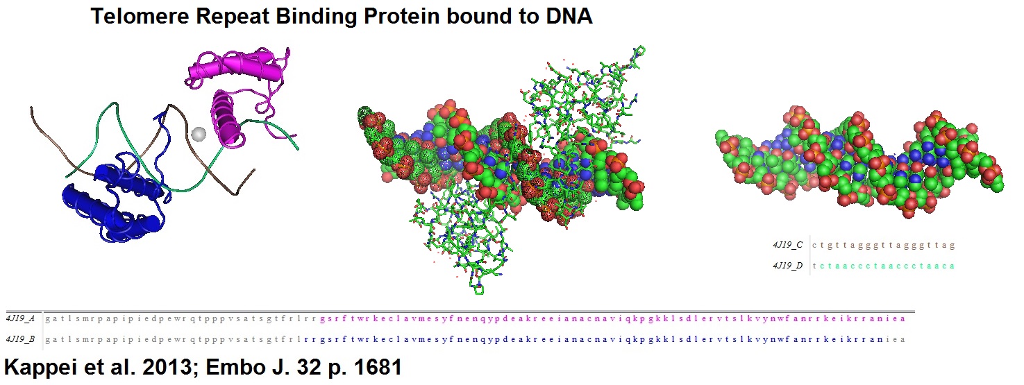
Figure 1:  Structural models of homeobox telomere-binding protein 1 (HOT 1), a mammalian direct telomere repeat-binding protein. HOT1 is a positive regulator of telomere length that supports telomerase-depending telomere elongation.
 

A universal priming probe was used for TRFanalysis


5’-(Phos)GACTCTCAACTATC+T+A-3’


+N represents the location of BNAs.

Universal BNA Priming Probe
-.-

BNA Probes and Oligonucleotides can be ordered from Bio-Synthesis Inc. 

-.-


BNA-digoxigenin-probes for enhanced telomere length analysis


BNA-oligonucleotide probes were designed to specifically bind to telomere repeats. For this, the researchers designed a non-radioactive labeling method that uses 3’ fill-in combined with lambda exonuclease digestion for the incorporation of one or more digoxigenin molecules into bridged nucleic acid (BNA)-containing oligonucleotides. Using this method, the researchers generated probes for the detection of both C- and G-rich telomeric DNA strands. The use of this type of probes enhanced the sensitivity of telomere length measurements significantly.

In humans, telomere lengths have been associated with cancer and age-related diseases. Telomeres are located at the ends of chromosomes and are composed of tandem 5’-TTAGGG-3’ repeats. Shelterin proteins associate with telomeres and play essential roles in telomere protection, telomerase regulation and the prevention of chromosome degradation. As humans age, telomeres gradually shorten in all dividing cells. The shortening triggers DNA damage responses and cellular senescence that can lead to genomic instability and cancer progression, especially if oncogenic changes in cells occur.

Telomere-specific probes used for Southern blotting in combination with a technique called “Terminal Restriction Fragment Analysis” enables the direct detection of different sizes of telomeres.

Terminal restriction fragment (TRF) pattern analysis, also known as “Terminal Restriction Fragment Length Polymorphisms (T-RFLP) analysis, is a recently developed PCR-based method. This technique also allows studying microbial community structure and dynamics. 

How does the technique work?

1.  T-RFLP analysis measures the size polymorphism of terminal restriction
      fragments from PCR amplified markers. 

2.  Primers needed for this technique are designed with the help of comparative
      genomics. 

3.  Primers are designed against the amplification product or amplicon. 

4.  PCR amplifies the signal from a high background of unreleated markers. 

5.  Subsequent digestion with correctly selected restriction endonucleases
      produce terminal fragments.

6.  Fragments are separated on high resolution sequencing gels.

7.  A digital output is generated if separation is done in a capillary electrophoresis
      system.

8.  The use of fluorescently tagged or labeled primers limits the analysis to only
      the terminal fragments of the digestion.

9.  Using internal size markers with a different fluorophore makes the sizing
      very accurate.


Outline of TRF analysis protocol


According to Lai et al. 2016.

Step 1

a.     Prepare template DNA for DIG-labeled telomere C-rich (TC) or
   G-rich (TG) probe synthesis.

b.    Anneal G-rich or C-rich template oligonucleotides to a universal
  priming oligonucleotide.


Templates


The 5’-phosphorylated template oligonucleotide begins with seven telomeric repeats followed by a short non-telomeric sequence.

G-rich template

5’-(Phos)CCC TAA CCC TAA CCC TAA CCC TAA CCC TAA CCC TAA CCC TAF ATA GTT GAG AGT C-3’

C-rich template

5’-(Phos)GGG TTA GGGTTA GGGTTA GGGTTA GGGTTA GGGTTA GGGTTA GAT AGT TGA GAG TC-3’


Universal priming


The universal priming oligonucleotide is phosphorylated at the 5’-end and contains a sequence complementary to the non-telomeric sequence in the template oligonucleotide with additional thymine (T) and adenine (A) at the 3’-end to ensure that it anneals at the correct spot.

Oligonucleotides modified with BNAs are used to increase resistance to nuclease digestion and the affinity for the target DNA or RNA.

Universal primer

5’(Phos)GAC TCT CAA CTA TC+T+A-3’; +N = BNAs


Use Exo- Klenow Fragment together with a dNTP mix containing DIG-11-dUTP (Roche Applied Sciences, Mannheim, Germany) for 3’ fill-in reactions.


Remove additional nucleotides from 3’-end

Step 2

Apply T4 DNA polymerase to remove additional nucleotides at the 3’ end from the template DNA generated by 3’ fill-in reactions. This increases the specificity of DIG-labeled telomere probes.

Step 3

Use λ exonuclease to digest the 5’-phosphorylated template oligonucleotide and non-telomeric sequence in the priming oligonucleotide (5’->3’ direction). Note: λ exonuclease is unable to degrade BNA-containing telomeric-specific single-stranded DNA.

Step 4 A

At this step a dot blot on a nylon membrane can be performed to check that the experiment worked. [See DIG Application Manual for filter hybridization.

Step 4B

Perform Southern blot analysis.


Southern Blot Analysis

Step 1

Digest DNA and DIG-labled molecular weight marker II.

Step 2

Separate on a 7% agarose gel.

Step 3

Depurinate, denature and neutralize the gel.

Step 4

Transfer DNA fragments onto a positive  charged nylon membrane using a vacuum blotting system.

Step 5

Fix the DNA fragments on the membrane by UV-crosslinking.

Step 6

Pre-hybridize.

Step 7

Hybridize with DIG Easy Hyb solution containing one of the DIG-labeled telomeric probes over night.

Step 8

Wash membrane.

Step 9

Detect chemiluminescence signals.

Step 10

Analyze.


See Supplement “Material and methods” from Lai et al. for more details.


Example of DIG-probe synthesis


Start with templates and universal BNA probe(s):


                 Tandem 5’-TTAGGG-3’ repeat

                        GGGTTAGGGTTAGGGTTAGGGTTA...

5’(Phos)GACTCTCAACTATCTA-3’

     3’-CTGAGAGTTGATAGATCCCAATCCCAATCCCAATCCCAAT...N-PHOS


->Anneal -> 3’fill-in (Exo- KF)  ->  DNA blunting (T4 DNA polymerase)  ->  5 ->3’ digestion (λ exonuclease)  -> DIG probes.


The use of fluorescently labeled oligonucleotide probes instead of 32P-labeled oligonucleotides makes this technique more convenient and less hazardous since many waste disposal and safety issues are associated with radioactivity.


Reference

DIG RNA Labeling Kit (SP6/T7): http://www.sigmaaldrich.com/catalog/product/roche/11175025910?lang=en&region=US

Kyung H. Choi, Amy S. Farrell, Amanda S. Lakamp, Michel M. Ouellette; Characterization of the DNA binding specificity of Shelterin complexes. Nucleic Acids Res. 2011 Nov; 39(21): 9206–9223. Published online 2011 Aug 18. doi: 10.1093/nar/gkr665, PMCID:  PMC3241663.

Raffaella Diotti, Diego Loayza; Shelterin complex and associated factors at human telomeres. Nucleus. 2011 Mar-Apr; 2(2): 119–135. doi: 10.4161/nucl.2.2.15135, PMCID:  PMC3127094.

Kong PL, Looi LM, Lau TP, Cheah PL.; Assessment of Telomere Length in Archived Formalin-Fixed, Paraffinized Human Tissue Is Confounded by Chronological Age and Storage Duration. PLoS One. 2016 Sep 6;11(9):e0161720. doi: 10.1371/journal.pone.0161720. eCollection 2016.

Aubert G, Lansdorp PM.; Telomeres and aging. Physiol Rev. 2008 Apr;88(2):557-79. doi: 10.1152/physrev.00026.2007.

Lai, Tsung-Po, Wright, Woodring E., and Shay, Jerry W. ; 2016. Generation of digoxigenin-incorporated probes to enhance DNA detection sensitivity. BioTechniques 60:306-309 (June 2016).

Marsh TL; Terminal restriction fragment length polymorphism (T-RFLP): an emerging method for characterizing diversity among homologous populations of amplification products. Curr Opin Microbiol. 1999 Jun;2(3):323-7.

Dennis Kappei, Falk Butter, Christian Benda, Marion Scheibe, Irena Draškovi?, Michelle Stevense, Clara Lopes Novo, Claire Basquin, Masatake Araki, Kimi Araki, Dragomir Blazhev Krastev, Ralf Kittler, Rolf Jessberger, J Arturo Londoño?Vallejo, Matthias Mann, Frank Buchholz; HOT1 is a mammalian direct telomere repeat?binding protein contributing to telomerase recruitment. The EMBO Journal (2013) 32, 1681-1701.

-.-

Why do we need cell line identification and authentication?

$
0
0

Why do we need cell line identification and authentication?


As reported in the literature, cell lines are frequently contaminated or misidentified. A high percentage of cell lines are cross-contaminated with cells from other cell lines. This phenomenon is known as “cell line cross-contamination” or (CLCC). According to MacLeod, CLCC occurs primarily during the establishment of a cell culture. Presumably, new cell cultures are contaminated during the early phases of their establishment. Cell line cross-contamination is a result of continuous cell line culture. To avoid misidentification quality controls need to be carried out to address this issue.

Sometimes, cross-contaminating cell lines can overgrow the original cell line. Cross-contamination has caused the number of misidentified circulating cell lines to be unacceptably high. In particular, the use of cancer cell line models may lead to wrong results if not identified or authenticated correctly. Therefore scientists have recommended that researchers should provide authenticities of all experimental cell lines used in experiments before publishing experimental results.

The reported misidentification of cell lines is estimated to be between 16 % to 36 %. These findings are based on the analysis of submitted cell lines. One of the best examples is the false description of a HeLa cervical cancer cell line. As a result, cell line identification has now become an essential method for the identification of human cell lines used in research. Cell lines are important reagents for experimental biomedical sciences. A large number of cell lines have been derived from patients. Among them are cell lines from patients with multiple myelomas, plasma cell leukemia, Plasmacytoma and many others. 

 Bio-Synthesis Inc. offers Custom Cell Line Identification

Cell lines can be tested and identified using multiallelic variable number of tandem repeats (VNTR). HLA typing and DNA fingerprinting using short tandem repeat (STR) and a variable number of tandem repeats for intra-species cross-contamination have been used for cell line identification. However, DNA fingerprinting hase become the method of choice for cell line identification.

Cell banks that archive and distribute cell lines are responsible for guaranteeing the authenticity of cell lines and will need to ensure cell line authenticity via DNA finger printing.

As pointed out by John M. Butler, STR typing enables the rapid discovery of cross-contamination between cell lines. Since STR typing is now used as the major tool for characterization of human cell lines this approach has also been called “cell culture forensics.”  The American Type Culture Collection (ATCC), the National Institutes of Biomedical Innovation containing the Japanese Collection of Research Bioresources (JCRB) Cell Bank, the “Deutsche Sammlung von Mikroorganismen und Zellkulturen GmbH” (DSMZ), as well as others, offer cell lines for research and have created databases containing information for many cell lines.


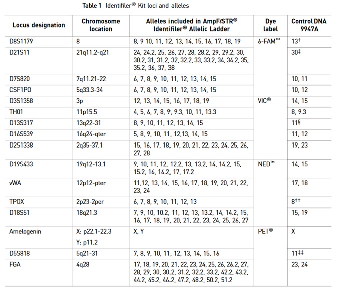
PCR-based identification kits use short tandem repeat (STR) amplifications assays. For example, the AmpFιSTR®PCR Amplification Kit is a short STR multiplex assay that amplifies 15 tetranucleotide repeat loci and the Amelogenin gender-determining marker in a single PCR amplification experiment. Fluorescent multi-color dye technology allows the analysis of multiple loci. Alleles with overlapping size ranges are also included. Locus-specific primers labeled with different colored dyes allow distinguishing overlapping loci.


STR markers are polymorphic DNA loci containing repeated nucleotide sequences. STR repeat units can range from two to seven nucleotides in length. However, the number of nucleotides per repeat unit is the same for a majority of repeats within an STR locus, but the number of repeat units at any STR locus may differ. Therefore alleles of many different lengths are possible. Polymorphic STR loci are very useful for human identifications as well as for cell line identification.


STR loci can be amplified using the polymerase chain reaction (PCR) process. PCR products are analyzed by electrophoresis to separate alleles according to size. Methods such as fluorescent dye labeling, silver staining, or fluorescent dye staining can be used for detection. 

The human genome contains approximately three (3) billion base pairs in a single copy.  Because of the Human Genome Project, a reference genome is now available.

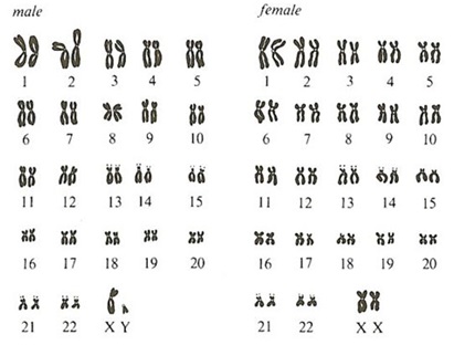
Genomic DNA found in the nucleus of a human’s cell is divided into chromosomes. In chromosomes, DNA is densely packaged around proteins called histones. Twenty-two (22) matching pairs of autosomal chromosomes and two (2) sex chromosomes make up the human genome. Hence, the cell of normal humans contains 46 different chromosomes.  Females are designated XX and males are designated XY, because they contain a single copy of an X and Y chromosome. Somatic chromosomes in cells in the human body are in a diploid state. They contain two sets of chromosomes. However, gametes (sperms or eggs) are in a haploid state. The combination of an egg cell with a sperm cell results again in a diploid state. DNA in chromosomes is divided into “coding” and “non-coding” sequence regions. Coding regions are known as “genes” and can contain a few thousand to tens of thousands base pairs. Since the finished Human Genome Project, we now know that the human genome codes for ~30,000 protein-coding genes. Therefore genes make up ~5% of human genomic DNA.

Human polymorphic markers are found in specific loci!

Markers useful for human identity or cell line identification testing are found in “non-coding” regions, either between genes or within genes (i.e. introns).
Polymorphic or variable markers are found throughout the non-coding regions of the human genome. The location or position of a gene or DNA marker is referred to as a “locus” (plural: loci). Thousands of these loci have now been mapped to specific regions of human chromosomes.
Pairs of chromosomes are called “homologous” since they are the same size and contain the same genetic structure. One chromosome in each pair is inherited from the mother and one from the father. Therefore the DNA in the two homologous chromosomes may not be identical since mutations could have occurred over time.
Alternative regions in homologous chromosomes are called “alleles.”
If DNA sequences in two alleles at a genetic locus on homologous chromosomes differ they are called “heterozygous,” but if they are similar they are called “homozygous.”  
A ‘genotype” refers to alleles that are present at a genetic locus. If two alleles are present at one locus, AA and aa, three genotypes are possible, AA, Aa, and aa. A “DNA profile” is the combination of genotypes obtained for multiple loci.
DNA profiling refers to the process of determining the genotype present at specific locations along the DNA molecule.     

A Brief Nomenclature for DNA Markers

Commonly used STR markers are found in sequence repeats present in satellites (~100 to 1000 repeated bases), minisatellites (VNTR = variable number tandem repeat; ~10 to 100 repeated bases), microsatellites (STR = short tandem repeat; 2 to 6 repeated bases. Most commonly used in forensics).

Examples:


STR markers within a gene
:  ( HUM)THO1


HUM = human; TH = thyrosine hydroxylase gene located on chromosome 11; 01= repeat region is located within intron 1.


STR markers outside gene regions
D16S539

D = DNA; 16 = chromosome; S = single copy sequence; 539 = 539th locus described on chromosome 16.


The vast majority of DNA molecules, approximately over 99.7%, are the same between humans. Only a small fraction, approximately 0.3% or close to 10 million nucleotides, differs between individual people. These variable regions provide the information for human or cell line identification tests.


Two forms of variation are possible at the DNA level (Butler, 2005):

1.     Sequence Polymorphisms, and

2.     Length polymorphisms.


Example 1:  Sequence polymorphism

-----AGACTAGACATT----

-----AGATTAGGCATT----

Example 2:  Length polymorphism

 ----(AATG)(AATG)(AATG)---- 3 repeats

 ----(AATG)(AATG)----       2 repeats


Example of the DNA sequence in a STR repeat region


---->

          1    2    3    4    5    6

5’-TTTCCC TCATTCATTCATTCATTCATTCAT TCACCATGGA-3’

3’-AAAGGG AGTA AGTA AGTA AGTA AGTA AGTA AGTGGTACCT-5’

                 6    5    4    3    2    1

                                                <----

Using the top strand versus using the bottom strand results in different repeat motifs and starting positions. TCAT repeat units are found in the top strand and TGAA repeat units are found in the bottom strand. According to guidelines issued by the International Society of Forensic Genetics (ISFH; http://www.isfg.org) the top strand should be used for the design of STR markers using alleles. 

For STR based typing usually length polymorphism based markers are used. As a rule of thumb: The more multiple markers are used for testing the greater the chance that two unrelated individuals will have different genotypes.

PCR base STR analysis

PCR-based STR analysis has become the method of choice for STR analysis because the small size of STR loci improves the chance of obtaining a good result, especially when small amounts of DNA or degraded DNA are in the sample. Because of the small size range of STR loci, co-amplification can be achieved, and typing is possible from a single PCR. Discrete sizes of STR alleles allow for easier interpretation of results. PCR-based tests are usually rapid, generating results within 24 hours, and lend themselves to automatization and standardization. Automation and standardization ensures that testing results in reproducible results. 

A typical protocol for cell line identification


1.    DNA is isolated from the cell pellets, tissues or cryovial (frozen cells) with
       a DNA extraction kit and purified with a Qiagen or Zymo DNA extraction kit.

2.    Quantitation of the extracts can be performed using a
       NanoDropTM spectrophotometer. 

3.    Usually, DNA amplification and fragment fluorescent labeling is
       performed using the PCR System thermal cycling instrument and
       the IdentifilerTM AmpF/STR® PCR kit. 

4.    Approximately 20.0 ng/µl of purified DNA is used for amplification. 

5.    Capillary electrophoresis and fragment detection are performed
       with a 310 Genetic Analyzer with Data Collection software v 3.0
       (Applied Biosystems). 

6.    GeneMapper ID® (Applied Biosystems) software is used to assign fragment
       repeat numbers which becomes collectively the STR type for the individual
       cell line identified.

The AmpFι STR®PCR Identifiler Kit uses the following STR marker for genotyping in a single reaction:


A brief glossary of STR testing


Humans = Homo Sapiens

Description

Genome

All the DNA chromosomes

22

Autosomal chromosomes

2

Sex chromosomes

Females

XX

Males

XY

genes

Coding regions

~30,000 genes

~5% in genomic DNA

Locus

Gene location

Homozygous

DNA sequences in two alleles at a genetic locus on homologous chromosomes are similar.

heterozygous

DNA sequences in two alleles at a genetic locus on homologous chromosomes are different.

Genotype

Genotype refers to alleles that are present at a genetic locus.

DNA profile

DNA profile refers to the combination of genotypes obtained for multiple loci.

DNA profiling

DNA profiling refers to the process of determining the genotype present at specific locations along the DNA molecule.   


Reference

ATCC cell lines: https://www.atcc.org/en/Products/Cells_and_Microorganisms/Cell_Lines.aspx

Butler, J.M.; Forensic DNA Typing.  2nd Edition.2005 Elsevier Academic Press.

BSI blog

https://blog-biosyn.com/2013/04/15/why-perform-a-cell-identification-check/

https://blog-biosyn.com/2013/02/18/cell-line-authentication-and-identification/

https://blog-biosyn.com/2013/06/27/why-should-i-test-my-cho-cell-culture-for-virus-infections/

Cabrera CM, Cobo F, Nieto A, Cortés JL, Montes RM, Catalina P, Concha A.; Identity tests: determination of cell line cross-contamination. Cytotechnology. 2006 Jun;51(2):45-50. doi: 10.1007/s10616-006-9013-8. Epub 2006 Aug 3.

Chatterjee R.; Cell biology. Cases of mistaken identity. Science. 2007 Feb 16;315(5814):928-31.
https://www.ncbi.nlm.nih.gov/pubmed/17303729

DNA Analyst Training – Laboratory Training Manual Protocol 5.02 PCR: Amplification and Electrophoresis of STRs. Presideint’s DNA Initiative. 
https://static.training.nij.gov/lab-manual/Linked%20Documents/Protocols/pdi_lab_pro_5.02.pdf.

Drexler HG, Dirks WG, MacLeod RA.; False human hematopoietic cell lines: cross-contaminations and misinterpretations. Leukemia. 1999 Oct;13(10):1601-7.

Hans G. Drexler, Roderick A. F. MacLeod, Willy G. Dirks;  Cross-contamination: HS-Sultan is not a myeloma but a Burkitt lymphoma cell line. Blood 2001 98:3495-3496; doi:10.1182/blood.V98.12.3495

Hashiyada, M.; DNA Biometrics. www.intechopen.comhttp://cdn.intechopen.com/pdfs/16506/InTech-Dna_biometrics.pdf

Masaki Hashiyada (2011). DNA biometrics, Biometrics, Dr. Jucheng Yang (Ed.), ISBN: 978-953-307-618-8, InTech, Available from: http://www.intechopen.com/books/biometrics/dna-biometrics

MacLeod RA, Dirks WG, Matsuo Y, Kaufmann M, Milch H, Drexler HG.; Widespread intraspecies cross-contamination of human tumor cell lines arising at source. Int J Cancer. 1999 Nov 12;83(4):555-63.

Roderick A.F. MacLeod, Wilhelm G. Dirks and Hans G. Drexler; One falsehood leads easily to another. Int. J. Cancer: 122, 2165-2168 (2008).

User Guide - AmpFιSTR® Identifiler® PCR Amplification Kit ABI – life technologies (PN: 4322288).


-.-


Carnosine (β-Ala-L-His)

$
0
0

Carnosine (β-Ala-L-His)


The dipeptide carnosine is a bioactive endogenously abundant peptide. High amounts of carnosine are found in muscle and brain tissues. A Russian scientist with the name of Vladimir Sergeevich Gulevish is credited to discover carnosine for the first time in mammalian muscle. Gulevich also discovered a number of new nitrogen compounds in muscle, including carnosine, carnitine, and anserine. Although carnosine is a natural dipeptide, the peptide can be synthesized using Fmoc-chemistry based solid phase peptide synthesis (SPPS). 


Carnosine is a zwitterion. Zwitterions are neutral molecules that have a positive and negative charge. Carnosine can be analyzed in an amino acid analyzer as the dipeptide or hydrolyzed into the two amino acids β–alanine and L-histidine. Mass spectrometry, especially LC-MS(MS) offers itself as well as a sensitive tool for its detection and analysis.


High rates of carnosine synthesis are thought to occur in glial cells, not in neurons. Carnosine is now thought of as an intracellular pH buffer modulator, Zn/Cu ion chelator, antioxidant, aldehyde-scavenger, anti-glycating and anti-crosslinking agent for proteins. Furthermore, in the central nervous system, the CNS, it appears to work as a multi-functionally homeostatic and protective molecule in neuronal and non-neuronal cells. Taken together, carnosine appears to protect against neurodegenerative conditions.

Carnosine together with homocarnosine and anserine have been shown to act as scavengers of hydroxyl radicals (•OH). Aruoma et al. in 1989 suggested that carnosine and anserine may act as an antioxidant at physiological levels.

Barski et al. in 2013 reported that carnosine inhibits atherogenesis by facilitating aldehyde removal from atherosclerotic lesions. Carnosine prevents oxidation of low-density lipoprotein (LDL) as well as the cytotoxicity of reactive aldehydes generated by lipid oxidation.

In mice, dietary intake of carnosine decreases the formation of atherosclerotic lesions. However, the role of LDL oxidation in atherogenesis remains unclear. Barski et al. suggested that treatment with carnosine may decrease atherosclerosis by preventing LDL oxidation and the cytotoxic effects of lipid peroxidation products. 

Atherogenesis is a disorder of the artery wall. Atherogenesis is characterized by the adhesion of monocytes, phagocytic white blood cells, lymphocytes, small white blood cells of the lymphatic system, to the endothelial cell surface, and the migration of monocytes into the sub-endothelial space, the differentiation into macrophages and the ingestion of low-density lipoproteins and modified or oxidized low-density lipoproteins by macrophages via several pathways. The result is an accumulation of cholesterol esters and the formation of “foam cells”. The foam cells together with T lymphocytes form the fatty streak. Migrating vascular smooth muscle cells into the intima proliferate and form the atherosclerotic plaques. The term intima refers to the innermost coating or membrane of a part or organ, in this case, a vain or artery. 

All steps of this process involve cell adhesion, migration, differentiation, proliferation and cell interaction with the extracellular matrix which are regulated by a complex network or cascade of cytokines and growth regulatory peptides. Therefore it is thought that atherosclerosis is a result of a inflammatory-fibroproliferative process which has developed into a chronic disease state.

Also in 2013, Aloisi et al. reported that carnosine inhibits Aβ1-42 fibrillogenesis in vitro. Published results showed an effective role of carnosine against Aβ1-42 aggregation. The data suggest that carnosine induces a less ordered Aβ1-42 amyloid aggregation causing a lesser growth of fibrils. Carnosine appears to operate as an interfering, anti-aggregating agent. To date, small oligomers are considered the major aggressive variant of the amyloid formations. The so-called “oligomer cascade hypothesis” is a key premise in structure-neurotoxicity relationship studies of amyloid formations. 

Alzheimer’s disease is the 3rd most costly disease and estimated to be the 6th leading cause of death. Researchers still search to identify the primary toxin that causes Alzheimer’s disease.

Reference

https://books.google.com/books?id=CTIOsAGBsi8C&pg=PA304#v=onepage&q&f=false

Aloisi, A., Barca, A., Romano, A., Guerrieri, S., Storelli, C., Rinaldi, R., & Verri, T. (2013). Anti-Aggregating Effect of the Naturally Occurring Dipeptide Carnosine on Aβ1-42 Fibril Formation. PLoS ONE, 8(7), e68159. http://doi.org/10.1371/journal.pone.0068159. 

Aruoma, OI; Laughton, MJ; Halliwell, B (1989). "Carnosine, homocarnosine and anserine: could they act as antioxidants in vivo?". The Biochemical Journal. 264 (3): 863-9. 
 doi:10.1042/bj2640863PMC 1133665PMID 2559719.

Bickford PC, Tan J, Shytle RD, Sanberg CD, El-Badri N, Sanberg PR.Nutraceuticals synergistically promote proliferation of human stem cells. Stem Cells Dev. 2006 Feb;15(1):118-23.

Ferreira, S. T., & Klein, W. L. (2011). The Aβ oligomer hypothesis for synapse failure and memory loss in Alzheimer’s diseas. Neurobiology of Learning and Memory, 96(4), 529–543.
 
http://doi.org/10.1016/j.nlm.2011.08.003

Gulewitsch, Wl.; Amiradžibi, S. (1900). "Ueber das Carnosin, eine neue organische Base des Fleischextractes". Berichte der deutschen chemischen Gesellschaft. 33 (2): 1902–1903. doi:10.1002/cber.19000330275.


Ross R, Agius L.; The process of atherogenesis--cellular and molecular interaction: from experimental animal models to humans.
 Diabetologia. 1992 Dec;35 Suppl 2:S34-40.

-.-

Explanation of Bioanalytical Methods and Analytical Services

$
0
0

Bioanalytical Methods and Analytical Services


To fully characterize biological samples or compounds such as amino acids, carbohydrates, vitamins, metabolites, oligonucleotides, peptides, and proteins a variety of specific bioanalytical methods are used including various molecular biology services. Specifically, bioanalytical methods include a variety of particular methods that are applied for quantitative measurements of analytes in a given biological matrix such as a formulation buffer or a natural sample matrix. Typical examples of samples are purified or formulated biosimilars, oligonucleotides, proteins, peptides, blood, plasma, serum, or urine. 

Bioanalytical methods used should be completely reliable and reproducible. To achieve reliable results, it is necessary to employ properly-characterized and validated bio-analytical methods. Often formulated proteins are formulated as lyophilized powders from sterile solutions. A typical formulation buffer is PBS, at pH 7.4. plus 5 % - 8 % trehalose and/or mannitol. 

However, for protein profiling methods newly developed methods are used.

Methods and Services


Method/Service

Result

Notes

AAA

Determination of free amino acids or of the protein concentration in a sample. Recovered amino acids are usually reported in picomoles and nanograms recovered.

Presently this is considered to be the most accurate method for the determination of amino acids and protein contents.

However, matrix effects due to interfering compounds in the formulation buffer are possible. In the case of formulated proteins, sometimes the method only works accurately if the protein is highly purified prior to the analysis.

Electroblotting

A picture of the membrane containing the protein band(s) is reported.

Needed for N-terminal Sequencing.

HPLC

Detection of the purity of a biological compound, small molecule, amino acid, peptide or protein often as a single peak. A typical HPLC report includes a chromatogram showing a single peak or, in the case when impurities are present, multiple peaks.

Analytical run to determine purity and to estimate amounts of peptides, proteins or other compounds of interest. However, for proteins, the measurement of exact concentrations is not possible unless a control protein with the exact sequence and protein folding and modification is available for establishing a calibration curve. Similarly, for other biomolecules an accurate standards are needed as well for a quantitative analysis.

LC-MS/MS

Protein sequence identification is done from gel pieces or protein pellets containing the protein(s) of interest via tryptic digest, and LC-MS/MS analysis followed by database searches. Often nano-spray-LC-MS/MS is used to achieve more sensitive results.

Protein identification via mass pattern matching. The N-terminal and C-terminal end may be present in the data. However it is possible that the N- and C-terminal are missing.

MALDI-MS

This type of analysis will result in a mass measurement of a compound such as a carbohydrate, lipid, oligo-nucleotide, peptide or protein. Mass spectra with the observed peaks are usually reported.

This analysis is more accurate than molecular weight determination via SDS-PAGE. Impurities originating from peptides and protein fragments or other sourcers are observed as well.

N-terminal sequencing

For X cycles. A typical report will contain the observed sequence plus the chromatogram of the cycles done.

This method will provide sequence information starting from the N-terminal end of the protein. Sometimes up to 40 or 45 cycles may be observed. However, in the earlier days of protein sequencer development up to 60 or even 75 cycles were reported.

SDS-PAGE

Usually a picture of a 1D gradient gel is reported showing the apparent molecular weights.

Removes PBS and sugars from oligonucleotides, peptides, and proteins.

UV absorbance at 280 nm

The absorbance of the protein at 280 nm is measured and reported. A BSA standard curve can be used to estimate the specific absorbance.

Unless the specific absorbance is known this will only give an estimate of the protein concentration.


-.-


Myristoylated peptides are candidates for drug design

$
0
0

Myristoylated peptides are candidates for drug design!


Myristoylation of proteins or peptides is a posttranslational modification found in eukaryotic and viral proteins in which the N-terminal end is acylated with myristic acid. Myristic acid, or Tetradecanoic acid, is a fatty acid with the molecular formula CH3(CH3)12COOH, and a molecular weight of 228.27 g/mol. The modification of the protein N-terminus with a myristoyl group [a (cis,cis-delta 5, delta 8)-tetradecadienoyl group =  myristoylation with 2 double bonds] adds an average mass of 206 dalton, and the myristoleylation (= myristoyl with one double bond) adds a mass of 208 daltons to the target protein. Modern peptide synthesis chemistry now allows for the production of modified peptides such as myristoylated or palmityolated peptides.

N-myristoylation refers to the acylation process specific to the N-terminal amino acid glycine in proteins. This process has been observed in hundreds of proteins in lower and higher eukaryotes. Proteins labeled with this modification are involved in oncogenesis, in secondary cellular signaling, in infectivity of retroviruses and other virus types. The cytosolic enzyme responsible for this modification is N-myristoyltransferase (NMT).

If NMT is selectively inhibited a dose-responsive loss of N-myristoylation on protein targets is observed. A recent proteomic study showed that NMT inhibition killed HeLA cells apparently through endoplasmic reticulum (ER) stress and the unfolding response signalling (UPR) pathways. 

Also, Rampoldi et al. in 2015 found that protein myristoylation is indispensable in T cell development and activation. A deficiency in N-myristoyl transferase (Nmt) 1 and 2 activity caused a defective transmission of TCR signals, a developmental blockage of thymocytes at the transition from double-negative 3 to 4 stages, and a reduction of all following stages. Further more, the study found that two main myristoylated kinases in T cells were mislocalized in the absence of Nmt activity, and the absence of myristoylation resulted in an immunosuppressive effect on T cells.


Figure 1:  Crystal structure based model of calcium-calmodulin (Ca2+-CaM) bound to a myristoylated peptide derived from the N-terminal domain of CAP-23/NAP-22.

 
More than 300 million people are infected worldwide with the hepatitis B virus. The hepatitis B virus (HBV) is a common cause of liver disease and liver cancer. Hepatitis B infections are caused by HBV affecting the liver. People infected with this virus are at risk of developing chronic liver disease, cirrhosis and hepatocellular carcinoma.  

Source: https://pixabay.com/en/hepatitis-b-virus-virus-3d-1186581/

König et al. in 2014 showed that N-terminal myristoylated lipopeptides, derived from the pre-S protein of the hepatitis B virus, can be used to study their binding kinetics to the human liver bile acid transporter Na+/taurocholate cotransporter. The ultimate goal of this study is the design of lipopeptide-based drugs useful for the management of hepatitis B infections.

According to König et al., the binding of myristoylated hepatitis B virus preS1 (myr-preS1) protein to the human liver bile acid transporter Na+/taurocholate cotransporter (NTCP) is necessary for a productive infection by the hepatitis B virus (HBV). The infection interferes with the physiological bile acid transport function of NTCP. The binding of the myr-preS1 peptide to NTCP initiates the infection. König et al. investigated the binding kinetics of myr-preS1 peptides to NTCP. The results of the study suggested that the myr-preS1 peptide inhibits bile acid transport in the cells. Cell lines used for the studies where primary Tupaia belangeri (PTH; northern treeshrew; an animal model) and human (PHH) hepatocytes as well as NTCP-transfected human hepatome HepG2 cells. The research group suggests that NTCP substrates may be useful tools for the design of NTCP-inhibiting drugs allowing effective management of HBV infections. 

Plants, more specifically maize, have been investigated to allow high level expression of Hepatitis B Surface Antigens for the development of plant-based oral HBV vaccines.


The myristoylated peptide used for the study was the following peptide

Myr-GQNLSTSNPLGFFPDHQLDPAFRANTANPDWDFNPNKDTWPDANKVG-C(Alexa594)-COOH


Many viral and signal transduction proteins are known to be myristoylated. The N-terminal myristoyl group is directly involved in protein-protein interactions. For some proteins, this modification appears to be essential for proper functioning of the modified proteins. For example, the activity of p60src from Rous sarcoma virus is dependent on its myristoylation. It is assumed that hydrophobic acyl groups, for example, myristoyl and palmitoyl groups, are often involved in protein-protein interactions.

The crystal structure of calcium-calmodulin (Ca2+-CaM) bound to a myristoylated peptide derived from the N-terminal domain of CAP-23/NAP-22 has been solved see figure 1). Cap-23/NAP-22 is a brain-specific protein kinase C substrate involved in axon regeneration that binds calmodulin with high affinity. A synthetic myristoylated peptide of nine amino acids and purified recombinant human CaM was used for hanging-drop vapor diffusion crystallization. The researchers were able to solve the structure of the complex from a single crystal at a resolution of 2.3 Å.

Reference


Boutin JA.; 
Myristoylation. Cell Signal. 1997 Jan;9(1):15-35.

P. GRIPON, J. LE SEYEC, S. RUMIN, C. GUGUEN-GUILLOUZO;
Myristylation of the Hepatitis B Virus Large Surface Protein Is Essential for Viral Infectivity. Virology. 1995 Nov 10;213(2):292-9.

König, Alexander et al.; Kinetics of the bile acid transporter and hepatitis B virus receptor Na+/taurocholate cotransporting polypeptide (
NTCP) in hepatocytes. Journal of Hepatology , Volume 61 , Issue 4 , 867 – 875.  https://www.ncbi.nlm.nih.gov/pubmed/24845614.

Matsubara M, Nakatsu T, Kato H, Taniguchi H.; Crystal
structure of a myristoylated cap-23/nap-22 n-terminal domain complexed with ca2+/calmodulin. Embo J. (2004) 23 p.712.

Francesca Rampoldi, Mahnaz Bonrouhi, Martin E. Boehm, Wolf D. Lehmann, Zoran V. Popovic, Sylvia Kaden, Giuseppina Federico, Fabian Brunk, Hermann-Josef Gröne and Stefan Porubsky; Immunosuppression and Aberrant T Cell Development in the Absence of N-Myristoylation. The Journal of Immunology November 1, 2015 vol. 195 no. 9 4228-4243.

Thinon E, Morales-Sanfrutos J, Mann DJ, Tate EW.;
N-Myristoyltransferase Inhibition Induces ER-Stress, Cell Cycle Arrest, and Apoptosis in Cancer Cells. ACS Chem Biol. 2016 Aug 19;11(8):2165-76. doi: 10.1021/acschembio.6b00371.  PMID: 27267252.

Wang, Miao, Kaufman, Randal J.; The impact of the endoplasmic reticulum protein-folding environment on cancer development. Nat Rev Cancer
14, 581-597 (2014) http://dx.doi.org/10.1038/nrc3800.

Chunyan Xu, Beatrice Bailly-Maitre, and John C. Reed; Endoplasmic reticulum stress: cell life and death decisions.
J Clin Invest. 2005 Oct 1; 115(10): 2656–2664.  doi:  10.1172/JCI26373 PMCID: PMC1236697.

-.-


Therapeutic nucleoside and nucleotide analogs

$
0
0

Since the development and introduction of solid phase oligonucleotide synthesis in the 1950s several nucleoside and nucleotide analogs have been identified as potent antiviral drugs. These compounds exhibit activities against poxviruses including variola, vaccinia, monkeypox, cowpox, molluscum contagiosum, and the orf virus. Nucleoside and nucleotide analogs including fluroescently labled nucleotides can be incorporated synthetically into oligonucleotides using modified phosphoramidites. Depending on application, short and long modifed oligos can be synthesized using RNA and/or DNA synthesis.

Biosynthesis Inc. offers automated custom synthesis for un-modified and modified short, median length, and long DNA and RNA oligos.

Molecular levels of the nucleoside system including 5’-nucleotidases (5’-NTs) and other nucleoside metabolic enzymes, as well as nucleoside transporters, and receptors, were found to be unevenly distributed in the brain. The nucleosides adenosine (Ado, A), guanosine (Guo, G), inosine (Ino, I) and uridine (Urd, U), are known to modulate both physiological and pathophysiological processes in the brain. We know now that these molecules are important players in the regulation of sleep, pain, memory, depression, schizophrenia, epilepsy, Huntington’s disease, Alzheimer’s disease and Parkinson’s disease.

5,6-dihydro-5-azacytosidine (DZCyt, or DHAC)


Figure 1:  Structures of 5,6-dihydro-5-azacytidine (DHAC) and its phosphoramidite.


Sheikhnejad in 1999 showed how 5,6-dihydro-5-azacytosidine (DZCyt or DHAC), a cytosine analog with a sp3-hybridized carbon (CH2) at position 6 and an NH group at position 5, can mimic the non-aromatic character of the cytosine ring in the transition state. The substitution of DZCyt or DHAC for target cytosines in C-G dinucleotides of single-stranded or double-stranded oligodeoxy-ribonucleotide substrates led to complete inhibition of methylation by the murine DNA (cytosine C5)-methyl-transferase (DNA C5-MTase). Substitution of DHAC for the target cytosine in G-C-G-C sites in double-stranded oligodeoxyribonucleotides had a similar effect on the methylation by HhaI methyltransferase (M. HhaI).

Oligodeoxyribonucleotides containing DZCyt can form a tight but reversible complex with the enzyme M. HhaI. The compounds are known to be potent inhibitors of DNA methylation. Methyltransferase inhibitors can restore transcriptionally silenced genes and are therefore important molecular tools for current therapies of myelodysplastic syndromes and certain types of leukemias.

A series of 5-azacytidine nucleosides have been investigated recently for their hypomethylation potential (Matoušová et al., 2011). Among these are nucleoside analogs including 5-azacytidine (AC), 2′-deoxy-5-azacytidine (DAC), its α-anomer (α-DAC), 5,6-dihydro-5-azacytidine (DHAC), 2′-deoxy-5,6-dihydro-5-azacytidine (DHDAC, KP-1212), its α-anomer (α-DHDAC), and a 2-pyrimidone ribonucleoside called zebularine. These compounds are now well established, preclinically tested inhibitors of DNA methylation. Protected phosphoramidite monomers are useful for the synthetic incorporation of these monomers into oligonucleotides of various lengths.

Zebularine (1-beta-D-ribofuranosyl-2(1H)-pyrimidinone)


Figure 1: Structures of Zebularine, 5-Me-Zebularine, and the phosphoramidite of 5-Me-Zebularine.


Zebularine (1-beta-D-ribofuranosyl-2(1H)-pyrimidinone) is another chemically stable, cytidine analog that displays anti-tumor properties. The molecule acts as a transition-state analog inhibitor of cytidine deaminase by binding to the active size as covalent hydrates. It was also shown to inhibit DNA methylation and tumor growth, both in vitro and in vivo.

The 5-methyl-pyrimidin-2-one, 2’-deoxynucleoside analog has been used as a molecular probe to study the initiation of cellular DNA repair. Zebularine-modified DNA cannot be methylated by the cytosine-[C5]-specific DNA methyltransferases (C5 MTases) M. MSP I and M. Hha I. The m5C-DNA methyltransferases catalyze the transfer of a methyl group from S-adenosyl-L-methionine to the C-5 position of cytosine within the recognition sequence of the substrate DNA. Hhal Methyltransferase modifies the internal cytosine residue (C5) of the sequence GCGC (GC{Me}GC).


The 5-Me-2'-deoxyZebularine-CE Phosphoramidite can be used for the synthesis of oligonucleotides containing this modification. Cytosine DNA methyltransferase inhibitors such as this molecule are known to restore androgen responsiveness in androgen-refractory tumor cells. The treated tumor cells are then sensitive to growth inhibition by anti-androgens.

Cytosine arabinoside (araC)


Figure 3: Structures for araC and its phosphoramidite.


Cytosine arabinoside (araC), a pyrimidine analogue, is one of the most effective agents or drugs used in the treatment of acute leukemia. araC is thought to exert its chemotherapeutic activity by the inhibition of the nuclear factor NF-κB. However, araC needs to be incorporated into chromosomal DNA to have a cytotoxic effect. The drug is converted to its “active” nucleoside triphosphate form by the pyrimidine salvage pathways. After integration into chromosomes, the arabinoside inhibits chain elongation and bypasses synthesis. In some cases, araC can induce DNA chain termination or duplication of DNA sequences.

However, the precise mechanism of araC-induced cell death may still need to be established. Apparently, treatment with this drug leads to reduced rates of DNA replication, DNA strand breaks, and chromosome fragmentation.

Reference

Susan D. Cline and Neil Osheroff; Cytosine Arabinoside Lesions Are Position-specific Topoisomerase II Poisons and Stimulate DNA Cleavage Mediated by the Human Type II Enzymes. Vol. 274, No. 42, Issue of October 15, pp. 29740 –29743, 1999.

De Clercq E, Neyts J.; Therapeutic potential of nucleoside/nucleotide analogues against poxvirus infections. Rev Med Virol. 2004 Sep-Oct;14(5):289-300.


Drugbank: 
http://www.drugbank.ca/drugs/DB03068

Kovács Z, Dobolyi A, Kékesi KA, Juhász G.; 5'-nucleotidases, nucleosides and their distribution in the brain: pathological and therapeutic implications.
Curr Med Chem. 2013;20(34):4217-40.

Kumar S, Cheng X, Klimasauskas S, Mi S, Posfai J, Roberts RJ, Wilson GG.; The DNA (cytosine-5) methyltransferases. Nucleic Acids Res. 1994 Jan 11;22(1):1-10.

Matoušová M, Votruba I, Otmar M, Tloušťová E, Günterová J, Mertlíková-Kaiserová H. 2′-deoxy-5,6-dihydro-5-azacytidine—a less toxic alternative of 2′-deoxy-5-azacytidine: A comparative study of hypomethylating potential. Epigenetics. 2011;6(6):769-776. doi:10.4161/epi.6.6.16215.

David P. Martin, Thomas L. Wallace, and Eugene M. Johnson, Jr.; Cytosine Arabinoside Kills Postmitotic Neurons in a Fashion Resembling Trophic Factor Deprivation: Evidence That a Deoxycytidine-Dependent Process May Be Required forNerve Growth Factor Signal Transduction. The Journal of Neuroscience, January 1990, IO(l): 184-193.

Gholamreza Sheikhnejad, Adam Brank, Judith K. Christman, Amanda Goddard, Estela Alvarez, Harry Ford Jr, Victor E. Marquez, Canio J. Marasco, Janice R. Sufrin, Margaret O'Gara and Xiaodong Cheng; Mechanism of Inhibition of DNA (Cytosine C5)-Methyltransferases by  Oligodeoxyribo-nucleotides Containing 5,6-Dihydro-5-azacytosine. J. Mol. Biol. (1999) 285, 2021-2034.

Singleton SF, Roca AI, Lee AM, Xiao J. Probing the structure of RecA-DNA filaments. Advantages of a fluorescent guanine analog. Tetrahedron. 2007;63(17):3553-3566.  doi:10.1016/j.tet.2006.10.092.

Zhou L, Cheng X, Connolly BA, Dickman MJ, Hurd PJ, Hornby DP. Zebularine: A Novel DNA Methylation Inhibitor that Forms a Covalent Complex with DNA Methyltransferases. Journal of molecular biology. 2002;321(4):591-599. doi:10.1016/S0022-2836(02)00676-9.

-.-

How is Extinction Coefficient Determined for Proteins

$
0
0

How are extinction coefficients determined for Proteins?

Extinction coefficients for proteins are determined at absorbance maxima near 280 nm.


Protein analysis is needed to determine if a sample solution contains the desired protein. For example, measuring the absorbance of a protein sample at 280 nm with a spectrophotometer is a rapid and straightforward method. In many bio-analytical applications, it is important to estimate or accurately determine the concentration of sample solutions containing purified biomolecules such as oligonucleotides, peptides or proteins. Often this information is needed or used for the design of down-stream experiments in analytical chemistry, biology, biochemistry, biophysics, medicinal chemistry, and pharmacy.

Absolute quantification of protein samples to determine accurate protein mass or concentration is often required to estimate recovery at different purification stages, to measure the specific activity of a protein, and to prepare known amounts of protein samples for analytical analysis. In general, calibrated sets of external standards containing known amounts of a specific protein such as bovine serum albumin (BSA) are often used to create calibration curves.

Commonly used methods for the quantification of proteins are:

  • Absorption of UV light by side chains and peptide bonds of a protein.
  • Chromogenic reactions involving complexes formed under alkaline conditions between protein and cuprous ions employing the biuret reaction. 
  • Binding of a chromophore to the protein.
  • Dot-blotting and staining.

Each method has advantages and limitations. Therefore amino acid analysis should also be considered for quantitative analysis of unknown proteins.

Amino acid analysis in combination with UV-absorbance measurements at 280 nm can be used to accurately determine protein concentrations as well as extinction coefficients in unknown protein samples. UV absorbance is the most popular method because it is fast, convenient, and reproducible. UV-absorbance measurements do not consume the protein and do not require additional reagents, standards or incubations. However, the measurement of protein solutions at 280 nm is not strictly quantitative for all proteins since the assay is based on the strong absorbance of tyrosine, tryptophan and phenylalanine residues. Protein concentrations can also be measured at 214 nm, and at 205 nm. 

No method of protein concentration determination is perfect because each method has different constrains. Interfering buffer components, contaminating proteins or reactivity of individual proteins and buffer components can influence the measurement and lead to false values. Therefore amino acid analysis is still considered to be the most accurate method for protein quantification, in particular when a highly purified protein sample is used. If accurate protein concentrations are critical, results from several methods or assays may need to be compared to trust the observed values. 

Proteins and peptides containing aromatic amino acid side chains exhibit strong UV-light absorption, and each protein has a distinct UV spectrum. Proteins and peptides absorb UV-light proportional to their amino acid content and total concentration. Different proteins may have widely varying extinction coefficients. If a protein does not contain any tyrosine, tryptophan, and phenylalanine residues, it will not be detected.

Optical spectroscopy is used in biochemistry for the observation and measurement of the absorption of oligonucleotide, peptide and protein solutions in the UV range. According to Planck’s law, E = hν, the absorbed energy must be related to the difference between energy levels and the linearity of absorbance is usually confined to dilute solutions governed by the Beer-Lambert law describing the absorption process:

A = log (I/I0) = ϵlc    => Beer-Lambert equation.


where A is the absorbance, I and I0 are the intensities of the transmitted and incident light beams, respectively, ϵ is the proportionality constant or specific extinction coefficient, l is the optical path length, and c is the concentration of the absorbing species usually reported in mol/L.

When light of a specific wavelength λ is passed through a solution layer at a path length L of the solution, a certain portion of the light is absorbed by the solution if the solution has a specific absorbance at this wavelength. The fraction of light transmitted is known as transmittance T. For homogenous samples, each successive layer of the solution will receive fractionally less light. Beer-Lamberts law mathematically describes this phenomenon. Beer-Lamberts law describes the exponential decay of the transmitted light versus path length and concentration.

The transmittance T decreases exponentially with respect to the concentration C of the compound and to the length of the light path, L.

T = 10-ϵcL  =>  -log10T = log10(1/T) = ϵcL =A    => Beer-Lambert equation.


Where ϵ is the absorption coefficient or absorptivity, characteristic for the compound measured at the particular wavelength of light under a defined set of conditions. The absorptivity is also known as extinction coefficient.

The absorbance A is defined as –log10T.  
Often absorbance is also called optical density (OD).


Extinction Coefficient


According to Merriam-Webster, the extinction coefficient refers to “a measure of the rate of transmitted light via scattering and absorption for a medium.” However, in analytical chemistry, the quantity ϵ (epsilon) is called the molar absorptivity (ϵmolar) or extinction coefficient. ϵ has the units M-1 cm-1. Molar absorptivity refers to the characteristics of a substance that tells how much light is absorbed at a particular wavelength.  Whereas the “specific absorption coefficient (a)” refers to the absorbance of light per unit path length, usually expressed in cm, and per unit of mass concentration. 

The “molar absorption coefficient (ϵmolar)” refers to the absorbance of light per unit path length and per unit of concentration expressed in “moles per liter.”

For proteins, an absorbance maximum near 280 nm (A280) in the UV spectra of a protein solution is mostly due to the presence of aromatic tryptophan and tyrosine residues, and to a minor portion phenylalanine. For a given protein, the A280 is proportional to its concentration of amino acids. However, corrections may be needed to calculate the accurate absorbance value, the type, and the environment the amino acids are in. Using the known amino acid sequence of a protein allows estimation of a sufficiently accurate extinction coefficient.

In general, a 1 mg/ml solution of most proteins has an A280 of ~ 1 ± 0.6. 


Since the introduction of the NanoDrop 1000 and the NanoDrop 8000 A280 instruments, these spectrophotometers are now often used for routine measurements of protein absorbance at 280 nm.  

Using a spectrophotometer such as the nanodrop instrument the concentration of a purified protein samples is determined according the Beer-Lambert equation [A = E * b * c ] which is used for all protein calculations to correlate absorbance with concentration. Where A = absorbance value (A), E = wavelength-dependent molar absorptivity coefficient (or extinction coefficient) with units of liter/mol-cm, b = the path length in centimeters, c = analyte concentration in moles/liter or molarity (M).

The equation for the Beer-Lambert law can also be written as [ Aλ = ϵλbc ] because A and ϵ depend on the wavelength of light. The greater the molar absorptivity, the greater the absorbance, and A and ϵ vary with the wavelength. Furthermore, the part of a molecule that is responsible for light absorption is called a chromophore. 

Each protein has a distinct UV spectrum as well as an extinction coefficient at 280 nm (ϵ280). The specific UV spectrum is based on its amino acid composition. Major contributions to the spectra stem from aromatic tryptophan (W) and tyrosine (Y) residues with high extinction coefficients of 5500 and 1490 M-1cm-1. Phenylalanine (F) absorbs maximally at 260 nm but little at 280 nm. Cystine (C) in disulfide bonds has a relatively low extinction coefficient of 125 M-1cm-1. The absorbance of reduced cysteine is negligible at wavelength above 260 nm.

If the number of absorbing side chains in the amino acid sequence of a protein is known the specific extinction coefficient at 280 nm can be estimated using the following formula:

ϵ280 = nW x 5,500 + nY x 1,490 + nC x 125

where ϵ280 is the molar extinction coefficient at 280 nm, and n is the number of corresponding residues present in the protein.

Molar concentration can be calculated as follows:

Molar concentrations = A280 x (dilution factor) / ϵ280

Concentrations [mg/ml] =  A280 x (dilution factor) x (moluclar weight in daltons  / ϵ280

When nucleic acids are present, the following correction can be used:

Protein concentration (mg/ml) = 1.55A280– 0.75A260

where A280 and A260 are the absorbance values of the protein solution at 280 nm and 260 nm.

A table of extinction coefficient values for selected proteins is shown in Table 1.

Table 1:  Absorbance and Extinction Coefficient Values for selected Proteins

Protein at 1 mg/ml

A0.1%280 value

Molecular Weight (Mw)

Molar extinction coefficient 280 nm


Bovine Serum Albumin (BSA)

0.63, 0.67 or 0.7

~ 66,400 dalton

~ 68,000 dalton

~43,824 M-1cm-1

IgG (bovine, rabbit, human)

1.38 or 1.37

~ 150,000 dalton

~210,000 M-1cm-1

ϒ-Globulin

1.38


Trypsin

1.6


Chymotrypsin

2.02


Ribonuclease A

0.77

~ 13,700 dalton


α-Amylase

2.42


Chicken Ovalbumin

0.7 or 0.79


Lysozyme

2.64


36,00 to 39,000 M-1cm-1

Enterotoxin

1.33


GST produced by most fusion vectors (Schistosoma Japonicum)

2.0


W, Trp, Tryptophan


5500 M-1cm-1

Y, Tyr, Tyrosine


1490 M-1cm-1

F, Phe, Phenylalanine


200 M-1cm-1

C, Cys, Cysteine disulfide bonds


125 M-1cm-1


Measuring Absorbance


Measurements of protein samples can be performed in a standard spectrophotometer with quartz or methacrylate cuvettes or, in the case of a Nanodrop instrument, by directly using a small aliquot of the solution, ~1.5 μl. To monitor the quality of the spectrum, a scan over a range of wavelengths should be performed to determine the maximum absorbance of the protein solution. For a protein solution, the maximum absorbance should occur near 280 nm. 

Before measuring the absorbance of the protein sample, a matching buffer or a water reference is scanned as a blank of baseline to correct for background absorbance. For accurate measurements, it is important to adjust the protein concentration to an absorbance value within the linear dynamic range of the spectrophotometer.

Dilute samples may need to be concentrated, and more concentrated samples must be diluted prior to measurements. Also,  accurate UV spectra of protein solutions depend on the absence of interfering substances that absorb at 280 nm or close to 280 nm. These include nucleic acids (DNA, RNA) or nucleotides (ATP, GTP, etc.), many small molecules (imidazole, nicotinamide adenine dinucleotide [NADH], and others), certain detergents (for example Triton X-100, Nonidet P-40), or proteins with prosthetic groups, such as heme, that absorb in the near-UV range.   

Conversions


The relationship between molar extinction coefficient (ϵmolar) and percent extinction coefficient (ϵ1%) is:

  (ϵmolar)*10 = (ϵ1%) x (molecular weight of protein)


Example 1
:  Determination of ϵ1% for a protein.

Molar extinction coefficient = 43,824 M-1cm-1.

Molecular weight (Mw) = 66,400 daltons.


   ϵ1% = (ϵmolar *10)/(Mw)

   ϵ1% = (43,824 *10)/(66,400)

   ϵ1% = 6.6


Example 2
:  Determination of ϵ1% for an IgG protein.

Molar extinction coefficient = 210,000 M-1cm-1.

Molecular weight (Mw) = 150,000 daltons.


  ϵ1% = (ϵmolar *10)/(Mw)

  ϵ1% = (210,000 *10)/(150,000)

  ϵ1% = 1.4


Example 3
:  Bovine serum albumin (BSA). NIST based solution at 2 mg/ml in 0.9% NaCl.

The product (BSA standard Nos. 23209 or 23210 from Pierce) is calibrated by absorbance at 280 nm to a BSA Fraction V standard from the National Institute of Standards and Technology (NIST) for which the reported percent solution absorbance (= ϵpercent ) is equal to 6.67.

The predicted absorbance at 280 nm for this standard solution at 2 mg/ml is:


   ϵpercent c L / 10 = A

   {(6.67)(2.00)(1)} / 10 = 1.334


If an absorbance reading of 1.346 is obtained relative to a water reference, the calculated concentration is:


(A/ ϵpercent)10 = cmg/ml

(1.346 / 6.67) 10 = 2.018 mg/ml 


Immunoglobulins


Most mammalian antibodies (i.e., immunoglobulins) have protein extinction coefficients (ε percent) in the range of 12 to 15. Therefore, for typical antibody solutions, the following numbers are assumed:


  A1%280nm = 14  or A0.1%280nm = 1.4.


For a typical IgG with MW = 150,000, this value corresponds to a molar extinction coefficient (ε) equal to 210,000 M-1cm-1. 


The typical ϵpercent or Apercent280nm used for the nanodrop for IgGs is A1%280 nm = 13.7  or A0.1%280nm = 1.37.


For unknown IgG samples the reference option is used to calculate protein concentrations using the mass extinction coefficient of 13.7 at 280 nm for a 1% (10 mg/ml) IgG solution.

Another useful conversion is a conversion from DNA units to protein units and vice versa:


   1kB of DNA ~ 333 amino acids ~ 3.7 x 10⁴ Mw

   10,000 Mw protein ~ 270 bp DNA


Reference

Bollag, Daniel M. and Edelstein, Stuart J.; Protein Methods. Wiley-Liss. 1991. 

Green, Michael R. and Sambrook, Joseph; Molecular Cloning – A Laboratory Manual. 4th Edition. Cold Spring Habor Laboratory Press. 2012.

Harris, Daniel C.; Quantitative Chemical Analysis. 5th edition. W.H. Freeman and Company, NY. 1998.

NanoDrop 1000 & 8000 T010-Technical Bulletin. Thermo Fisher Scientific.

Ninfa, Alexander and Ballou, David P.; Fundamental Laboratory Approaches for Biochemistry and Biotechnology. Fitzgerald Science Press, Inc. Bethesda, Maryland.  1998.

Simpson, Richard J., Adams, Peter D. , and Golemis, Erica A.; Basic Methods in Protein Purification and Analysis – A laboratory manual. CSH Press. 2009.

TECH TIP #6 Extinction Coefficients Thermo Scientific 

The Maillard reaction and Amadori rearrangement

$
0
0

The Maillard reaction


The Maillard reaction is a complex set of chemical reactions between amines and carbonyl compounds such as sugars to ultimately form Amadori products.

The following scheme shows a simplistic view of the Maillard reaction:

Aldose + amino compounds -> N-substituted glycosylamines -> Amadori and fission products.

However, the complex Maillard reaction involves multiple reaction steps as will be discussed below.

Major parts of the chemistry of the Maillard reaction have been unraveled in the last decades and much of the complex reactions of the Maillard reaction is now known. However, to understand the impact of Maillard reaction products (MRPs) in human health and disease more research will need to be conducted. Close to 25 MRPs have already bene observed in body tissues and have been isolated and structurally characterized.

The Maillard reaction is a ‘non-enzymatic browning” reaction involving reduced sugars with compounds possessing free amino groups. A reactive sugar, such as glucose, can react with amino groups in amino acids, peptides, and proteins as well as with other molecules that contain free amino groups. In 1912, the French scientists Louis-Camille Maillard described the reaction between amino acids and reducing sugars during heating. The reaction generated a discolored (browning) reaction mixture. The multitude of complex reactions between amino acids and reducing sugars is now known as the Maillard reaction. 

The Maillard reaction became recognized as part of the browning reactions taking place in food and beverages. A complex Maillard reaction is known to occur in virtually all heat processed and stored foods, in papers, textiles, in biopharmaceutical formulations, in the soil, as well as in glycation reactions in the mammalian body, including in the aging human body. The reaction of glucose or its autoxidation products with amines, amino acids, peptide and proteins in the human body is considered to be the first step of this complex glycation reaction leading to the formation of sugar-derived protein adducts and crosslinks in later stages. The resulting products are known as advanced glycation end-products (AGEs) observed in pathogenic stages of chronic diseases such as diabetes. 

Analysis of advanced glycation end-products (AGEs) 

AGE-modified proteins are usually detected and analyzed using traditional approaches such as high-performance liquid chromatography (HPLC) and capillary electrophoresis (CE), and more recently with sensitive mass spectrometry-based methods.  Early studies of sugars and their derivatives by electron impact mass spectrometry were limited to volatile derivatives such as trimethylsilyl ethers, acetal derivatives, as well as acylated and methylated derivatives. However, with the development of so-called “soft” ionization techniques such as chemical ionization, field-desorption, fast-atom bombardment, and more recently electro-spray ionization and laser-desorption time-of-flight mass spectrometry, it became possible to study unmodified sugars as well as complex oligosaccharides, carbohydrates, nucleotides, peptides, proteins and related modified molecules. As a result, it is now possible to study glycoproteins as well as their oxidation products as found in AGEs.   

The mutarotation reaction of sugars is the key for the initial reaction step of the Maillard reaction.

Mutarotation of glucose in aqueous solution

Freshly prepared solutions of glucose in water gradually change in optical rotary power. The cause is a mutarotation reaction in which the dissolved glucose undergoes a transformation from one form to another. Figure 1 shows the mutarotation between the a-anomer and the b-anomer of glucose. 

Figure 1: Mutarotation of glucose. Mutarotation is a characteristic of the cyclic hemiacetal forms of glucose. Aldehydes cannot undergo mutarotations. Mutarotation occurs by the opening of the pyranose ring to the free aldehyde form. This reaction is a reversal of a hemiacetal formation reaction. A rotation of 180° of the carbon-carbon bond to the carbonyl group allows reclosure of the hemiacetal ring via the reaction of the hydroxy group at the opposite site of the carbonyl carbon. In the glucose molecule, the two pyranose forms are interconverted. However, other carbohydrates can undergo more complex mutarotations. For example, D-fructose can mutarotate into pyranose and furanose forms.

Amadori Product Formation

Schiff base formation and Amadori rearrangement

Primary amines can react with aldehydes or ketones to form imines. This reaction is known as Schiff base formation.

Schiff base forming reaction:

R3-NH2 + R1HCO (or R1R2CO) -> R1HC=N-R3 (or R1R2C=N-R3)

The Amadori rearrangement occurs during cross-linking reactions often observed in collagen and protein glycosylation reactions. Chemically, the Amadori rearrangement refers to the conversion of N-glycosides of aldoses to N-glycosides of the corresponding ketoses. The reaction is catalyzed by acids or bases.


Steps of the Maillard reaction according to the Hodge Diagram.

1.    Initial reaction between a reducing sugar and an amino group forms
       an unstable Schiff base.

2.    The Schiff base slowly rearranges to form Amadori products.

3.    Amadori products degrade. 

4.    Formation of reactive carbonyl and dicarbonyl compounds.

5.    Production of Strecker aldehydes from amino acids and aminoketones.

6.    Production of aldol condensation products of furfurals, reductions, and
       aldehydes produced during steps 3, 4 and 5.

7.    Melanoidin formation: Furfurals, reductones, and aldehydes produced in
       steps 3, 4, and 5 react with amino compounds to form melanoidines.

8.    Free radicals can mediate the formation of carbonyl fission products
       resulting from reducing sugars.


Chemistry of the Maillard Reaction and Formation of Amadori Product


Reaction between an aldehyde group on a glucose molecule and a free amino group.


Dehydration reaction to form a Schiff base via β-elimination.


Formation of Amadori products. 

Figure 2:  Reaction between glucose and the amino group of amino acids, proteins or peptides. The nucleophilic attack by a free amino group on the aldehyde of glucose initially forms a carbinolamine. The carbinolamine subsequently dehydrates to a Schiff base. Next, the Schiff base undergoes a slow rearrangement to form the Amadori product. Only one Amadori product is shown here, however, due to the complexity of the Maillard reactions a mixture of several isoforms of Amadori products are generated during any Maillard reaction. Next, oxidative decomposition of Amadori products can lead to the formation of a wide range of reactive carbonyl and dicarbonyl compounds.  


The Schiff base, or imine, formation is catalyzed by acids, and the dehydration of the carbiolamine is the rate-limiting step of imine formation. Imine formation is a sequence of two reactions, namely, carbonyl addition followed by β-elimination.

According to Hodge et al., browned flavors generated by the Maillard reaction are essential for the recognition and taste of many processed foods.

Browned flavors include:

(1)    Food aromas that are described as toasted, baked, nutty, or roasted.

(2)    Corny and amine-like aromas from cooked grains and meals. This includes
         desirable and undesirable burnt aromas, bitter tastes, roasted malt, nuts,
         coffee, chicory, cocoa, meats, fruits, and vegetables.

Flavor compounds isolated from browning reactions allowed correlation of aromas to chemical structures. It was found that many of these flavor compounds were formed through sugar-amines condensations followed by Amadori rearrangement at lower temperatures. The Amadori compounds 1-amino-1-deoxy-2-ketoses are important nonvolatile precursor molecules originating from Maillard reactions.


Figure 3:  Maillard reaction and flvor formation in foods.

Maillard reaction products can have positive and negative effects on health. Maillard reaction products can act as antioxidants, bactericidal compounds, as antiallergic and antibrowning molecules, as prooxidants, and even carcinogens. The type of food processing appears to determine which properties are produced. It has been observed that acrylamides are formed in many foods via the Maillard reaction at high temperature.  

Reference

http://sphx.col.ynu.edu.cn/myfoodweb4/foodchemistry/maillard.html

Ames, J.M.; Dietary Maillard reaction products: Implications for human health and disease. Czech J. Food Sci. 2009, (27)  S66-S69. http://www.agriculturejournals.cz/publicFiles/07583.pdf

Hodge, J. E. (1953). "Dehydrated Foods, Chemistry of Browning Reactions in Model Systems". Journal of Agricultural and Food Chemistry. 1 (15): 928–43. doi:10.1021/jf60015a004.

Hodge, J.E., Mills, F.D., and Fisher, B.E.; Compounds of browned flavor derived from sugar-amine reactions. Cereal Science Today, 1972, vol. 17, No. 2, 34-40. https://naldc.nal.usda.gov/download/31078/PDF.

Loudon, Marc: Organic Chemistry. 5th edition. Roberts and Company Publishers. 2009. http://www.macmillanlearning.com/Catalog/product/organicchemistry-sixthedition-loudon

Maillard LC. Action of amino acids on sugars. Formation of melanoidins in a methodical way. Compt Rend 1912; 154:66–68.

Tamanna, N., and Mahmood, N.; Food processing and Maillard reaction products: Effect on human health and nutrition. International Journal of Food Science. 2015. 1-6. https://www.hindawi.com/journals/ijfs/2015/526762/.

Zhang, Q., Ames, J.M., Smith, R. D., Baynes, J.W., and Metz, T. O.; A perspective on the Maillard reaction and the analysis of protein glycation by mass spectrometry: probing the pathogesis of chronic disease. J Proteome Res. 2009, 8(2): 754-769. https://www.ncbi.nlm.nih.gov/pmc/articles/PMC2642649/

What are Signature Peptides

$
0
0

What are Signature Peptides?

Signature peptides are unique tags or biomarkers, detected as molecular markers or as unique sequence tags. Signature peptides are useful tools for biomarker discovery and measurements.

Proteomic research involves the large-scale study of proteins in living organism. One important area of proteomics is the quantitative determinations of the protein content at a certain developmental or disease stage of an organism, including the human proteome. For this, absolute quantification is needed. Recent advancements made in mass spectrometry-based technologies has now enabled targeted protein quantification. However, many proteomic studies report only relative quantification and many methods for relative quantification now exist.

Absolute quantification is needed for biomarker analysis and system biology research. Typically quantitative proteomic approaches involve mass spectrometric determination of signature peptides which are usually enzymatically derived together with their isotope-labeled analogs. In general, tryptic peptides of target proteins are used. Unique peptide sequences are important for protein identification and selected signature peptides can be used as peptide or protein biomarkers.

A web tool called Unimap allows in-silico searching for signature peptides to find

(i)    a given molecular mass that is a unique molecular mass present or found in
       one human protein,

(ii)  a given peptide sequence or sequences found exclusively in one human protein,
       and 

(iii)  a specific protein for which unique masses or peptide sequences exist.

Already many novel protein candidates associated with various diseases have been identified. But because of the complexity of biological systems, the heterogeneity of human samples, and the lack of universal standardized quantitative technologies, biomarker validations have been challenging.

The human genome sequencing project has transformed biomedical research in the last decade. Also, a draft map of the human proteome was published in 2004 (Kim et al.). Proteomic profiling of 30 histological normal human samples resulted in the identification of 30,057 proteins encoded by 17,294 genes. A large number of peptides sequences were identified. These genes accounted for approximately 84% of the total annotated protein-coding genes in humans. The resulting peptide data is available as an interactive web-based resource. This data set is thought to complement human genome and transcriptome research which hopefully accelerates biomedical research in health and disease possibly leading to new and better therapeutic approaches.

For the experimental identification of signature peptides, data-dependent mass spectrometry experiments are performed. Different mass spectrometry platforms or workflows can be used. A typical setup consists of an on-line nanoLC chromatography system coupled to a mass spectrometer. The following platforms are examples: micro- or nano-LC systems coupled to Orbitrap type mass spectrometers (Thermo), to QTOF mass spectrometers (Agilent and Waters), to ion-trap mass spectrometers (Bruker), to TripleTOF mass spectrometers (Sciex), or to  MALDI-TOF/TOFs (ABI) or similar MALDI-MS instruments.

An example of this approach is the empirical peptide selection work flow for robust protein quantification reported by Fu et al. in 2015 (on-line publication). The research group compared the relative SRM signal intensity of 12 uromodulin-derived peptides between tryptic digests of 9 urine samples. Absolute quantification was performed using stable isotope–labeled peptides as internal standards. A standard curve needed to be prepared from a tryptic digest of purified uromodulin. The research group showed that the comparison of the peptide abundance of several peptides derived from the same target protein allows selection of signature peptides to detect and quantify proteins in biological samples, in this case, uromodulin. Also, the research group showed that one cannot take shortcuts in peptide selection if the development of a robust assay is desired.  

Uromodulin, UMOD or Tamm-Horsfall glycoprotein, was selected because it is the most abundant protein in healthy human urine. The uromodulin protein is encoded by the UMOD gene. Under physiological conditions, uromodulin is the most abundant protein in the mammalian urine. Uromodulin is thought to act as an inhibitor of calcium crystallization in renal fluids and its excretion in urine provides defense against urinary tract infections caused by uropathogenic bacteria. Gene defects of the UMOD gene are associated with the renal disorders medullary cystic kidney disease-2 (MCKD2), glomerulocystic kidney disease with hyperuricemia and isosthenuria (GCKDHI), and familial juvenile hyperuricemic nephropathy (FJHN). The gene is alternatively spliced. 

Fu et al. argue that exact quantification of urinary uromodulin can act as a biomarker for susceptibility to chronic kidney disease and hypertension. Uromodulin signature peptides can be potentially used as future diagnostic biomarkers for monitoring blood pressure-lowering treatments.


Uromodulin signature peptides selected by Fu et al. (2016) as biomarker peptides.


Peptide

Sequence

M/z mono

M/z average

DWVSV

DWVSVVTPAR

[M]    1,128.59280

[M+H]+  1,129.60008

[M]    1,129.28105

[M+H]+ 1,130.28832

YFIIQ

YFIIQDR

[M]   953.49711

[M+H]+  954.50439

[M]  954.09353

[M+H]+  955.10080

TLDEY

TLDEYWR

[M]   981.45564

[M+H]+  982.46291

[M]  982.06056

[M+H]+ 983.06784

FVGQG

FVGQGGAR

[M]   790.40863

[M+H]+  791.41591

[M]   790.87720

[M+H]+  791.88448


M/z values were calculated with the fragment ion calculator from the proteomicsToolkit
However, a researcher should always check experimentally how these peptides are detected in each individual mass spectrometer system used for the analysis. Methionine containing peptides where excluded because different levels of oxidation were observed during the study. According to Fu et al. purification of the digested peptides on an HLB microplate gave the best recoveries as validated with stable isotope-labeled peptides. The “Oasis HLB” resin from Waters consists of a strongly hydrophilic, water-wettable polymer with a unique hydrophilic-lipophilic balance. A typical workflow for the identification of signature biomarker peptides is shown below.

A Typical Peptide Selection Workflow

Theoretical = “in-silico”

In silico digestion.

Select peptides with 6 to 21 amino acids.

Identify constrained peptides for PTM and isoforms.

Eliminate peptides with methionines and cysteines.

Empirical and Experimental

Optimize trypsin digestion and peptide cleanup.

Assay 10 to 20 peptides by SRM in 10 to 20 biological samples.

Correlate (r2) peak areas for all pairs of peptides.

Select peptides with high correlation, strong signals, high signal to noise ratio, and sequences unique to the protein of interest.

Quantitative Assay

Synthesize or purchase 15N-labled internal standard peptides.

Optimize LC and SRM parameters.

Determine LLDQ and ULOQ with purified recombinant proteins.

Determine reproducibility.

Evaluate recovery.

Abbreviations: SRM,  selected reaction monitoring; MS, mass spectrometry; ARIC, Atherosclerosis Risk in Communities; SIL, stable isotope–labeled; LLOQ, lower limit of quantification.


Reference

Anastasia Alexandridou, George Th. Tsangaris, Konstantinos Vougas, Konstantina Nikita, George Spyrou; UniMaP: finding unique mass and peptide signatures in the human proteome. Bioinformatics 2009; 25 (22): 3035-3037. doi: 10.1093/bioinformatics/btp516.

Fu, Qin, Grote, Eric, Zhu, Jie, Jelinek, Christine, Köttgen, Anna, Coresh, Josef, Van Eyk, Jennifer E.; An Empirical Approach to Signature Peptide Choice for Selected Reaction Monitoring: Quantification of Uromodulin in Urine. Clinical Chemistry 2016, 62, 1, 198-207. http://clinchem.aaccjnls.org/content/62/1/198.abstract

Geng M, Ji J, Regnier FE.; Signature-peptide approach to detecting proteins in complex mixtures. J Chromatogr A. 2000 Feb 18;870(1-2):295-313.

Grant RP, Hoofnagle AN. From lost in translation to paradise found: Enabling protein biomarker method transfer by mass spectrometry. Clin Chem 2014;60:941-4.

Kim MS, Pinto SM, Getnet D et al., A draft map of the human proteome. Nature. 2014 May 29;509(7502):575-81. doi: 10.1038/nature13302.

Lee JW, Devanarayan V, Barrett YC, Weiner R, Allinson J, Fountain S, et al. Fit-for-purpose method development and validation for successful biomarker measurement. Pharmaceutical research 2006;23:312-28.

MacLean B, Tomazela DM, Shulman N, Chambers M, Finney GL, Frewen B, et al. Skyline: An open source document editor for creating and analyzing targeted proteomics experiments. Bioinformatics 2010;26:966-8.

Sheng Pan, Ruedi Aebersold, Ru Chen, John Rush, David R. Goodlett, Martin W. McIntosh, Jing Zhang, and Teresa A. Brentnall; Mass spectrometry based targeted protein quantification: methods and applications.  J Proteome Res. 2009 February ; 8(2): 787–797. doi:10.1021/pr800538n

Uromodulin gene info: https://www.ncbi.nlm.nih.gov/gene/7369.

Viewing all 591 articles
Browse latest View live