Quantcast
Channel: Latest Articles of Bio-Synthesis Inc.
Viewing all 591 articles
Browse latest View live

The Genetic Code and its Nomenclature

$
0
0
The Genetic Code and its Nomenclature


The genetic code is a set of rules mapping codons to amino acids. This is the alphabet used to encode genetic information for the synthesis of proteins. 

•    There are 64 codons. Each is a triplet of nucleotides.

•    Only twenty (20) amino acids are used, called standard amino acids.

•    XYU and XYC always code the same amino acid.

•    XYA and XYG often code the same amino acid.

•    In 8 out of 16 possible cases, XY• encodes a single amino acid, where • represents any
      of the four bases.

•    The code is nearly universal. It appears that the vast majority of living organisms on
      Earth us this code. This is known as the Canonical Genetic Code.

•    There appears to be an underlying order. For example, all codons with a U in the
      second place code for hydrophobic amino acids. 

The information for protein synthesis is stored in genomic deoxyribonucleic acid (DNA). However, ribonucleic acid (RNA) carries out the instructions encoded in DNA. Proteins carry out most biological activities. For cells to function well, proteins need to be accurately synthesized. The linear order of amino acids in each protein determines its function. Therefore, mechanisms that maintain the synthesis order during protein synthesis are critical. Many textbooks covering molecular cell biology describe the central dogma in detail and are available for review. Numbering conventions are used for the chemical description of nucleic acids, the building blocks of oligonucleotides.

In cells, the synthesis of DNA, RNA and protein is circular. The flow from nucleic acids to proteins has been called the central dogma of molecular biology. This information flow can be written as follows:

1.    DNA directs the synthesis of RNA,

2.    RNA directs the synthesis of proteins,

3.    Proteins catalyze the synthesis of both RNA and DNA.


The premises are:    DNA encodes mRNA, and mRNA, protein

The conclusion is that    “Genes are the blueprint for life”


In past years, the central dogma has guided research to determine the causes of diseases and phenotypes, and also guided the development for tools that allowed theses scientific studies to occur. The final relay and expression of genetic information in a time-dependent manner depend on molecular nano-machines present in cells. Many of these function as nucleic acid translocases and many molecular machines have now been studied and are described in detail. The majority appear to function as enzymes that couple a thermodynamically spontaneous chemical reaction such as nucleotide hydrolysis to a mechanical task.


Figure 1: The central dogma of molecular biology
               with expanded functions is illustrated in this figure.


Four general rules have emerged from the review of experimental data:

1.  Proteins and nucleic acids are made up of a limited number of different subunits.

2.  Subunits are added one at a time.

3.  Each chain has a specific starting point. Growth proceeds in one direction to a
     fixed terminus.

4.  The primary synthetic product is usually modified.


The canonical genetic code


(Ref.: Harvey Lodish, Arnold Berk, S Lawrence Zipursky, Paul Matsudaira, David Baltimore, and James Darnell. Molecular Cell Biology, 4th edition. Molecular Cell Biology, 4th edition. New York: W. H. Freeman; 2000. ISBN-10: 0-7167-3136-3
).


RNA to Amino Acids


First Position (5’ end)

Second Position

Third Position (3’ end)


U

C

A

G


U


Phe (F)

Phe (F)

Leu (L)

Leu (L)

Ser (S)

Ser (S)

Ser (S)

Ser (S)

Tyr (Y)

Tyr (Y)

Stop (och)

Stop (amb)

Cys (C)

Cys (C)

Stop

Trp (W)

U

C

A

G


C


Leu (L)

Leu (L)

Leu (L)

Leu (L)

Pro (P)

Pro (P)

Pro (P)

Pro (P)

His (H)

His (H)

Gln (N)

Gln (N)

Arg (R)

Arg (R) Arg (R) Arg (R)

U

C

A

G


A


Ile (I)

Ile (I)

Ile (I)

Met (Start)

Thr (T)

Thr (T)

Thr (T)
Thr (T)

Asn (N)

Asn (N)

Lys (K)

Lys (K)

Ser (S)

Ser (S)

Arg (R)

Arg (R)

U

C

A

G


G


Val (V)

Val (V)

Val (V)

Val (V) (Met)

Ala (A)

Ala (A)

Ala (A)

Ala (A)

Asp (D)

Asp (D)

Glu (E)

Glu (E)

Gly (G)

Gly (G)

Gly (G)

Gly (G)

U

C

A

G


Note: “Stop (och)” stands for the ochre termination triplet, and “Stop (amb)” stands for the amber, named after the bacterial strains in which they were identified. AUG is the most common initiator codon. 


There are three different stop codons in the standard genetic code:


DNA

TAG ("amber")

TAA ("ochre")

TGA ("opal" or "umber")

RNA

UAG ("amber")

UAA ("ochre")

UGA ("opal")


[For more info on codons and their variation see: https://en.wikipedia.org/wiki/Genetic_code, and
https://en.wikipedia.org/wiki/Genetic_code#Variations_to_the_standard_genetic_code]


IUPAC Codes used for DNA

[INTERNATIONAL UNION OF PURE AND APPLIED CHEMISTRY]


Recommendations on Organic & Biochemical Nomenclature, Symbols & Terminology etc.

Nucleic Acids, Polynucleotides and their Constituents

Amino Acids and Peptides

Nucleotide Code

Base

Mnemonic

A

Adenine


C

Cytosine


G

Guanine


T

Thymine


U

Uracil


R

A or G

puRine

Y

C or T

pYrimidine

S

G or C

Strong interaction

W

A or T

Weak interaction

K

G or T

Keto group

M

A or C

aMino group

B

C or G or T

Not A

D

A or G or T

Not C

H

A or C or T

Not G

V

A or C or G

Not T/U

N

any base

aNy

. or -

gap


IUPAC amino acid code

Three letter code

Amino acid

A

Ala

Alanine

C

Cys

Cysteine

D

Asp

Aspartic Acid

E

Glu

Glutamic Acid

F

Phe

Phenylalanine

G

Gly

Glycine

H

His

Histidine

I

Ile

Isoleucine

K

Lys

Lysine

L

Leu

Leucine

M

Met

Methionine

N

Asn

Asparagine

P

Pro

Proline

Q

Gln

Glutamine

R

Arg

Arginine

S

Ser

Serine

T

Thr

Threonine

V

Val

Valine

W

Trp

Tryptophan

Y

Tyr

Tyrosine


However, due to our new understanding of molecular processes taking place in a cell, the definition of the central dogma is expanding. A new synthesis of the central dogma is emerging. In this new view genetic information moves within and between different networks without strict directionality. The notion is that information now flows within and between genomic, transcriptomic, metabolomic, and proteomic networks in the cell.


Selected References


Bustamante C, Cheng W, Meija Y. Revisiting the Central Dogma One Molecule at a Time. Cell. 2011;144(4):480-497. doi:10.1016/j.cell.2011.01.033.


Johnson, A.D.; An extended IUPAC nomenclature code for polymorphic acids. Bioinformatics 2010, 26(10): 1386 – 1389. [http://www.ncbi.nlm.nih.gov/pmc/articles/PMC2865858/]


Franklin
S, Vondriska TM. Genomes, Proteomes and the Central Dogma. Circulation Cardiovascular genetics. 2011;4(5):576. doi:10.1161/CIRCGENETICS.110.957795.


Koonin EV. Does the central dogma still stand? Biology Direct. 2012;7:27. doi:10.1186/1745-6150-7-27.


McManus J, Cheng Z, Vogel C. Next-generation analysis of gene expression regulation – comparing the roles of synthesis and degradation. Molecular bioSystems. 2015;11(10):2680-2689. doi:10.1039/c5mb00310e.


Piras V, Tomita M, Selvarajoo K. Is central dogma a global property of cellular information flow? Frontiers in Physiology. 2012;3:439. doi:10.3389/fphys.2012.00439.


Wright LK, Fisk JN, Newman DL. DNA → RNA: What Do Students Think the Arrow Means? Campbell AM, ed. CBE Life Sciences Education. 2014;13(2):338-348. doi:10.1187/cbe.CBE-13-09-0188.


Woese CR, Dugre DH, Saxinger WC, Dugre SA. The molecular basis for the genetic code. Proceedings of the National Academy of Sciences of the United States of America. 1966;55(4):966-974.


Young E, Alper H. Synthetic Biology: Tools to Design, Build, and Optimize  Cellular Processes. Journal of Biomedicine and Biotechnology. 2010;2010:130781. doi:10.1155/2010/130781.


-.-


Applications for synthetic gene fragments

$
0
0
Applications for synthetic gene fragments


Since the development of modern molecular biology, cells, and their structures have been studied in detail. It has now become apparent that highly organized molecular structures act as 
molecular machines or nano-machines within cells. DNA, RNA polymerase, the spliceosome, aminoacyl-tRNA synthetases, and the ribosome are examples that participate in transcription and translation of genetic information into proteins. Other examples are the flagellum, found in bacteria, the cilium, present in eukaryotic cells, and the blood clotting cascade, antibodies, myosin, kinesin, the proteosome, the kinetochore, as well as many others. These molecular complexes carry out complex physicochemical processes in the living organism. 

Figure 1:  A RNA Polymerase II Transcriptional Machine. 

Different views of a molecular model illustrate the architecture of the RNA polymerase II-Mediator core transcription initiation complex (4V1O).
Plaschka et al. in 2015 reconstituted an active 15-subunit core Mediator (cMed) complex with all essential Mediator subunits from Saccharomyces cerevisiae and used cryo-electron microscopy to determine the structure of cMed bound to a core initiation complex at a resolution of 9.7 Å. The co-activator complex Mediator enables regulated transcription initiation by RNA polymerase (Pol) II. Mediator is a large, multisubunit RNA polymerase II transcriptional regulator component of the Pol II transcriptional machinery. This molecular complex has critical roles in multiple stages of transcription.


Many innovations in synthetic chemistry and instrumentation have already enabled their study and new developments in the field are speeding up investigations of how these machines work.

Gene Knockouts using RNAi


Genes can now be knocked down or down-regulated using RNA interference (RNAi). RNAi can induce rapid degradation of the transcript of a gene by down-regulating its translation. Synthetic double-stranded RNA duplexes have been shown to be useful tools for the design of RNAi screens for the study of gene expression or gene expression pathways.

Long noncoding RNAs (LncRNAs) 


Since the finish of the human genome project, now known as the postgenomic era, non-coding RNAs have been recognized as important players in cell regulation with diverse functions in health and disease. It appears that non-coding RNAs, once called “junk” RNA, when misregulated are at the cause of cancer and other metabolic diseases. For example, it was recently discovered that estrogen receptors can cause the androgen-signaling pathway to promote prostate cancer growth. The long noncoding RNA (lncRNA) NEAT1 acts as a modulator of prostate cancer. The estrogen receptor alpha (ERα) can be activated by NEAT1. NET1 was observed to be expressed in cancerous prostate cells. Recent findings indicate that deregulated lcnRNAs may impact a diverse array of human cancers and their progression. Furthermore, many scientists now suspect that lncRNAs are the missing link to cancer, especially the treatment of cancers that are refractory to classical existing therapeutics. Therefore, lncRNAs now represent new biomarkers and targets for drug development for many cancers.

Biosynthesis Inc. can synthesize long RNAs using chemical synthesis or in vitro transcription to obtain long RNA transcripts with the help of bacteriophage polymerases (T7 RNA polymerase) in high-quality.


Chromatin immunoprecipitation methods reveal existence of noncoding RNAs (ncRNAs) 


Recent advances in RNA sequencing technologies in combination with chromatin immunoprecipitation (ChIP) methods have now revealed the existence of thousands of noncoding RNA species. Often, these lncRNAs are also associated with histone modifications.

Earlier studies of RNA in mammalian cells or tissue involved incorporating radioactive nucleotides into cellular RNA, extraction of the RNA, examination of the RNA for size, determination of base composition, detection of chemically modified bases, and specific measurements of the relative synthesis rate. Early on, this was done using cDNAs in a run-on analysis. This way heterogeneous nuclear RNA (hnRNA), now also called pre-mRNA, was discovered. One of the first discovered lncRNA is Xist RNA. Xist RNA is involved in X-chromosome repression in mammals, including humans.The availability of synthetic oligonucleotides, native or modified, together with many innovative improvements made during the last decades in analytical methodologies allow now studying gene expression and its modulation by targeting RNA or the genome itself. Advancements in nucleic acid chemistry drive the development of new generations of therapeutics. Artificial nucleic acids such as bridged nucleic acids (BNAs) are poised to play major roles in these developments.

Applications


Typical applications for the use of double-stranded gene fragments (ds gfDNA or gfRNA or gXNA) are:


  • CRISPR/Cas9
  • qPCR Controls
  • Gene Construction or Assembly
  • The use of genomic DNA or RNA fragments (gXNA) for the production of recombinant antibodies
  • As Enzyme Substrates
  • For in vitro transcription approaches
  • Gene Assembly
  • Affinity capture probes


CRISPR-Cas


The increase in publications using the CRISPR Cas systems indicates that this technology offers itself for the development of new innovative applications in biology and possibly in bio-medicine. The CRISPR Cas system in now used in many labs worldwide. Just recently, Kiani et al., in 2015, demonstrated that Cas9 guide RNA (gRNA) system engineering can be utilized for genome editing, activation, and repression. The altering of the length Cas9-associated guide RNA allowed the control of Cas9 nuclease activity. Also, the researchers report that they were able to perform genome editing and transcriptional regulation using a single Cas9 protein simultaneously.

CRISPRs (Clustered regularly interspaced short palindromic repeats) are segments of prokaryotic DNA containing short repetitions of oligonucleotide sequences found in bacteria and archaea as a defense and regulatory mechanisms allowing them to react to various environmental stressful situations, such as a virus attack. CRISPR-Cas functions as a prokaryotic immune system that confers resistance to exogenous genetic elements such, for example from plasmids and phages, by providing a form of acquired immunity. The CRISPR Cas system has two novel features allowing the host to incorporate specifically short sequences from invading genetic elements into a region of its genome. This genomic region is distinguished by clustered regularly interspaced short palindromic repeats (CRISPRs). These sequences are transcribed and precisely processed into small RNAs to guide a multifunctional protein complex (Cas proteins) to recognize and cleave incoming foreign genetic material. Hence, exogenous DNA is cleaved into smaller pieces by the Cas proteins and inserted into CRISPR locus. When expressed, CRISPR loci guide Cas proteins to silence exogenous genetic elements.


The CRISPR Cas system is thought to be an adaptive immunity system that uses a library of small noncoding RNAs as a powerful weapon against fast-evolving viruses and as a regulatory system by the host cells.

RNA- guided endonucleases (RGENs) adapted from the CRISPR/Cas system can now be used as engineering tools. The Cas9 protein forms a sequence-specific endonuclease when in complex with two RNAs. Already, RGENs enable genome editing by delivering purified recombinant Cas9 proteins together with guide RNAs into cultured mammalian cells and fibroblasts. 

CRISPR interference (CRISPRi) uses a catalytically inactive version of the Cas9 (dCas9) endonuclease and a chimeric small guide RNA (sgRNA) to bind DNA at either promoter or coding regions to regulate genes by repressing transcription initiation or elongation.

In vitro transcription templates for sgRNA can be synthesized by annealing and extension of two complementary synthetic oligonucleotides.

Already, various cloning vectors, recombinant Cas proteins, and synthetic DNA and RNA-based oligonucleotides are available as toolkits for genetic engineering. The further development together with new and improved experimental and computational methods using these systems promise to accelerate our understanding of a biological system and enable the design of new biological systems for the regulation and modulation of gene expression.


BSI can synthesize customer tailored genomic DNA or RNA fragments (gfXNAs). These are useful tools for many applications such as the one listed above.


qPCR Controls


For the validation of PCR-based or qPCR-based assays, control samples are needed. The use of proper controls allows for the validation of biochemical, biological, molecular biology, and molecular medicine techniques and methods. Controls allow verification of analytical results to ensure these are accurate and that the assays used work as indented. The following controls should are recommended for their use in PCR or qPCR reactions. Many of these can be generated synthetically.  


Negative Controls


No Template controls (NTCs)

NTCs omit any DNA or RNA template from a reaction and serve as a general control for extraneous nucleic acid contamination. The detection of an amplification signal in this control sample indicates a potential contamination or the formation of primer dimers. Background PCR signals may be ignored if the difference in Cq value between the NTC and the sample with the highest Cq value is sufficiently large. 


No reverse or minus reverse transcriptase control (NRT or MRT)

An NRT control is used to assure the absence of contaminating genomic DNA (gDNA) for qPCR-based gene expression analysis. However, a  reverse transcription step of a qRT-PCR experiment in the absence of reverse transcriptase needs to be performed. An efficient strategy for the removal of contaminating DNA is to include an enzymatic DNase treatment step. Unfortunately, this treatment may not be 100% effective, and a careful check for the absence of DNA will be needed as well. This control helps to assess the amount of DNA contamination present in an RNA preparation.


No amplification control (NAC) or no enzyme control (NTC) 

The no amplification control (NAC) omits the enzyme, the DNA polymerase, used for the assay from the PCR reaction. This control checks for background fluorescence that is not a result of the PCR. This type of fluorescence is typically caused by the use of a degraded, dual-labeled probe. However, when utilizing SYBR-Green probe chemistries, this control is unnecessary. 


Positive Controls


Positive controls allow for the verification of negative amplification results. The use of appropriate positive controls in qPCR experiments is recommended to determine false positive signals.


External or exogenous positive control (EPC)

External or exogenous positive controls are one or more separate samples that contain a control template. This can be any synthetic DNA or RNA sequence.Usually, positive controls are assayed in separate reaction wells or tubes in addition to the experimental samples. EPCs serve as controls to establish that the reverse transcription and or PCR reaction conditions work optimally in the assay. External DNA or RNA positive controls can be spiked into experimental samples. If assayed in parallel or multiplex format with the targets of interest these controls allow verification whether samples contain any components that inhibit reverse transcription and or PCR.


Internal or endogenous positive controls (IPC or EPC)

Internal positive controls can be used together with templates and primers as control targets. IPCs are included in the reaction along the target of interest. The control target should be easily distinguished, for example, by electrophoretic migration or Tm, from the target of interest. The use of IPCs do not requiring a separate reaction and allow to pinpoint problems that are intrinsic to the sample reaction. In addition, these controls are often used for the normalization of reactions, and for corrections of quantity and quality differences between different samples.


Positive PCR Controls (PPCs) 


Positive PCR controls can also contain a plasmid with a primer assay that allow detection of a sequence it produces thereby confirming the performance of the PCR. 

DNA extraction controls (DECs) and RNA extraction controls (RECs)


In addition, DNA extraction controls (DECs) and RNA extraction controls (RECs) can be used to verify the efficiency of the PCR assay as a whole. The use of these controls more closely mimics the test samples, as compared to the other control types. 


Gene Construction and Assembly


Assembly of large fragments of DNA encoding whole genes for biochemical pathways or part of a genome still presents a significant experimental challenge. Different strategies that utilize cloning The DNA assembler method introduced by Shao et al., in 2009, allow researchers to assemble DNA or gene fragments or blocks in sizes of ~9 kb up to ~19 kb. These pieces of DNA can contain up to eight genes. The one-step method uses in vivo homologous recombination in yeast (S. cerevisiae) for the assembly and integration of a biochemical pathway. This technique is thought to enable the insertion of exogenous DNA molecules into the cell to potentially establish whole, large biochemical pathways or even a whole genome. Nowadays, these techniques and methodologies are heavily used in synthetic biology.

Typically, for the assembly or the construction of genes, gene fragments are synthesized, either chemically or synthetically, or by using combinations of both methodologies. Next, the correctly assembled gene fragments are selected, and gene assemblies with deletions are removed as well as other unwanted modifications.

Finally, the sequence of the inserted fragments will need to be confirmed. In general electrophoresis methods such as polyacrylamide gel electrophoresis (PAGE), capillary electrophoresis (CE), mass spectrometry (MS), and/or Sanger Sequencing are used.

BSI can synthesize custom gfXNAs, as dsDNA or RNA, or ssDNA or RNA fragments, which are useful tools for many applications such as the ones listed above.


Use of gene blocks (gXNA) for the production of recombinant Antibodies


Monoclonal antibodies (mAbs) are now used for many diagnostics and therapeutics. The demand for these proteins has sparked the development of large-scale manufacturing technologies to allow production in gram if not kg amounts. Viruses, yeast, or cell cultures such as Chinese hamster ovarian (CHO) cells can be used to create or engineer recombinant antibodies. Recent advances in molecular biology now allow the synthesis of antibodies de novo in vitro without the use of animals. Because complete mouse antibodies have a deleterious effect in humans, generation of humanized or fully human monoclonal antibodies is now the ultimate goal. For the production of mAbs useful for human therapeutics the following steps need to be taken in account: (1) humanization of mouse monoclonal antibodies, (2) human antibody isolation using transgenic mouse systems or models, (3) application of phage display in generating human antibodies and/or antibody fragments in vitro, and (4) production and manufacturing of humanized and/or human antibodies. Historically, transgenic mouse systems such as phage display methods were the primary technologies used for the production of recombinant mAbs. The techniques rely on rapid cloning of immunoglobulin gene segments for the creation of antibody libraries with slightly different amino acid sequences. Antibodies with desired specificities can then be selected. For the production of recombinant antibodies usually recombinant DNA sequences are translated and displayed on the surfaces of cells or phage particles. However, newer gene editing tools, such as the CRISPR-Cas system, zinc finger nucleases (ZFNs) based systems or transcription activator-like effector nucleases (TALENs), together with large synthetic DNA or RNA gene blocks have the potential to speed up this process. 


Enzyme Substrates


Synthetic DNA and RNA fragments or blocks can be used to study the specificity, selectivity, and kinetics of various oligonucleotide binding enzymes that cleave or repair DNA or RNA. For example, two non-radioactive assays can be used to evaluate mispaired heteroduplex DNA cleavage. These are a PCR amplification method and an oligonucleotide-based assay. Using these methods, Langhans and Palladino in 2009 demonstrated that most restriction endonucleases are capable of site-specific double-strand cleavage when heteroduplex mispaired DNA substrates were used. They also showed that certain mispaired substrates inhibit cleavage to undetectable levels. Various forms of synthetic oligonucleotides in different sizes can be prepared as substrates for the study of DNA and RNA-binding enzymes. Synthetic gene fragments modified or non-modified can be used as single-stranded DNA or double-stranded DNA for the study of single-strand breaks, double strand breaks, base modifications and their repair. In addition, synthetic gene fragments in various sizes are useful tools for the study of replication mechanism. These can be modified as well. Various modifications can be selectively incorporated into oligonucleotide substrates. Synthetic oligonucleotides can be used as tools for the study of DNA repair diseases at the molecular level, such as amyotrophic lateral sclerosis  (ALS), Ataxia, Alzheimer’s, Huntington’s disease and others.


In vitro transcription (IVT)


In vitro transcription (IVT) refers to the synthesis of RNA using a T7 promoter and purified enzymes. This procedure allows for the template-directed synthesis of RNA oligonucleotides. Short oligonucleotides and large oligonucleotides can be synthesized in µg to mg quantities. IVT allows for the synthesis of RNA in the laboratory. IVT can be used to synthesis RNA probes, useful for hybridization and nuclease protection assays. RNA transcripts containing modified nucleotides such as BNAs can also be synthesized to be used as probes or tools for the study of various biochemical and biological pathways. Other applications are the amplification of artificial RNA, expression studies, as well as structural and mechanical studies involving RNA molecules of different sizes and structure. 

Bio-Synthesis provides long synthetic RNA oligonucleotides and controls using chemically synthesized RNA oligonucleotides, or Long RNA Transcription Synthesis generated from a template containing a bacteriophage promoter, a plasmid or PCR product, and cDNA.

Synthesis of long RNA by in vitro transcription is an alternative to chemically synthesized RNA. Long RNA transcripts are obtained with the help of bacteriophage polymerases (T7 RNA polymerase) in high-quality.


Affinity Capture Probes


RNA affinity capture probes can be used for the study of transcription and translation events during physiological or pathophysiological stress conditions. For example, RNA-affinity chromatography facilitates isolation and characterization of various RNA-binding proteins. In the past decades, affinity chromatography has been used to purify diverse sets of biological molecules. For example, poly-A tailed RNA-bound proteins can be isolated using poly-U Sepharose. The development of more sensitive analytical methods in more recent years now allows for the analysis and characterization of smaller quantities of biological materials isolated from cells or even in vivo studies. Hybridization capture probes that contain a conjugated biotin moiety can be used to pull out or detect specific sequence regions. Many target enrichment strategies using hybridization probes have been developed to expand sequencing technologies.

Next generation sequencing (NGS) is an emerging diagnostic tool that utilizes amplification-based target enrichment strategies for the genomic profiling of cancer tissue. Different types of capture probes have already been designed and utilized for the specific enrichment of genomic regions to allow for multiplex analysis of tens of thousands of genomic loci. Among them are annealing probes such as padlock probes, molecular inversion probes, connector inversion probes, FRET-probes, or molecular beacons, as well as others. Artificially modified oligonucleotides, for example, using bridged nucleic acids (BNAs) allow for the synthesis of dual labeled qPCR primers and RNA probes use in 5' nuclease assays. Spiking oligonucleotides with BNA base in the probes or primers increase sensitivity in real-time quantitative PCR assays. Specific BNA probes allow for distinguishing DNA base-pair mismatches with increased sensitivity and are commonly used for SNP genotyping assays.


BSI custom-synthesizes various forms of DNA or RNA oligonucleotides that are useful tools for many applications in genomics and molecular biology.


Glossary


BNA

Bridged Nucleic Acid

Cas:

CRISPR-associated genes, located in the vicinity of CRISPR array and are necessary for the silencing of invading nucleic acid.

Cas9t:

Cas9–crRNA–tracrRNA ternary complex: Functions as an RNA-guided DNA endonuclease and mediates site-specific DNA cleavage.

Cas9mRNA


Catalytically dead Cas9 (dCas9)

dCas9 is a catalytically dead version of Cas9 endonuclease.

Clustered regularly interspaced short palindromic repeat (CRISPR):

An array of short conserved repeat sequences interspaced by unique DNA sequences of similar size called spacers that often originate from phage or plasmid DNA. CRISPR array together with Cas genes form the CRISPR Cas system, which functions as an adaptive immune system in prokaryotes.

CRISPR RNA (crRNA):

small RNA molecule generated by transcription and processing of the CRISPR array. crRNA is composed of a conserved repeat fragment(s) and a variable spacer sequence, which matches the complimentary sequence in the invading nucleic acid.

CRISPR interference (CRISPRi)

CRISPRi uses a catalytically dead version of the Cas9 (dCas9) endonuclease and a chimeric small guide RNA (sgRNA) to bind DNA at either promoter or coding regions to regulate genes by repressing transcription initiation or elongation.

gDNA

 Genomic DNA.

Homologous repair (HR):

Error-free DNA repair pathway that seals the broken DNA molecule using a homologous sequence (template).

hnRNA

Heterogeneous nuclear mRNA. hnRNAs are the original RNA transcripts found in eukaryotic nuclei before post-transcriptional modifications. Many RNA types are found in the nucleus, including mRNA precursors (pre-mRNA) and other types of RNA.

lncRNA

Long noncoding RNA.

lncRNA database

http://rnacentral.org/expert-database/lncrnadb

mRNA

Messenger RNA

miRNA

MicroRNA

Non-homologous end joining (NHEJ):

A pathway that repairs DNA double strand breaks (DSB) in the absence of a homologous template; usually leads to small insertions or deletions.

Protospacer adjacent motif (PAM):

A short conserved nucleotide stretch located in the vicinity of a protospacer in the target DNA and necessary for DNA cleavage by Cas9t.

Protospacer:

A fragment in the target DNA, which matches a spacer sequence in the CRISPR array.

RNA- guided endonucleases (RGENs)

RGENs are adapted from the CRISPR/Cas system and are now used as genetic engineering tools. The Cas9 protein forms a sequence-specific endonuclease when in complex with two RNAs.

Scaffold RNAs (scRNA)

ScRNAs use fusions to the dCas9 or the sgRNA to localize functional protein domains. For example, effector proteins that can additionally regulate gene expression.

Single guide RNA (sgRNA):

RNA hairpin obtained by connecting crRNA and tracrRNA into a single molecule.

Short guide RNA (sgRNA)

sgRNA directs the Cas9 nuclease to a cleavage site in the genome. sgRNA must match a 20 nt target sequence or protospacer sequence in the genomic DNA and must be followed by a protospacer adjacent motif (PAM) sequence of NGG. The NGG motif is essential for DNA cleavage. The PAM site is not included in the sgRNA sequence. The 12nt sequence preceding the PAM is called the "seed" sequence. It is necessary for efficient cleavage. A perfect match between the seed sequence and non-target loci should be avoided when designing sgRNAs. Mismatches close to PAM site usually abolish DNA cleavage.

Single-stranded oligonucleotides (ssODNs)

ssODNs can be used as repair templates. These are usually 120- 200 nt in size. Synthetic desalted ssODNs are used. The use of PAGE-purified oligos is not recommended since these can introduce trace chemical contaminants that can reduce embryo viability.

Protospacer adjacent motif (PAM)

PAM or NGG motif essential for DNA cleavage.

tmRNA

Transfer-messenger RNA, also known as 10Sa RNA or SsrA, is a bacterial RNA molecule having dual tRNA-like and messenger RNA-like properties. It forms an RNA-protein complex with ribosomal S1, eleongationfactur Tu, and the protein SmpB.  http://bioinformatics.sandia.gov/tmrna/

RNA database

RNA central. http://rnacentral.org/ 

Transcription activator-like effector nuclease (TALEN):

An artificial nuclease obtained by fusing Xanthomonas transcription activator-like effector (TALE) DNA binding domains to the nonspecific nuclease domain.

Trans-acting CRISPR RNA (tracrRNA):

Trans-encoded small RNA molecule that forms a duplex with a repeat fragment of crRNA.

Triple helix forming oligonucleotide (TFO):

An artificial oligodeoxynucleotide, which binds to the polypurine sequences of the double-stranded DNA forming DNA triple helix.

Zinc finger nuclease (ZFN):

An artificial nuclease created by fusing zinc finger motifs, which serve as DNA recognition modules, to a nonspecific DNA cleavage domain of the FokI restriction endonuclease.


Reference


BeckertB, Masquida B.; Synthesis of RNA by in vitro transcription. Methods Mol Biol. 2011;703:29-41. doi: 10.1007/978-1-59745-248-9-3.


BSI TEW:http://www.biosyn.com/tew/template-optimization-using-in-vitro-transcription.aspx?_escaped_fragment_=#!


Dimple Chakravarty, Andrea Sboner, Sujit S. Nair, Eugenia Giannopoulou, Ruohan Li, Sven Hennig, Juan Miguel Mosquera, Jonathan Pauwels, Kyung Park, Myriam Kossai, Theresa Y. MacDonald, Jacqueline Fontugne, Nicholas Erho, Ismael A. Vergara, Mercedeh Ghadessi, Elai Davicioni, Robert B. Jenkins, Nallasivam Palanisamy, Zhengming Chen, Shinichi Nakagawa, Tetsuro Hirose, Neil H. Bander, Himisha Beltran, Archa H. Fox, Olivier Elemento & Mark A. Rubin; The oestrogen receptor alpha-regulated lncRNA NEAT1 is a critical modulator of prostate cancer. NATURE COMMUNICATIONS | 5:5383 | DOI: 10.1038/ncomms6383 | www.nature.com/naturecommunications. http://www.nature.com/ncomms/2014/141121/ncomms6383/pdf/ncomms6383.pdf


James Chappell, Kyle E Watters, Melissa K Takahashi, Julius B Lucks; A renaissance in RNA synthetic biology: new mechanisms, applications and tools for the future. Current Opinion in Chemical Biology, Volume 28, October 2015, Pages 47–56.


Ronald C. Conaway, Joan Weliky Conaway; Origins and activity of the Mediator complex. Seminars in Cell & Developmental Biology, Volume 22, Issue 7, September 2011, Pages 729–734.


CRISPRs: http://principlesofmolecularvirology.blogspot.com/2013/12/crisprs-clustered-regularly-interspaced.html


Darnell, James; RNA –Life’s indispensable molecule.


D’haene, B., and Hellemans, J.; The importance of quality control during qPCR data analysis. International DrugDiscovery August/September 2010.


Kim S, Kim D, Cho SW, Kim J, Kim J-S. Highly efficient RNA-guided genome editing in human cells via delivery of purified Cas9 ribonucleoproteins. Genome Research. 2014;24(6):1012-1019. doi:10.1101/gr.171322.113.


A. Rita Costa, M. Elisa Rodrigues, Mariana Henriques, Joana Azeredo, Rosário Oliveira; Guidelines to cell engineering for monoclonal antibody production. European Journal of Pharmaceutics and Biopharmaceutics 74 (2010) 127–138.


Langhans MT, Palladino MJ. Cleavage of mispaired heteroduplex DNA substrates by numerous restriction enzymes. Current issues in molecular biology. 2009;11(1):1-12.


Eva Nogalesand Nikolaus Grigorieff; Molecular Machines. Putting the Pieces Together. The Journal of Cell Biology, Volume 152, Number 1, January 8, 2001 F1–F10.  http://jcb.rupress.org/content/152/1/F1.full


Plaschka, C., Lariviere, L., Wenzeck, L., Seizl, M., Hemann, M., Tegunov, D., Petrotchenko, E.V., Borchers, C.H., Baumeister, W., Herzog, F., Villa, E., Cramer, P.; Architecture of the RNA Polymerase II-Mediator Core Initiation Complex. (2015) Nature 518: 376.


Shao Z, Zhao H, Zhao H. DNA assembler, an in vivo genetic method for rapid construction of biochemical pathways. Nucleic Acids Research. 2009;37(2):e16. doi:10.1093/nar/gkn991.


-.-


HaloTag® fusion proteins and conjugates for cell imaging

$
0
0
HaloTag® fusion proteins and conjugates for cell imaging

By Klaus D. Linse 


The HaloTag® fusion protein technology can be used to design and prepare fusion proteins, fusion protein conjugates, and protein conjugates useful as tools for cell imaging.


Several approaches allow tethering of organic probes directly to specially designed reporter molecules. Many reporter proteins, such as the green fluorescent protein, are conjugated to affinity probes. Affinity probe can be a peptide, a protein or an oligonucleotide-based hybridization probe. However, any other affinity tag may be used as well. Often, proteins fused to selected tags are expressed in living cell. Many expressed proteins are found mainly in the inclusion bodies and require refolding before their use. Often, refolding is not successful. The HaloTag® fusion proteins are more soluble and, therefore, provide a means for the production of soluble recombinant proteins.


Typical applications for HaloTag fusion proteins and conjugates are:


1.      Affinity capture of various biomolecules;

2.      Chromatin immunoprecipitation;

3.      Fluorescent dye-based detection of biomolecules;

4.      Hydrophobic tagging induced protein degradation;

5.      Protein pull-downs;

6.      Protein purification for mammalian proteins;

7.      Protein purification of e. coli proteins or from other cell cultures;

8.      Protein solubilization during expression;

9.      Protein arrays;

10.    Protein-oligonucleotide conjugates for capture or detection probes;

11.    Quantum dot based detection of biomolecules;

12.    etc.


The HaloTag® fusion protein technology, developed by scientists at Promega Inc., offers a versatile tool for the study of molecular interaction within cells. To achieve optimal results the HaloTag® fusion protein technology allows for imaging of fixed or live cells. The key to the HaloTag® approach is the covalent tethering of organic probes directly to a specially designed reporting protein that can be expressed in living cells. The HaloTag® approach allows the introduction of genetically-encoded fusion proteins into living cells for imaging with the help of chemical conjugation probes. The model of a HaloTag® fusion protein is shown in figure 1.


Figure 1:Model of the HaloTag® protein. The binding pocket for covalent interaction with the HaloTag® ligands is shown to scale. (Source: © Promega Corporation. Reused with permission of Promega Corporation).


The HaloTag® reporter protein can be conjugated to other molecules such as receptor proteins, peptides or oligonucleotides. Various cloning strategies allows designing fusion proteins that may contain whole proteins, protein domains, protein binding motifs, or peptides as well as linkers and spacers. For example, the tobacco etch virus (TEV) protease cleavage site can be incorporated between the HaloTag® protein and the selected fusion protein (Figure 2). The incorporation of this cleavage site allows releasing the fusion protein from the tag. This is a very useful feature if the fusion protein needs to be purified. For the conjugation of specific probes, such as cyclic peptides or single-stranded and double-stranded oligonucleotides useful for RNAi experiments, chemical conjugation is needed. To achieve this, a correctly designed HaloTag® reporter protein containing a functional group accessible for conjugation reactions can be used.


Figure 2: Model of a HaloTag® fusion protein. The linker with the TEV site and the binding pocket for covalent interaction with the HaloTag® ligands is shown to scale. (Source: © Promega Corporation. Reused with permission of Promega Corporation).


Biomolecular imaging of living cells allows for the localization and quantification of biological events in cells. Using this technology as well as other similar approaches allows scientists to generate a more realistic view of the dynamics of cell metabolism and cell biology. Increasingly, it has become important to understand the functional roles of proteins, peptide, oligonucleotides such as various RNA molecules, carbohydrate structures, and other biological molecules in the cell and how they interact with each other within cells.


Improvements made in microscopy-based cell imaging technologies and instrumentation have enabled the study of molecules at the nanoscale level in more detail and higher resolution than ever before. Cell imaging of living cells using specifically labeled probes such as proteins, peptides or oligonucleotides can reveal detailed information about molecular functions, dynamics and their location of targeted biomolecules in cells. In the past decades, new innovative methods and technologies have been developed to enable specific labeling of diverse molecules for their use as molecular probes. A wide range of compounds with different optical properties and functionalities can be designed.


The HaloTag® methodology can be used for profiling of drugs and lead compounds by screening a wide array of cellular pathways. This approach allows for the identification of on-target and off-target activities of drugs in mammalian and human cells. Using this approach together with high-content, cell-based methods allow for drug discovery within living cells. The use of such methods is thought to allow for a better understanding of the nature of cell signaling, cellular networks, and drug effects influencing those networks. 


Fluorescent dyes and quantum dots conjugated to a chloroalkane group have already been used for fluorescence imaging of cells and cell compartments. This approach combines a genetically encoded tag, the HaloTag protein, with covalent labeling of the tag using ligands. Furthermore, the use of multicolor imaging of cellular proteins enables the imaging of different proteins in the same cell. This is possible if several different fusion tags are used in the same cell.


Liu et al. in 2013 reported the development of a method for targeting quantum dots (QD) to proteins in living cells. This QD targeting method is based on E. coli lipoic acid ligase (LplA) ligation of a haloalkane to a ligase acceptor peptide (LAP) fusion protein, followed by detection with HaloTag®-conjugated QDs. 


Figure 3: Schematic representation of HaloTag® -TMR ligand binding to the active site in the protein. Schematic representation of HaloTag® -TMR ligand binding to the active site in the protein. HaloTag® ligands with different functional groups are shown. Functional groups such as surfaces, e.g. beads, fluorescent dyes or reactive groups can be modified with the constant binding group, the chloroalkane ligand group. Depending on the nature of the functional group multiple functions supporting imaging, immobilization and other, can be added to a HaloTag® fusion protein. The final protein construct can be used in a number of in vitro and in vivo assay. (Source: © Promega Corporation. Reused with permission of Promega Corporation).


Furthermore, HaloTag® fusion proteins can be expressed and purified by affinity chromatography or by using a combination of chromatographic methods to achieve a highly pure protein. If the HaloTag® is fused to a target protein as a reversible tag the HaloTag® approach can be used to pull out proteins or protein complexes that bind to the selected fusion protein or probe from cells using affinity capture. One elegant approach to capture proteins and protein complexes is the use of biotinylated ligands together with solid supports such as streptavidin-coated particles or beads. If the HaloTag® is fused to a single- or double-stranded oligonucleotide an affinity capture probe can be designed, for example, to pull-down miRNA and miRNA-protein complexes.


For protein expression, a HaloTag pHT2 Vector is used. Many other vectors for the encoding of fusion and tag proteins and modified ligands have now been developed. Both, vectors and HaloTag® Ligands are available from Promega. 


A general process for cell-based applications includes the following steps:


(1)    Make a vector encoding a fusion of the HaloTag® protein to a protein,
         a protein-domain, or peptide sequence of interest. A cleavage site or
         linker peptide may also be included;

(2)    Express the fusion chimera in cells;

(3)    Label the cells with the HaloTag ligand;

(4)    Image the sample, either as live or fixed cells.

(5)    If desired, purify the protein from cell cultures.

HaloTag® protein engineering


The HaloTag® protein is an engineered, catalytically inactive derivative of a bacterial hydrolase. A hydrolase acts on halide bonds in carbon-halide compounds. The native hydrolase has a molecular weight of approximately 33 kDa and exists as a monomer. The enzyme cleaves carbon-halogen bonds in aliphatic halogenated compounds. The reaction involves a hydrolytic triad at the active site of the protein.


R-Cl + Enzyme + HOH  -> R-Enzyme + Cl- +HOH -> R-OH + H+ + Cl-


The native enzyme catalyzes the reaction by forming an enzyme-substrate complex. The reaction involves a nucleophilic attack involving Asp106 to form an ester intermediate with the halide group in the catalytic center. His272 activates H2O that hydrolyzes the intermediate. Finally, the product is released from the catalytic center. A His272Phe substitution in the protein impairs the hydrolysis step. However, a covalent bond between protein and ligand can be formed. The ligand can contain a functional reporter group useful for imaging applications. Further optimization of the amino acid sequence of the protein provided a better access to the active site by different modified ligands. This resulted in a dramatic increase in the ligand binding rate by several thousand-fold. An almost immediate binding of the HaloTag® TMR Ligand binding to GST-HaloTag® fusion protein is reported.


Haloalkane Dehalogenases


Haloalkane dehalogenases are known to catalyze the hydrolytic cleavage of carbon-halogen bonds. This is a key step to mineralization of many pollutants. The molecular model of the HaloTag 7 haloalkane dehalogenase at a high resolution is illustrated in figure 4. The sequence annotated with secondary structure information is shown as well.


Figure 4: Model of HaloTag 7 haloalkane dehalogenase.

Lahoda et al. in 2014 reported the design of a haloalkane dehalogenase variant Dha A31 that showed increased catalytic activity toward 1,2,3-trichloroporpane (TCP), and important toxic pollutant. This enzyme could be used for the bio-remediation of TCP. The model of this structure is shown in figure 5.

Figure 5: Molecular models of haloalkane dehalogenase variant Dha A31.


Figure 6: Molecular models of a haloalkane dehalogenase complexed with halid ions.


Reference


HaloTag® Technology: Focus on Imaging. Technical Manual. Promega. TM260.

http://www.promega.com

http://www.promega.com/resources/vector-sequences/halotag-vectors/


Lahoda
, M.,Mesters, J.R., Stsiapanava, A., Chaloupkova, R., Kuty, M., Damborsky, J., Kuta Smatanova, I.; Crystallographic analysis of 1,2,3-trichloropropane biodegradation by the haloalkane dehalogenase DhaA31. (2014) Acta Crystallogr.,Sect.D 70: 209-217 PubMed: 24531456 Search on PubMedDOI: 10.1107 / S1399004713026254  Primary Citation of Related Structures: 3RK4, 4FWB, 4HZG.

Daniel S. Liu
, William S. Phipps, Ken H. Loh, Mark Howarth, and Alice Y. Ting; Quantum Dot Targeting with Lipoic Acid Ligase and HaloTag for Single Molecule Imaging on Living Cells. ACS Nano. 2012 December 21; 6(12): 11080–11087. doi:10.1021/nn304793z.


Scott N. Peterson and Keehwan Kwon;
The HaloTag: Improving Soluble Expression and Applications in Protein Functional Analysis. Current Chemical Genomics, 2012, 6, (Suppl 1-M2) 8-17.


Sun
et al. (2015), HaloTag is an effective expression and solubilisation fusion partner for a range of fibroblast growth factors. PeerJ 3:e1060; DOI 10.7717/peerj.1060


Urh
, M. and Rosenberg, M.; HaloTag, a Platform Technology for Protein Analysis. Current Chemical Genomics, 2012, 6, (suppl 1-M8) 72-78.

-.-

Chemically Modified Nucleic Acids for CRISPR-Cas

$
0
0

Chemically Modified Nucleic Acids for CRISPR-Cas


Chemically modified nucleosides are useful for the enhancement of genome editing efficiency when using the CRISPR-Cas systems. Recently scientists studying the CRISP Cas system type II showed that artificially modified nucleic acids can be successfully incorporated into CRISPR guide RNA (gRNA).

Recently, Hendel et al. in 2015 reported the use of chemically modified and protected nucleoside phosphoramidites for the synthesis of single guide RNAs (sgRNAs) to enhance genome editing efficiency in human primary T cells, CD34+ hematopoietic stem and progenitor cells. The researchers argued that co-delivery of chemically modified sgRNAs with Cas9 mRNA or protein is an efficient RNA- or ribonucleoprotein (RNP)-based delivery method for the CRISPR-Cas system. According to Hendel et al., this approach is a simple and effective way for the development of new genome editing methods. This technique is thought to potentially accelerate the development of a wide array of biotechnological and therapeutic applications of the CRISPR-Cas technology.

Hendel et al. used three different modifications for the synthesis of sgRNAs. The modified nucleosides 2’-O-methyl (M), 2’-O-methyl-3’-phosphorothioate (MS), and 2’-O-methyl-3’-thiophosphonoacetate (MSP) were incorporated as protected nucleoside phosphoramidites. However, other modified nucleosides, such as bridged nucleic acids, can be used as well. The modified nucleic acids were incorporated at the 5′ and 3′ terminal positions of the synthetic sgRNAs.


Figure 1: Structures of chemical modifications that can be incorporated into RNAs, in this case into sgRNAs. Structures of RNA dimers and modified RNA chimeras are shown.


Many scientists now use the CRISPR-Cas system for targeted gene editing. Because of its ease of use CRISPR-Cas appears to have become the gene editing tool of choice. The increasing number of papers investigating the CRISPR-Cas systems and their use illustrate this nicely. Figure 1 shows the number of published papers as a result of a Pubmed search.


Figure 2: Numbers of published CRISPR-Cas papers in Pubmed.


The CRISPR (clustered regularly interspaced short palindromic repeat) loci together with a diverse cassette of CRISPR-associated (Cas) genes provide a sophisticated adaptive immune system to bacteria and archaea. The pre-crRNA is encoded in the CRISPR locus. This locus consists of repeat and spacer sequences. In some cases, the repeat sequences fold into stem-loop structures. The spacer sequences originate from previously cell attacking invader DNA. CRISPR locus transcription starts from a leader region yielding the pre-crRNA. The pre-crRNA is processed to generate crRNAs. Each crRNA is specific for one invader. (Yingjun Li, Saifu Pan, Yan Zhang, Min Ren, Mingxia Feng, Nan Peng, Lanming Chen, Yun Xiang Liang, and Qunxin She; Harnessing Type I and Type III CRISPR-Cas systems for genome editing Nucl. Acids Res. first published online October 13, 2015 doi:10.1093/nar/gkv1044).

Figure 2: The CRISPR locus. The CRISPR locus encodes the pre-crRNA that consists of repeat and spacer sequences. Some of the repeat sequences fold into stem-loop structures. Spacer sequences are derived from invader DNA from previous cell attacks. CRISPR locus transcription starts from the leader region (black arrow) generating pre-crRNA. The pre-crRNA is subsequently processed into crRNAs, and each crRNA is specific for one invader. (Maier L-K, Fischer S, Stoll B, et al. The immune system of halophilic archaea. Mobile Genetic Elements. 2012;2(5):228-232. doi:10.4161/mge.22530).


The CRISPR Cas base immune defense proceeds in three stages: 


(1) Adaptation

Invading nucleic acid of the invading element enters the cell. This is immediately recognized as a foreign element. A piece of the invader DNA (the protospacer) is selected and integrated into the CRISPR locus as a new spacer. The protospacer as part of the invading DNA sequence is called a spacer after integration into the CRISPR locus. Selection of a new spacer depends on the presence of a specific neighboring sequence, the protospacer adjacent motif (PAM). This has been shown to be the case for CRISPR-Cas systems type I and type II.

(2) Expression

The CRISPR locus is expressed. A pre-crRNA is generated and subsequently processed to short crRNAs. Each crRNA is specific for a single invader sequence. 

(3) Interference

Cas proteins together with crRNA recognize the invader during the defense reaction. The spacer sequence of the crRNA form base pairs with the invader sequence from which it was derived and hybridizes with it. This makes the defense sequence specific.


CRISPR-Cas immune systems are classified into three main types and eleven or more subtypes. All CRISPR-Cas systems operate through three stages: acquisition, CRISPR RNA (crRNA) biogenesis, and target interference. CRISPR-Cas based genome editing relies on guide RNAs (gRNAs) that direct site-specific DNA cleavage. The Cas endonuclease facilitates the cleavage. CRISPR-derived RNAs (crRNAs) together with Cas proteins capture and degrade invading genetic materials in prokaryotes. 

Genome editing using CRISPR systems

$
0
0
 Genome editing using CRISPR systems


The CRISPR systems based gene editing method has been named the breakthrough technology of the year 2015 by the Journal Science. This gene editing method known as “clustered regularly interspaced short palindromic repeats, or short, CRISPRs”, is now ” ..poised to revolutionize research”, as was pointed out by Marcia McNutt, Editor-in-Chief, Science Journals. CRISPR systems are now heralded as the most promising revolutionary gene-editing methods useful for the discovery of new drugs or medicines.

CRISPR systems are now moving closer to therapeutic uses and raise high expectations for the discovery of new types of drugs and treatments. However, before these promises can become real and move on to human therapeutic uses, CRISPR systems need to be better understood. For example, more efficient and reliable tools will need to be developed. Specifically, CRISPR tools that allow selective, accurate and specific integration of gene fragments without off-target effects are needed. More control of the repair mechanism in the targeted cells, as well as quantitative delivery of CRISPR tools into the targeted cell, is also needed. Many research groups are now working to address these issues. To address these issues Kleinstiver et al. in early 2016 describe SpCas9-HF1, a high-fidelity CRISPR Cas9 nuclease variant harbouring alterations designed to reduce non-specific DNA contacts.The research group reported that SpCas9-HF1 retains on-target activities comparable to wild-type SpCas9 with >85% of single-guide RNAs (sgRNAs) as tested in human cells. However, sgRNAs targeting to standard non-repetitive sequences using SpCas9-HF1 rendered all or nearly all off-target events undetectable usiing genome-wide break capture and targeted sequencing methods. The SpCas9-HF1 CRISPR Cas9 nuclease with its precision now provides an alternative to wild-type SpCas9 for research and therapeutic applications. The researchers suggest that this approach is a general strategy for optimizing genome-wide methods using other CRISPR-RNA-guided nucleases. However, because of the nature of this new technology, ethical questions about the implications of these techniques are now also being discussed in the public realm. You may want to read Jennifer Doudna's article in the Journal Nature to review her reflections on CRISPRs and how it influenced her life in 2015.


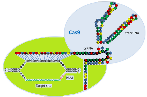
Figure 1:  Schematic model of the naturally occurring RNA-guided nuclease systems.


This model of the naturally occurring dual RNA-guided Cas9 nuclease illustrates the interaction of crRNA with the complementary strand of the DNA target site harboring an adjacent PAM sequence, shown as green and red text. TracrRNA base pairs with the crRNA, and the overall complex is recognized and cleaved by Cas9 nuclease shown in light blue color. Folding of the crRNA and tracrRNA molecules was predicted by the program Mfold and the association of the crRNA to the tracrRNA is partially based on the model proposed by Jinek et al. (2012).


Potential applications for the CRISPR systems

  • Cell engineering,
  • Drug Discovery,
  • CRISPR gRNA screening,
  • siRNA screens,
  • shRNA screens,
  • Cell-based assays, 
  • Cell-line identification assays, 
  • Stem cell screening assays,
  • CRISPR-based fine screening of the Human Genome, 
  • Epigenome editing, 
  • Libraries for next-generation sequencing (NGS), 
  • Determination of essential genes (House Keeping Genes), 
  • Development of better cell and animal models to test new drugs and therapies, 
  • New types of immuno-oncology treatments for cancer, Patient-derived animal models, 
  • Cancer and tumor screening and modeling, 
  • Transplantation models, 
  • Knockouts, 
  • Knockins, 
  • Tagging, Chromosomal rearagments, 
  • Transcriptional interference or activation, 
  • Genetic screening, 
  • And many more.

How do CRIPSRs work?


For CRISPR Cas9 to work, researchers can design a “guide RNA” (gRNA or crgRNA) to match the sequence of a specific target gene. The RNA guides the Cas9 enzyme to the desired target, where it then cuts the DNA. This cut inactivates the gene. Alternatively, CRISPR Cas9 allows replacing the sequence section adjacent to the cut with a different version of the gene. The CRISPR Cas9 system can be used in a variety of applications. Researchers now hope to use CRISPR Cas systems for the repair of defective genes that cause disease. To improve the system chemically modified RNA may be used in the usual guide RNA.

Every recently published paper covering advances made in gene editing methods based on CRISPRs suggest that this technology is a new game-changing technology. It appears that the CRISPRs are surpassing the zinc fingers and transcription activator-like effector nucleases (TALENS) as gene editing tools because of their ease-of-use and versatility. The use of the CRISPR technology offers the potential to pinpoint and repair genetic mutations, for example in the chimeric antigen receptor (CAR) T-cell field, by employing various gene editing strategies for the development of toolkits and therapies.

Presently, the CRISPR Cas system is considered to be the system of choice for gene editing. This gene-editing method now allows for a variety of promising applications useful as a genomic cut-and-paste method. Already the technique has been used to create “gene drives”, heralded as a method to eliminate pests or diseases they carry, and the first editing of the DNA of human embryos. This notion has prompted headlines and concerns and ethical considerations discussing possible human gene editing using CRISPRs.

The rapid pace of development in CRISPR-based gene editing methods has already started a debate if, how and when the technology should be used in human germline cells or embryos. On the positive site, this type of gene editing promises the elimination of devastating diseases. On the negative site, it is possible that tinkering with the genome crosses a moral boundary, specifically since all the mechanisms how the technique works are not well understood at this point in time. Also, the occurrence of possible off-target effects will need to be avoided for this gene editing method to work specifically.

The discovery of an unusual segment of neighboring DNA originally without any known biological function consisting of short repeating nucleotide sequences flanked by short unique segments let to a system, the bacterial CRISP Cas system, that now allows for easy manipulation of many genomes (Ishino et al. 1987). However, it took more than 20 years for scientists to figure out the significance of this system. Ultimately, the CRISPR Cas9 type II system that needs only one protein for it to function has become the system most used. The CRISPR Cas9 type II system uses a single endonuclease, Cas9. This enzyme together with guide RNA locates and cleaves invading DNA at specific sites containing conserved sequences called proto-spacer adjacent motifs (PAMs).

The CRISPR Cas systems allow detection of and protection against genetic elements. The type II system uses a single endonuclease, Cas9, that acts together with guide RNA to locate and cleave invading DNA at specific sites that are separated or distinguished from other sequence loci called proteo-spacer adjacent motifs (PAMs). The formation of the DNA targeting complex requires Cas9 and two distinct RNA transcripts, CRISPR RNA (crRNA) and trans-acting CRISPR RNA (tracrRNA).

Cleavage of specific DNA sequences

Researchers discovered that S. pyrogenes Cas9 nuclease, when used with a small CRISPR associated RNA (crRNA) and a separate transactivating RNA (tracerRNA), can be directed to cleave specific DNA sequences. (Jinek et al., 2012). The active complex contains a protein, Cas9 nuclease, and two RNA molecules, crRNA and tracrRNA.

Selective disruption of specific genes

Genes can be disrupted selectively as was shown by Sato et al., in 2015. The disruption of selected genes was achieved by combined injection of guideRNA and human Cas9 mRNAs. The research group reported the disruption of a gene (GGTA1) encoding the α-1,3-galactosyltransferase that synthesizes the α-Gal epitope using parthenogenetically activated porcine oocytes. They describe how it was done as follows: “After electric activation of in vitro-matured oocytes, these were cytoplasmically injected with a solution (~2 pL) containing in vitro synthesized hCas9 mRNA (2 ng/μL), gRNA (2 ng/μL; specific to GGTA1 exon 4), and EGFP mRNA (2 ng/μL) and were then cultured in vitro until blastocyst formation for approximately seven days.”


Repair of T cells

Schuman et al., also in 2015, reported that the delivery of Cas9 protein pre-assembled with guide RNAs enabled successful Cas9-mediated homology-directed repair in primary human T cells.

Targeted gene regulation

Qi et al., in 2013, showed that a catalytically dead Cas9 without endonuclease activity can be coexpressed with a guide RNA to generate a DNA recognition complex. This complex can specifically interfere with transcriptional elongation, RNA polymerase binding, or transcription factor binding. This system is called CRISPR interference (CRISPRi) and can efficiently repress expression of targeted genes in Escherichia coli. No off-target effects were detected. The CRISPRi system can be used to repress multiple target genes simultaneously in a reversible manner.

Mammalian cell engineering with Cas9 protein transfection

Liang et al, in 2015 described methods for the rapid synthesis of gRNA and the delivery of Cas9 protein/gRNA ribonucleoprotein complexes (Cas9 RNPs) into a variety of mammalian cells. Delivery into cells was achieved through liposome-mediated transfection or electroporation. The delivery of Cas9 and synthesis of guide RNA (gRNA) are steps that can limit the overall efficiency and ease of use of CRISPR Cas systems. The researchers reported nuclease-mediated indel rates of up to 94% in Jurkat T cells and 87% in induced pluripotent stem cells (iPSC) for a single target. An “indel” refers to the insertion or deletion of bases in DNA sequences. Key features of these methods are the turn-around time. The design to analysis can be done within 3 days using in vitro transcribed gRNA and cas9 protein or mRNA. Effective transfection of Cas9 mRNA or RNPs is achieved using Lipofectamine® 3000 or RNAiMax. Electroporation of cas9 RNPs is done in difficult cell lines (Jurkat, iPSC, CD34+). Cas9 RNPs enables multiple and simultaneous targeting of loci. These methods are reported to allow for high throughput set up and transfection in multi-well plates.


RNA library preparation


RNA library preparation protocols require small RNAs with 5’P and 3’OH groups. Therefore when using CRISPR Cas based systems for library constructions the right choice of protocol is crucial since certain subtypes of CRISPR Cas produce crRNAs with different chemical groups at their 5’ and 3’ ends. Pre-crRNA cleavage products generated by Cas6 endoribonucleases, such as Cas6c, Casgf (type I) and Cas6 (type III) contain 5’ hydroxyl and 2-3’ cyclic phosphate ends. These crRNA pools are not accessible for 5’-linker ligation and cannot be extended by poly(A) polymerase. A treatment with poly nucleotide kinase (PNK) is needed prior to the cDNA preparation step for capture in RNA-sequencing libraries.


Example of a cell engineering workflow

(Liang et al., 2015):


Day 1

CRISPR Targets:

Preparation involves the design of CRISPR targets, preparation or ordering of primers, and cell seeding.

Day 2

gRNA:

Make gRNA. Assemble gRNA template. Perform IVT reaction and purify gRNA. A synthetic gRNA free of truncation and interfering byproducts can be used.

Delivery:

Deliver gRNA and Cas9 to cells. Incubate cells with gRNA-Cas9 protein complex with lipid based delivery or electroporation for 24 to 48 hours.

Day 3 to 4

Analyze results:

Analyze editing efficiency by PAGE, PCR and/or sequencing.


Parts of the CRISPR Cas system - Abbreviations

Below is a list of tools or parts the CRISPR systems need to function. Most of them can now be generated synthetically or via cloning of the various nucleases.

CRISPR Cas Tools

or Parts

Short Description

Synthetic

Cas

CRISPR-associated genes are located in the vicinity of CRISPR array and are necessary for the silencing of invading nucleic acid.


Cas9 nuclease

Cas9 (CRISPR associated protein 9). A RNA-guided DNA endonuclease enzyme associated with the CRISPR (Clustered Regularly Interspaced Short Palindromic Repeats) adaptive immunity system in Streptococcus pyogenes and other bacteria. The Cas9 protein in its active form can modify DNA utilizing crRNA as its guide. Many variants exist with differing functions such as single strand nicking, double strand break, or DNA binding due to Cas9's DNA site recognition function.

mRNA encoding different Cas nucelases. Can be injected into cells.

hCas9 mRNA.

Cas9t

Cas9–crRNA–tracrRNA ternary complex, which functions as an RNA guided DNA endonuclease and mediates site-specific DNA cleavage.


Clustered regularly interspaced short palindromic repeat (CRISPR)

An array of short conserved repeat sequences interspaced by unique DNA sequences of similar size called spacers. They often originate from phage or plasmid DNA. CRISPR array together with Cas genes form the CRISPR Cas system, which functions as an adaptive immune system in prokaryotes.


CRISPR RNA (crRNA)

Small RNA molecule generated by transcription and processing of the CRISPR array. crRNA is composed of a conserved repeat fragment(s) and a variable spacer sequence, which matches the complimentary sequence in the invading nucleic acid. Contains a target-specific protospacer of 20 to 40 bases and a repeat motif that is recognized and bound by the tracrRNA. Recognition and binding is usually occurring at a hairpin loop forming the active complex with Cas9.


gRNA or guide RNA

During gene editing, guideRNAs (gRNAs) can be synthesized to perform the function of the tracrRNA:crRNA complex. Recognizing gene sequences need to have a PAM sequence at the 3'-end.

gRNAs allow transportation of Cas9 to anywhere in the genome during gene editing. However, no editing can occur at any site other than the one at which Cas9 recognizes PAM.


Homologous repair (HR)

Error-free DNA repair pathway that seals the broken DNA molecule using a homologous sequence (template).


Indel

The term indel refers to the insertion or the deletion of bases in DNA sequences of an organism.

Wiki


Non-homologous end joining (NHEJ)

A pathway that repairs DNA double strand breaks (DSB) in the absence of a homologous template; usually leads to small insertions or deletions.


PAM or protospacer adjacent motif

A short conserved nucleotide stretch located in the vicinity of a protospacer in the target DNA and necessary for DNA cleavage by Cas9t. A DNA sequence immediately following the DNA sequence targeted by the Cas9 nuclease. PAM is part of the invading virus or plasmid DNA but not a component of the bacterial CRISPR locus. If the target DNA sequence is not followed by the PAM sequence Cas( will not be able to successfully bind to and cleave the target DNA.


Protospacer

A fragment in the target DNA, which matches a spacer sequence in the CRISPR array.


sgRNA

Single guide RNAs are a combined RNA consisting of a tracrRNA and at least one crRNA. RNA hairpin obtained by connecting crRNA and tracrRNA into a single molecule.

Synthetic sgRNA can be used for CRISPR experiments if they

tracrRNA or trans-activating crRNA

A small trans-encoded RNA. TracrRNA is a complementary RNA sequence to and base pairs with a pre-crRNA forming an RNA duplex. This duplex is cleaved by RNase III, an RNA-specific ribonuclease, forming a crRNA/tracrRNA hybrid. This hybrid acts as a guide for the endonuclease Cas9 that cleaves the invading nucleic acid.


Transcription activator-like effector nuclease (TALEN)

An artificial nuclease obtained by fusing Xanthomonas transcription activator-like effector (TALE) DNA binding domains to the nonspecific nuclease domain.


Trans-acting CRISPR RNA (tracrRNA)

Trans-encoded small RNA molecule that forms a duplex with a repeat fragment of crRNA.


Triple helix forming oligonucleotide (TFO)

An artificial oligodeoxynucleotide, which binds to the polypurine sequences of the double-stranded DNA forming DNA triple helix.


Zinc finger nuclease (ZFN)

An artificial nuclease created by fusing zinc finger motifs, which serve as DNA recognition modules, to a nonspecific DNA cleavage domain of the FokI restriction endonuclease.


Specific Primers and adaptors useful for CRISPR experiments


The CRISPR systems can be studied using RNA-sequencing techniques. Several studies have already used RNA-sequencing for crRNA analysis. Using this approach, primary and processed transcripts can be distinguished and transcriptional start sites can be annotated fro whole genomes. Also, CRISPR can be used for the typing of Myobacterium tuberculosis complex (MTC) and Salmonella enteric serotypes. During MTC spoligotyping a single couple of primers (DRa-DRb) allows amplification of a set of overlapping fragments that can be detected by hybridization by PCR. For the detection, one primer is biotinylated and a detectable reporter is added. Usually streptavidin-peroxydase using chemoluminescence detection or streptavidin-phytoerythrin using microbead-base laser detection is use for this approach. There is no end in sight for new types of CRISPR based applications. Below is a list of oligonucleotide sequences as examples for CRISPR based applications.


Primers or adaptors

Sequence

Use & Notes


cDNA Library Preparation

Heidrich et al. 2013

Repair template

DNA that guides the cellular repair process allowing insertion of a specific DNA sequence.

Insertion of DNA sequence.

RNA adaptor

5’-UUU CCC UAC ACG ACG CUC UUC CGA UCU-3’

5’ Illumina sequence adapter ligated to the 5’-phosphate of RNAs.

Oligo(dT)-adapter primer

5’-GTG ACT GGA GTT CAG ACG TGT GCT CTT CCG ATC TTT TTT TTT TTT TTT TTT TTT TTT TVN-3’

3’ Illumina sequencing adapter primer used for cDNA synthesis together with M-MLV reverse transcriptase.

Tru-Seq-Sense_Primer

5’-AAT GAT ACG GCG ACC ACC GAG ATC TAC ACT CTT TCC CTA CAC GAC GCT CTT CCG ATC T-3’

Adapter sequence flanking cDNA inserts.

Tru-Seq-Antisensce_Primer plus 6-mer barcode

5’-CAA GCA GAA GAC GGC ATA CGA GAT NNN NNN GTG ACT GGA GTT CAG ACG TGT GCT CTT CCG ATC (dT25)-3’

Adapter sequence flanking cDNA inserts.

CRISPR loci (CRISPR2 repeat sequence)

5-CGG TTT ATC CCC GCT GGC GCG GGG AAC AC-3’

29 bp in Salmonella


MTC-Spoligotyping


DRa

5’-GGT TTT GGG TCT GAC GAC-3’

MTC-Spoligotyping

DRb

5’-CCG AGA GGG GAC GGA AAC-3’

MTC-Spoligotyping

DRSTMA

5’-CCG CTG GCG CGG GGA ACA-3’

STM-CRISPOL


Priming


Priming Protospacer pg8_F

5’-ATGTTG TCT TTC GCT GCT GAG GGT GAC GAT CCC GC-3’

Plasmid generation.

Priming Protospacer pg8_R

5’-GCGGGATCG TCA CCC TCA GCA GCG AAA GAC AAC AR-3’

Plasmid generation.


Substitutions at the seed region (CIT) are highlighted in bold. And the functional ATG PAM sequences are underlined.


Oligonucleotide T4B_7-F

5’-TTT TTGGATCCG CGA CTT TAC CAG CGA STG-3’, BamHI restriction site

T4 phage insertion

Oligonucleotide T4B_7-R

5’-TT TTGAGCTCG GTA ATG CAG CTT CAG GAA AA-3’, ,SacI restriction site

T4 phage insertion


CRISPR I repeat spacer


g8-repeat

5’-CTG TCT TTC GCT GCT GAG GGT GAC GAT CCC GC-3’


CRISPR expansion check sequence


Promoter-g8 spacer Ec_LDR-F

5’-AAG GTT GGT GGG TTG TTT TTA TGG-3’


Promoter-g8 spacer

M13_g8

5’-GGA TCG TCA CCC TCA GCA GCG-3’


Molecular marker data

SITVIT Database

Mycobacterium tuberculosis


http://www.pasteur-guadeloupe.fr:8081/SITVITDemo


 E. coli strains targeting phage

5’ -   Protospacer Sequences plus PAM  -3’


 through CRISPR

CCA TAC CAA ACG ACG AGC GTG ACA CCA CGA TGA AG

pT7blue nP vector

{CRISPR, Methods and Protocols. 2015 MMB 1311}

TAT ATA TGA GTA AAC TTG GTC TGA CAG TTA CCA AG

pT7blue P vector


TTG GCC GCA GTG TTA TCA CTC ATG GTT ATG GCA AG

pT7blue P vector


TCA TTC TGA GAA TAG TGT ATG CGG CGA CCG AGA AG

pT7blue P vector


TGC TCA TCA TTG GAA AAC GTT CTT CGG GGC GAA AG

pT7blue P vector


AAA GAA GAC  GTA TTC AAC CCG GAT ATG CGA ATA AG

T4 P


ACC CGA CTA GAT GGG GAT ATG AAG ATA ATC TCA AG

T4 P


GAA CCA CGA TAT ATT CAT TCG TGC ATC TAT TTA AG

T4 P


ATG CTA TTG AAC ACA TTC CGG TAT CAG GAA CAA AG

T4 P


CAA ATC CTT TCC TTT AAC CCC ACG AAT AAT TTA AG

T4 P


ATA ACA CTT GAA TCA TTC  ATC TAT TTT AAC CTT AG

T4 P


Note 1: Results from clones containing CRISPR cassettes expanded by a single repeat-spacer unit. . The sequence including the PAM and the source of the spacer is listed. (For example, T4 insert of the pT4acq plasmid). The location of the PAM is highlighted in red. CRISPR cassettes expanded by single repeat unit. CRISPR cassettes indicate dynamics of CRISPR-phage interactions in metagenomes.


Note 2:  HLA-DR: HLA-DR is an MHC class II cell surface receptor. This receptor class is encoded by the human leukocyte antigen complex on chromosome 6 region 6p21.31. The complex of HLA-DR (Human Leukocyte Antigen - antigen DRelated) with its ligand is a ligand for the T-cell receptor (TCR). The HLA-DR ligands are peptides of 9 amino acids in length or longer.


Note 3:   CRISPR cassettes are transcriped as long pre-crRNAs and are processed into short crRNAs by one of the Cas proteins. crRNAs contain variable central sequences corresponding to CRISPR spacer which are flanked by fragments of CRISPR repeat sequences. crRNAs are bound by Cascade. Cascade is a complex of several Cas proteins. The Cascade-crRNA complex recognizes dsDNA containing a protospacer matching the crRNA spacer. The presence of the PAM sequence increases the strength of the interactions in vitro. Artificial nucleic acdis, such as bridged nucleic acids (BNAs), can be inserted into the PAM region of synthetic crRNAs to increase the binding strength of the crRNA. When the target is recognized an R-loop containing an extended RNA-DNA heteroduplex is formed. The heteroduplex involves the entire length of the spacer-protospacer. Successful target recognition leads to target cleavage.


Reference


Brouns SJ (2012). "A swiss army knife of immunity.". Science 337 (6096): 808–9. doi:10.1126/science.1227253. PMID 22904002.


Deltcheva E, Chylinski K, Sharma CM, Gonzales K, Chao Y, Pirzada ZA, et al. (2011). "CRISPR RNA maturation by trans-encoded small RNA and host factor RNase III.". Nature 471 (7340): 602–7. doi:10.1038/nature09886. PMC 3070239. PMID 21455174.


Gogleva, A. A., Gelfand, M. S., & Artamonova, I. I. (2014). Comparative analysis of CRISPR cassettes from the human gut metagenomic contigs. BMC Genomics, 15(1), 202. http://doi.org/10.1186/1471-2164-15-202


Ishino Y, Shinagawa H, Makino K, Amemura M, Nakata A (December 1987). "Nucleotide sequence of the iap gene, responsible for alkaline phosphatase isozyme conversion in Escherichia coli, and identification of the gene product". Journal of Bacteriology169 (12): 5429–5433. PMC 213968. PMID 3316184.


Jinek M, Chylinski K, Fonfara I, Hauer M, Doudna JA, Charpentier E.; A programmable dual-RNA-guided DNA endonuclease in adaptive bacterial immunity. Science. 2012 Aug 17;337(6096):816-21. doi: 10.1126/science.1225829. Epub 2012 Jun 28.


Jinek M, Chylinski K, Fonfara I, Hauer M, Doudna JA, Charpentier E (2012). "A Programmable Dual-RNA-Guided DNA Endonuclease in Adaptive Bacterial Immunity.". Science 337 (6096): 816–21. doi:10.1126/science.1225829. PMID 22745249.

Kleinstiver, Benjamin P., Pattanayak, Vikram, Prew, Michelle S., Tsai, Shengdar Q., Nguyen, Nhu T., Zheng, Zongli, Keith Joung, J.; High-fidelity CRISPR–Cas9 nucleases with no detectable genome-wide off-target effects. Nature 2016/01/06/online, advance online publication. 1476-4687. http://dx.doi.org/10.1038/nature16526., 10.1038/nature16526.

Le Rhun A, Charpentier E (2012). "Small RNAs in streptococci.". RNA Biol 9 (4): 414–26. doi:10.4161/rna.20104. PMID 22546939.

Xiquan Liang,Jason Potter, Shantanu Kumar, Yanfei Zou, Rene Quintanilla, Mahalakshmi Sridharan, Jason Carte, Wen Chen, Natasha Roark, Sridhar Ranganathan, Namritha Ravinder, Jonathan D. Chesnut; Rapid and highly efficient mammalian cell engineering via Cas9 protein transfection. Journal of BiotechnologyVolume 208, 20 August 2015, Pages 44–53. http://www.sciencedirect.com/science/article/pii/S016816561500200X


Lundgen, Charpentier, Fineran (ed.); CRISPR, Methods and Protocols. 2015 Methods in Molecular Biology 1311. Springer Series. Humana Press.


Marraffini, L. A., & Sontheimer, E. J. (2010). CRISPR interference: RNA-directed adaptive immunity in bacteria and archaea. Nature Reviews. Genetics, 11(3), 181–190. http://doi.org/10.1038/nrg2749.


Masahiro Sato, Miyu Koriyama, Satoshi Watanabe, Masato Ohtsuka, Takayuki Sakurai, Emi Inada, Issei Saitoh, Shingo Nakamura and Kazuchika Miyoshi; Direct Injection of CRISPR/Cas9-Related mRNA into Cytoplasm of Parthenogenetically Activated Porcine Oocytes Causes Frequent Mosaicism for Indel Mutations. Int. J. Mol. Sci. 2015, 16, 17838-17856; doi:10.3390/ijms160817838


Pougach, K., Semenova, E., Bogdanova, E., Datsenko, K. A., Djordjevic, M., Wanner, B. L., & Severinov, K. (2010). Transcription, Processing, and Function of CRISPR Cassettes in Escherichia coli. Molecular Microbiology, 77(6), 1367–1379. http://doi.org/10.1111/j.1365-2958.2010.07265.x


Ekaterina Savitskaya, Ekaterina Semenova, Vladimir Dedkov, Anastasia Metlitskaya & Konstantin Severinov (2013) High-throughput analysis of type I-E CRISPR/Cas spacer acquisition in E. coli, RNA Biology, 10:5, 716-725, DOI: 10.4161/rna.24325. http://dx.doi.org/10.4161/rna.24325.


Kathrin Schumanna, Steven Linc, Eric Boyera, Dimitre R. Simeonova, Meena Subramaniame,f, Rachel E. Gatee,f, Genevieve E. Haliburtona,b, Chun J. Yee , Jeffrey A. Bluestonea , Jennifer A. Doudnac, and Alexander Marsona; Generation of knock-in primary human T cells using Cas9 ribonucleoproteins. PNAS 2015, 112, 33, 10437-10442. http://www.pnas.org/content/112/33/10437


Terns MP, Terns RM (2011). "CRISPR-based adaptive immune systems.". Curr Opin Microbiol 14 (3): 321–7. doi:10.1016/j.mib.2011.03.005. PMC 3119747. PMID 21531607.


UC San Diego Health https://health.ucsd.edu/news/releases/Pages/2015-11-16-RNA-Based-Drugs-Give-More-Control-Over-Gene-Editing.aspx
 

A sensitive diagnostic test for the detection of mutations in chronic lyphocytic leukemia

$
0
0
A sensitive diagnostic test for the detection of mutations in chronic lyphocytic leukemia


Recently a research group at the NIH in collaboration with NeoGenomics Lab in California developed a sensitive diagnostic method for the detection of multiclonal mutations in patients with chronic lymphocytic leukemia (CLL) that were treated with Bruton’s tyrosine kinase (BTK) inhibitors.

Patients with CLL that develop resistance to BTK inhibitors are typically positive for mutations in BTK or phospholipase c gamma 2 (PLCγ2). BTK mutations present at the C481S mutation hotspot are known to alter the active site of the mutant BTK. This mutant binds Ibrutinib in a reversible manner.

To better understand the development of resistance mechanisms in patients with CLL, Albitar and others developed a high sensitivity (HS) mutation assay using bridged nucleic acids (BNAs). The very sensitive assay was used in combination with Sanger sequencing and next generation sequencing (NGS) to test cellular DNA and cell free DNA (cf-DNA) from patients with CLL.

The results of this assay suggested that ibrutinib-naïve patients with CLL do not have BTK or PLCγ2 mutations. The researchers suggested that assaying cfDNA using this method may allow detection of mutations much earlier as compared with assaying cellular DNA. This may allow assaying for these mutations in patients with lymphoma that have only a few circulating lymphoma cells.

What is chronic lymphocytic leukemia?


Chronic lymphocytic leukemia
 (CLL) is a type of cancer that starts from cells develop into a type of 
white blood cells called lymphocytes in the bone marrow. The cancer (leukemia) cells originate in the bone marrow but then migrate into the blood. In CLL, leukemia cells often develop slowly over time. Many people don't have any symptoms for at least a few years. However, in time, the cancer cells can spread to other parts of the body, including the lymph nodes, liver, and spleen

Links to the publications and BTK inhibitors

https://ash.confex.com/ash/2015/webprogram/Paper83847.html

http://www.bloodjournal.org/content/126/23/716?sso-checked=true


BTK inhibitors

Ibrutinib -  https://en.wikipedia.org/wiki/Ibrutinib                                            

Acalabrutinib -  https://en.wikipedia.org/wiki/Acalabrutinib 

BTK inhibitor review -  
http://www.nature.com/nrc/journal/v14/n4/full/nrc3702.html


What is Carrier-Induced Epitope-Specific Suppression

$
0
0

Conjugated Antigens

The problem of Carrier-Induced Epitope-Specific Suppression (CIESS)


“Carrier-Induced Epitope-Specific Suppression is a hapten-specific immune suppression that is induced following carrier-priming. This phenomenon has remained unexplained. The phenomenon relies on the observation that the production of hapten-specific antibodies (Abs) is reduced in mice that have been primed previously against the carrier.”


Because small compounds like peptides and oligosaccharides are poorly immunogenic and cannot induce an immune response by themselves, they usually need to be covalently linked to a carrier, which usually are large proteins that are highly immunogenic. This protein carrier can bind to an antigen presenting cell (APC) and provide T-cell epitopes via MHC Class II for presentation to T-helper cells; this way increasing the immune response and inducing an anamnestic response. While proteins, like KLH, tetanus toxoid (TT) and non-toxic bacterial toxin mutants, are used as effective carriers to make haptens-like peptides immunogenic, there is the potential problem of Carrier-Induced Epitope-Specific Suppression or CIESS associated with their use. In this phenomenon, the anti-peptide antibody response is suppressed, as the carrier protein induces an antibody response that competes with the anti-peptide one; a problem that may be aggravated if the animals have been exposed to that carrier before. This situation may become problematic in the case of vaccines that use the same carrier protein, like tetanus toxoid TT, where previous immunization with TT or a conjugate like TT-Ay, will most likely result in suppression of the immune response to a conjugate like TT-Ax.  These results underline the problems concerning the use of carrier proteins in conjugate vaccines. Indeed, under those circumstances there will be a need to substitute the carrier by a new protein, to which the individual or animal has not been exposed before. 


A less known fact is that cross-linking agents having aromatic groups and/or maleimide, which is commonly used to link peptides to carriers via a thioether, can be very immunogenic; causing the immune response to be focused on the linker rather than the peptide, potentially contributing to aggravate CIESS. Indeed, as maleimide is highly immunogenic, if possible at all it should be avoided to prepare conjugates for immunization; replacing it with a linker where the reactive maleimide has been substituted by an iodoacetyl group; the resulting covalent bond would be still a thioether, but without the maleimide group. However, maleimide cross-linkers may be used for the preparation of the same peptide-conjugate but as a detector, like in an ELISA assay, to eliminate the false positive reactions due to the linker. However, the intricacies of the immune response to conjugated vaccines are demonstrated by the fact that maleimide conjugation enhances the antibody as well as T-cell responses of idiotype-KLH vaccines. However, in the case of Id-KLH vaccines, the conjugated antigen is a whole IgG with B and T-cell epitopes, rather than a small peptide that frequently is lacking T cell epitopes. While it is possible to lessen the carrier-induced suppression, another complication is that this suppression is strain-dependent, i.e. it varies according to the recipient’s genotype. This is a complex situation, as optimization of a conjugate for one animal strain may not work well in other strains, the wild type or a different species; which may result in a widespread distribution of antibody titers. While this phenomenon would not impact most research carry out in single animal strains, it would affect the results of studies using animals with diverse genotype, like in the case of human and veterinary vaccines. Hence, evaluation of conjugated vaccines may require careful studies to make sure that they will be equally effective in different animal populations over extended periods of time. 


To avert CIESS, a strategy effectively used with some peptide vaccines has been to replace the carrier protein with a well-defined T-cell epitope (Th epitope), linked to the peptide in question made only of B-cell epitopes. Yet, the success of this approach may be restricted to peptides having only B-cell epitopes, very unlikely with most peptides that will doubtless have both T and B-cell epitopes. Hence, while hapten-conjugates can be valuable research tools as well as vaccines, special attention should be paid to the conjugate design to avoid the potential complications described here.

Contact us for more info

Messenger RNA in Gene expression

$
0
0
 Messenger RNA in Gene expression


Gene expression refers to the conversion of genetic information from genes via messenger RNA to proteins. The entire set of genes in an organism is called the genotype. This set of genes includes alleles that when expressed determine the trait or phenotype of an organism. According to the central dogma of molecular biology this conversion occurs via transcription to generate mRNA followed by translation to produce the gene product, usually a protein. The result of the total sum of expressed genes in an organism manifests the phenotype of the individual organism. However, gene expression of genes that do not code for proteins only involves transcription. This is the case for ribosomal RNA (rRNA) and transfer RNA (tRNA), as well as other RNA molecules involved in regulatory pathways. Messenger RNA is the key molecule to enable gene expression for the production of proteins. A structural model of a mature messenger RNA is illustrated in figure 1. 


Figure 1:  Mature messenger RNA.


Messenger RNA (mRNA) is the molecule that links genes to proteins. Efficient and smooth interactions of the molecules of life allow us humans to function well. Major parts of a human body, as well as any mammalian body, visible to human eyes, are made up of proteins. However, for a human cell to function, more than proteins are needed. The coordinated interplay of nano-machines consisting of proteins, DNA, RNA, metal ions and other metabolic molecules are needed for a cell to function. On the molecular level, most of a human’s daily functions are executed by proteins. For the production of proteins in cells, messenger RNA is needed. According to the “central dogma of molecular biology” double-stranded DNA (dsDNA) located in the cell nucleus is transcript into RNA complementary to the DNA template it originates from and is further translated into protein sequences made up of amino acids to produce functional proteins. This process is called gene expression. As humans and their cells age body protein synthesis change with changes occurring in their metabolism. The rate of protein synthesis per kg total body weight is known to decline with growth and development within a species.


Figure 2:  The central dogma of molecular biology as stated by Francis Crick explains the flow of genetic information within a biological system. Simplistically it states: “DNA makes RNA and RNA makes protein.” However, it has become apparent in recent years that this simplistic view is only part of the story of gene expression.


Gene expression is known to occur via a two-stage process – transcription and translation. 


Transcription


Transcription generates a single-stranded RNA (ssRNA) identical to one sequence stretch in the strands of duplex DNA. However, different types of RNA are generated by transcription. Protein synthesis in cells involves three principal classes: messenger RNA (mRNA), transfer RNA (tRNA), and ribosomal RNA (rRNA).


Translation


Translation converts the nucleotide sequence of RNA into the sequence of amino acids needed for the synthesis of a protein. An mRNA is translated into a protein sequence. tRNA and rRNA are other needed parts for the protein synthesis apparatus. However, the entire length of the mRNA is not translated. Each mRNA contains, at least, one coding region that is related to a protein sequence of the genetic code. Each nucleotide triplet called codon of the coding region represents one amino acid. Only one strand of a DNA duplex is transcribed into a messenger RNA molecule. The DNA strand that directs the synthesis of mRNA is called the template or antisense strand. mRNA synthesis occurs via complementary base pairing. The term “antisense” is used to describe a sequence of DNA or RNA that is complementary to mRNA. The DNA strand that contains the same sequence as the mRNA is called the coding strand or sense strand. However, it possesses T instead of U.


The genetic code is read on the mRNA which is usually described in terms of the four bases present in RNA. These are U, C, A, and G. Messenger RNA is translated by ribosomes. The ribosome catalyzes the translation of mRNA into polypeptide chains. Historically ribosomes are described by their sedimentation constant S. This constant S (for Svedberg) indicates the rate of sedimentation for the ribosome investigated. A greater rate of sedimentation indicates a larger mass. The ribosome is a ribonucleoprotein particle consisting of two subunits that work together as part of the complete ribosome.


Advances made in next-generation DNA sequencing and proteomics now provide the ability to survey mRNA and protein abundances. These proteome-wide surveys now enable determination of the extent to which different aspects of gene expression help regulate cellular protein abundances. Therefore, it has now become clear that substantial regulatory processes occur after mRNA is made. Recent studies indicate that post-translational, translational and protein degradation based regulation events control the steady-state of protein abundances. The steady-state of protein abundance refers to the condition of a cell or biological system in which the abundance of proteins stays constant or changes only negligibly over time.

The biological concentrations of proteins in cells are regulated by interactions of transcription, translation, mRNA and protein degradation. In 2011 Schwanhausser et al. measured concentrations of mRNAs and proteins and the corresponding degradation rates for >5,000 genes in mouse cells. Using a mathematical model, the researchers described the dynamics that govern mammalian protein production (see figure 3 below). 


Figure 3:  Revised description of the central dogma of molecular biology as first described by Francis Crick.


Reference


Lewin, Benjamin; Genes VII, 2000, Oxford University Press ISBN 0-19-979276-X (Hbk). pp 119 – 190.


VERNON R. YOUNG, WILLIAM P. STEFFEE, PAUL B. PENCHARZ, JOERG C. WINTERER& NEVIN S.SCRIMSHAW; Total human body protein synthesis in relation to protein requirements at various ages. Nature 253, 192 - 194 (17 January 1975); doi:10.1038/253192a0.


Vogel, Christine, and Edward M. Marcotte. “Insights into the Regulation of Protein Abundance from Proteomic and Transcriptomic Analyses.” Nature reviews. Genetics 13.4 (2012): 227–232. PMC. Web. 28 Jan. 2016.

Schwanhausser, Bjorn, Busse, Dorothea, Li, Na, Dittmar, Gunnar, Schuchhardt, Johannes, Wolf, Jana, Chen, Wei, Selbach, Matthias; Global quantification of mammalian gene expression control. Nature 2011/05/19/print 473, 7347, 337-342.


Drug Conjugation Synthesis Strategies

$
0
0

Drug Conjugation Synthesis Strategies


One of the most effective ways to deliver cytotoxic drugs is by conjugation, where a relatively small molecular weight drug is covalently linked to usually a protein, which recognizes a specific cellular receptor that is expressed de novo or over-expressed by the targeted cells. In fact, some of the most useful proteins are the monoclonal antibodies (mAbs), which can be selected to recognize a single specific epitope on the cell surface. Yet, proteins like transferrin, hemoglobin, albumin and others are also being used to prepare these conjugates. Because of the recognition site on a protein, the essential pharmacophores on the drug responsible for its pharmacological activity, preparation of a conjugate needs to address preserving the binding capacity of the protein and most likely the release of the drug once it has entered the cell so that it can find its therapeutic target.

Hence, the design of a drug conjugate would require identifying functional groups in the protein that are far from its binding site, so binding of the drug does sterically hinders that site. If the protein is produced by recombinant DNA technology, the appropriate groups like thiol group can be introduced in the protein amino acid sequence; otherwise, there will be a need to introduce new functional groups by selective modification of certain groups in the protein. For instance, thiolation of amino groups using either N-acetyl homocysteine thiolactone (SATA) (Fig. 1) or S-acetylmercaptosuccinic anhydride (SAMSA) is a common approach modifying proteins.


Fig.1. Modification of amino groups with SATA


The success of the modifications would depend on their selectivity and if the reactive groups are far from the protein’s binding site. In some cases, if the ligand is a carbohydrate or other non-peptide compound, it would be possible to saturate the binding site before modification to protect the active site. Once the modification has taken place, the ligand can be removed from the binding site by extensive dialysis or similar procedures. An alternative method would be the reaction of the terminal amino group of a protein with an aldehyde carrying bifunctional linker. This method takes advantage of the lower pKa of the terminal α-amino group (pKa 7.6-8) than that of the lysine ε-amino groups (pKa ~ 9.4). Thus by selecting the pH of the reaction, the aldehyde group will form an imine with the terminal α-amino group, which may be reduced to a stable secondary amine with sodium cyanoborohydride. Evidently, this method would be useful only in proteins where the terminal amino group is not present at the binding site. While modifications of the carboxyl groups are possible, its selectivity would be less than the above-described methods and may lead to cross-linking with neighboring proteins; unless there is evidence that no carboxyl groups are present at the binding site. 

In the case of conjugates with mAbs, one of the most used methods is to reduce one of the two disulfide bonds linking together the two IgG heavy chains to form two thiol groups. Under these conditions, the formed thiol groups are present in the Fc region and away from the IgG recognition sites formed by the heavy and light chains. These two thiols can them react with a maleimide of an iodoacetyl group to form a stable thioether; using this approach it is possible to introduce one or two drug molecules per IgG molecule. A complete reduction of the two disulfide bonds may deliver two half-IgG molecules, which can be linked to a drug molecule. However, after reduction of both disulfide bonds, in some cases the two half IgGs would not separate, which may interfere with the conjugate formation. While the reduction of the disulfide bond is quite effective for IgGs, it is doubtful that it may be used with other proteins, a disulfide bond usually stabilizes the three-dimensional structure of a protein. 

Because the conjugated drug usually needs to interact with a cellular target after intracellular delivery, it would need to be released from the protein to which it is linked. To achieve this goal, the drug is covalently linked to the protein via a cleavable linker, i.e. a chemical structure that upon exposure to an enzyme, low pH, excess of reducing thiol groups, will split leaving the drug free and able to interact with its target. There are several types of cleavable linkers, and some of the most frequently used are those having a hydrazone group, which is cleaved at acid pH, < 5, in the endolysosome compartment, after uptake of the conjugate by endocytosis (Fig, 2). Another type of cleavable linker is the peptide pro-drug linker (self immolatory) having a Val-Cit-PAB-PNP sequence. This type of linker has two cleavage sites; site “a” specific for the enzyme cathepsin B cleavage, and site “b” that is the (1,6)-fragmentation site of the self-immolative link. To protect the drug, the self-immolatory spacer is located between the site of enzymatic cleavage and the drug; subsequent to the enzymatic cleavage, there is an extensive chemical rearrangement, a 1,6 elimination, that results in the drug release. Self-immolatory linkers are cleaved at the lysosome by proteases, after uptake of the conjugate by endocytosis. (Fig. 3). Although some cleavable linkers containing disulfide bonds have been described, they are not too stable and may be reduced in circulation by compounds like cysteine, glutathione, and other thiols.


Fig. 2. Cyclohexyl-aryl hydrazone cleavable linker


Fig. 3. Peptide pro-drug linker (self immolatory): Mal-Val-Cit-PAB-PNP


The other crucial component of these protein-conjugates, is the drug; which usually is linked by one of its functional groups that is not an essential for its pharmacological activity. Special care should be taken to avoid any significant structural changes that may alter the interactions of the drug with its target. Because the activity would be dependent on the amount of drug, it is important to determine the composition of the conjugate and to have a relatively homogeneous conjugate population. 

Reference


Miguel Castro and Dante Marciani; Ebr Bioconjugates January 2012, 44-50. www.samedanltd.com

Ducry, L., Stump, B.; Antibody-drug conjugates: linking cytotoxic payloads to monoclonal antibodies. Bioconjug Chem. 2010 Jan;21(1):5-13. doi: 10.1021/bc9002019.

Roger L. Lundblad; Chemical Reagents for Protein Modification, Fourth Edition Fourth Edition, 2014. CRC Press.

-.-


The role of mRNA in Gene Expression

$
0
0
The role of mRNA in Gene expression


Gene expression refers to the conversion of genetic information from genes via messenger RNA (mRNA) to proteins. The entire set of genes in an organism is called the genotype. This set of genes includes alleles that when expressed determine the trait or phenotype of an organism. According to the central dogma of molecular biology this conversion occurs via transcription to generate mRNA followed by translation to produce the gene product, usually a protein. The result of the total sum of expressed genes in an organism manifests the phenotype of the individual organism. However, gene expression of genes that do not code for proteins only involves transcription. This is the case for ribosomal RNA (rRNA) and transfer RNA (tRNA), as well as other RNA molecules involved in regulatory pathways. Messenger RNA is the key molecule to enable gene expression for the production of proteins. A structural model of a mature messenger RNA is illustrated in figure 1. 


Figure 1:  Mature messenger RNA.


Messenger RNA (mRNA) is the molecule that links genes to proteins. Efficient and smooth interactions of the molecules of life allow us humans to function well. Major parts of a human body, as well as any mammalian body, visible to human eyes, are made up of proteins. However, for a human cell to function, more than proteins are needed. The coordinated interplay of nano-machines consisting of proteins, DNA, RNA, metal ions and other metabolic molecules are needed for a cell to function. On the molecular level, most of a human’s daily functions are executed by proteins. For the production of proteins in cells, messenger RNA is needed. According to the “central dogma of molecular biology” double-stranded DNA (dsDNA) located in the cell nucleus is transcript into RNA complementary to the DNA template it originates from and is further translated into protein sequences made up of amino acids to produce functional proteins. This process is called gene expression. As humans and their cells age body protein synthesis change with changes occurring in their metabolism. The rate of protein synthesis per kg total body weight is known to decline with growth and development within a species.


Figure 2:  The central dogma of molecular biology as stated by Francis Crick explains the flow of genetic information within a biological system. Simplistically it states: “DNA makes RNA and RNA makes protein.” However, it has become apparent in recent years that this simplistic view is only part of the story of gene expression.


Gene expression is known to occur via a two-stage process – transcription and translation. 


Transcription


Transcription generates a single-stranded RNA (ssRNA) identical to one sequence stretch in the strands of duplex DNA. However, different types of RNA are generated by transcription. Protein synthesis in cells involves three principal classes: messenger RNA (mRNA), transfer RNA (tRNA), and ribosomal RNA (rRNA).


Translation


Translation converts the nucleotide sequence of RNA into the sequence of amino acids needed for the synthesis of a protein. An mRNA is translated into a protein sequence. tRNA and rRNA are other needed parts for the protein synthesis apparatus. However, the entire length of the mRNA is not translated. Each mRNA contains, at least, one coding region that is related to a protein sequence of the genetic code. Each nucleotide triplet called codon of the coding region represents one amino acid. Only one strand of a DNA duplex is transcribed into a messenger RNA molecule. The DNA strand that directs the synthesis of mRNA is called the template or antisense strand. mRNA synthesis occurs via complementary base pairing. The term “antisense” is used to describe a sequence of DNA or RNA that is complementary to mRNA. The DNA strand that contains the same sequence as the mRNA is called the coding strand or sense strand. However, it possesses T instead of U.


The genetic code is read on the mRNA which is usually described in terms of the four bases present in RNA. These are U, C, A, and G. Messenger RNA is translated by ribosomes. The ribosome catalyzes the translation of mRNA into polypeptide chains. Historically ribosomes are described by their sedimentation constant S. This constant S (for Svedberg) indicates the rate of sedimentation for the ribosome investigated. A greater rate of sedimentation indicates a larger mass. The ribosome is a ribonucleoprotein particle consisting of two subunits that work together as part of the complete ribosome.


Advances made in next-generation DNA sequencing and proteomics now provide the ability to survey mRNA and protein abundances. These proteome-wide surveys now enable determination of the extent to which different aspects of gene expression help regulate cellular protein abundances. Therefore, it has now become clear that substantial regulatory processes occur after mRNA is made. Recent studies indicate that post-translational, translational and protein degradation based regulation events control the steady-state of protein abundances. The steady-state of protein abundance refers to the condition of a cell or biological system in which the abundance of proteins stays constant or changes only negligibly over time.

The biological concentrations of proteins in cells are regulated by interactions of transcription, translation, mRNA and protein degradation. In 2011 Schwanhausser et al. measured concentrations of mRNAs and proteins and the corresponding degradation rates for >5,000 genes in mouse cells. Using a mathematical model, the researchers described the dynamics that govern mammalian protein production (see figure 3 below). 


Figure 3:  Revised description of the central dogma of molecular biology as first described by Francis Crick.


Reference


Lewin, Benjamin; Genes VII, 2000, Oxford University Press ISBN 0-19-979276-X (Hbk). pp 119 – 190.


VERNON R. YOUNG, WILLIAM P. STEFFEE, PAUL B. PENCHARZ, JOERG C. WINTERER& NEVIN S.SCRIMSHAW; Total human body protein synthesis in relation to protein requirements at various ages. Nature 253, 192 - 194 (17 January 1975); doi:10.1038/253192a0.


Vogel, Christine, and Edward M. Marcotte. “Insights into the Regulation of Protein Abundance from Proteomic and Transcriptomic Analyses.” Nature reviews. Genetics 13.4 (2012): 227–232. PMC. Web. 28 Jan. 2016.

Schwanhausser, Bjorn, Busse, Dorothea, Li, Na, Dittmar, Gunnar, Schuchhardt, Johannes, Wolf, Jana, Chen, Wei, Selbach, Matthias; Global quantification of mammalian gene expression control. Nature 2011/05/19/print 473, 7347, 337-342.

Vaccine adjuvants that induce an immune response

$
0
0

Inducing a deliberate immune response with vaccine adjuvants 


Despite all of the recent advances in innate immunity and immune modulators or vaccine adjuvants, the induction of an immune response remains by a long way an empirical issue. While many vaccine adjuvants have been elucidated structurally and mechanistically, the common practice is to divide these compounds in two groups, pro-inflammatory Th1 and anti-inflammatory Th2 vaccine adjuvants. Yet, Th1-only adjuvants do not exist, as they always induce concomitant Th1 and Th2 immunities, however, some vaccine adjuvants may induce either a stronger or weaker Th2 immunity. A review of the known Th1/Th2 adjuvants, shows that the induced immunoresponse is different from adjuvant to adjuvant and that therefore we cannot assume that the immune response induced by complete Freund’s adjuvant (CFA) is the same as that induced by saponins like QS-21 or Quil A. Indeed, immunization using CFA in some cases rather than protecting against a pathogen potentiates infection, while immunization with Quil A results in a protection; despite the fact that both are Th1/Th2 adjuvants.

Hence, while use of “generic” vaccine adjuvants, like CFA, to elicit antibody production for research purposes may be fine, that may not be the case when the goal is to develop a protective immune response against a pathogen or malignant cell, as in the case of vaccine development. Hence, in vaccine development, using CFA as the gold standard for immunity may lead to the wrong conclusions, i.e. an immune potentiating response against a pathogen, which may be protective when using a different vaccine adjuvant. Moreover, that different adjuvants that are ligands for toll-like-receptors (TLRs) of innate immunity show either synergistic or antagonistic effects, confirms the fact that these ligands are not all created equally, but that are quite different both structurally and functionally, a situation that may be used to modulate in a specific way immunity. Perhaps, it should be indicated that CFA’s adjuvanticity is due to a mixture of innate immunity ligands derived from Mycobacterium tuberculosis, suspended in an oil/water emulsion, yet as indicated above many of these ligands can be agonistic or antagonistic, which may explain some of the results obtained with CFA.

While there is a widespread assumption that the vaccine adjuvants’ mechanisms are still a mystery, the reality is very different. Since the discovery of innate immunity and saponin adjuvants the structure-function of these modulators has been well established. Indeed, we know their structures, receptors and in most instance the pharmacophores responsible for their immunomodulatory activity. Thus, in the case of vaccine development it is feasible to select the appropriate adjuvants to obtain a desired immune response. Moreover, we should realize that in inducing an immune response, the vaccine adjuvant is always in the driver’s seat particularly with protein antigens. Indeed, regardless of the antigen(s), adjuvants induce a systemic immunity that spreads through the body by means of cytokines and other immunological mediators; a result of the fact that an immunoresponse is largely caused by an infection, which seldom are localized in one part of the body. Yet, there have been attempts to use Th1/Th2 vaccine adjuvants in combination with peptide antigens lacking T-cell epitopes, with the expectation that the response would be an anti-inflammatory one; an assumption that ignores the adjuvant’s capacity to induce a systemic immunity that will take place despite the fact that the co-administered antigen lacks T-cell epitopes. Due to evolutionary reasons, adjuvants mainly induce an inflammatory Th1/Th2 immunity, which is the right response to identify and destroy foreign pathogens as well as intra-cellularly infected and malignant cells.

However, in some cases like in proteinopathies, e.g. Alzheimer’s disease, multiple sclerosis (MS) and others, a Th1/Th2 inflammatory response is damaging, destroying organs in an irreversible way. In such a case, the desired immunity would be an anti-inflammatory one Th2-only. But, in contrast to vaccine adjuvants inducing Th1/Th2 immunity, which are common, those that induce Th2-only are quite rare; indeed the only one being used now is alum. But alum is a poor vaccine adjuvant in the very young and the elderly, as shown by the poor efficacy of the flu vaccine in the older population, i.e. while the efficacy is near 90 percent in the population around 20 years old, it decreases to 40 percent or lower in those older than 65. The reasons for the poor availability of Th2 adjuvants are evolutionary; while an inflammatory immunity designed to kill foreign invaders was developed quite early by multi-cellular organisms, the anti-inflammatory immunity was developed hundreds of millions of years after the inflammatory immunity. Indeed, these anti-inflammatory Th2 modulators appeared when parasites, besides unicellular become multi-cellular or metazoan parasites like helminths; i.e. destruction of these parasites will cause damage to both the parasite and the host. Hence, the milder humoral Th2 immunity appeared to help to heal the damage caused by the parasites, this way establishing an immunity that minimized damage to the host and the parasite.

Evolutionarily, the metazoan parasites developed compounds that mimic those produced by mammals to prevent rejection of the fetus, indeed agents that inhibited the inflammatory response while inducing an anti-inflammatory response. Their recent development, less than 400 million years would explain why these anti-inflammatory agents are not so common as compared to those for innate immunity. In fact, those compounds are based on 2 pharmacophores, phosphorylcholine (PC) and fucose. The PC is responsible for the immune modulatory effects of sphingosine-1-phosphate (S1P), which is produced by phosphorylation of sphingolipids by two sphingoside kinases and it is essential for immune-cell trafficking; i.e. S1P seems to favor Th2 and Th17, while dampening Th1 immunoresponses. In contrast to PC, fucosylated glycans act at the antigen presenting cells (APCs) level, i.e. dendritic cells (DCs) and macrophages, via the cell lectin DC-SIGN, which is also a receptor for HIV, Ebola and other viruses. In fact, DC-SIGN can accept to kinds of different ligands, those carrying either mannose or fucose, yet, the induced immune response is different with each ligand. Upon binding to the lectin, mannose ligands bias the immune response toward a pro-inflammatory one, while fucose ligands bias it toward an anti-inflammatory response. Hence, induction of Th2 immunity will require alum, a poor adjuvant, or vaccine adjuvants with either PC or fucose.

From the practical point of view, while CFA can be used to stimulate Th1 immunity with a useful antibody production for research purposes, such a response would not be satisfactory for vaccine purposes where the immune response needs to be more define. Indeed, preliminary vaccine studies with CFA may deliver misleading results that cannot be extended to other Th1 adjuvants. Thus, in vaccine development new vaccine adjuvants, e.g. TLR ligands should be considered (Table 1). An advantage of these vaccine adjuvants is that they can show a synergistic effect, which results in an immune response that it is much better than that the sum of the responses induced by the separate adjuvants. Yet, some of these adjuvants may also show antagonistic effects on the immune response. 

  Table 1.  Properties of some vaccine adjuvants

Adjuvant

Immunitya

TLRb

Alum

Th2

N/A

QS-21c

Th1/Th2

N/A

Quil Ac

Th1/Th2

N/A

Cholera toxin

Th1/Th2

N/A

MPLd

Th1/Th2

TLR-4

CpGe

Th1/Th2

TLR-9

Flagellin

Th1/Th2

TLR-5

dsRNAf

Th1/Th2

TLR-3


a   Defines the stimulated immunity.

bToll Like Receptor

c  These adjuvants belong to the Quillaja saponins group.

d  MPL: monophosphoryl lipid A

e   CpG DNA

f  double stranded RNA

N/A: Non applicable

Zika Virus Update February 2016

$
0
0

Zika Virus – an update


The Obama administration requested $1.8 billion in emergency funds to combat the Zika virus in early 2016 according to the journal CEN (CEN.ACS.ORG February 15, 2016). This money is indented to be used for the accelerated development of vaccines and diagnostics, to expand laboratory and testing capacity, and to boost mosquito control programs. Even though many people infected with the virus have no symptoms it is thought that the number of babies with unusually small heads and brain damage in brazil may be caused by the Zika virus.

The Zika virus (Zika) is now spreading in multiple countries and territories.


Health officials expect the virus to spread to nearly all countries in the Americas and expand warnings for pregnant women.


Expectant mothers are now adviced not to visit Barbados, Bolivia, Ecuador, Guadaloupe, St. Martin, Guyana, and Samoa until the risk for infection by the Zika virus is lower or over. 


Below is information that can be found at the CDC website [CDC = Centers for Disease Control and prevention].  

“Zika virus is spread to people primarily through the bite of an infected Aedes species mosquito (A. aegypti and A. albopictus). To date, there have been no reports of Zika being spread by mosquitoes in the continental United States. However, cases have been reported in travelers to the United States, as well as cases of sexual transmission. With the recent outbreaks in the Americas, the number of Zika cases among travelers visiting or returning to the United States will likely increase. CDC is not able to predict how much Zika virus would spread in the continental United States. Many areas in the United States have the type of mosquitoes that can become infected with and spread Zika virus. However, recent outbreaks in the continental United States of chikungunya and dengue, which are spread by the same type of mosquito, have been relatively small and limited to a small area.” 

Source: http://content.govdelivery.com/accounts/USCDC/bulletins/1373311

Sexual Transmission

Zika virus can be sexually transmitted by a man to his sex partners. Not having sex is the best way to prevent sexually transmitted Zika. If a person is sexually active, using condoms the right way every time they have sex can reduce the chance they can get Zika through sex. 
 

Prevention/Treatment

There is no specific medicine or vaccine for Zika virus.

How can people protect themselves against Zika?

The best way to prevent Zika is to prevent mosquito bites. Here’s how

  • Wear long-sleeved shirts and long pants.
  • Stay in places with air conditioning or that use window and door screens to keep mosquitoes outside.
  • Use Environmental Protection Agency (EPA)-registered insect repellents (bug spray). Always follow the instructions on the label and reapply every few hours.
  • Eliminate mosquito breeding sites, like containers with standing water.

Visit CDC’s website for more information about preventing mosquito bites.

Symptoms


The most common symptoms of Zika virus disease are

  • Fever

  • Rash

  • Joint pain

  • Conjunctivitis (red eyes)

Other symptoms include

  • Muscle pain

  • Headache


Most people infected with Zika virus won’t even know they have the disease. The sickness is usually mild with symptoms lasting for several days to a week. People usually don’t get sick enough to go to the hospital, and they very rarely die of Zika.


The following steps can reduce the symptoms of Zika:

  • Get plenty of rest.

  • Drink fluids to prevent dehydration.

  • Take medicine such as acetaminophen to reduce fever and pain. 

  • Do not take aspirin or other non-steroidal anti-inflammatory drugs. 

  • If you are taking medicine for another medical condition, talk to your healthcare provider before taking additional medication.


    To learn more, please visit CDC's Zika virus page.

Zika Virus Genome and Infections

$
0
0

Zika Virus Genome and Infections


The recent outbreak of the Zika virus in Brazil and its projected spread into other countries has put the spotlight on this virus. Since this is a newly emerged virus vaccines and fast and sensitive diagnostic tools are not yet available. However, this could change fast in the near future. Specific primers for development of diagnostic tools for the detection of the Zika virus will be needed. 

Zika virus is a member of the Flaviviridae family transmitted to humans by mosquitoes. It is related to other flaviviruses including dengue, West-Nile and Japanese encephalitis viruses. It produces a comparatively mild disease in humans.

See viral zone for more info:  
http://viralzone.expasy.org/all_by_species/43.html

Flaviviruses are small, enveloped animal viruses containing a single positive-strand genomic RNA. (Chambers et al. 1990; Flavivirus genome organization, expression, and replication.)

Since 2007 Zika virus has caused several outbreaks in the Pacific, and further spread in the Americas since 2015. These were the first documented transmissions outside of its traditional endemic areas in Africa and Asia. Zika virus is considered an emerging infectious disease with the potential to spread to new areas where the Aedes mosquito vector is present. There is no evidence of transmission Zika virus in Europe to date.


2016 Zika outbreak time line  http://www.healthmap.org/zika/#timeline

Mosquito Distripution in Europe 

Sources: http://ecdc.europa.eu/en/healthtopics/zika_virus_infection/Pages/index.aspx#sthash.YKl7UHeS.dpuf,

http://ecdc.europa.eu/en/healthtopics/zika_virus_infection/Pages/index.aspx

Journey of adaptation of the Plasmodium falciparum malaria parasite to New World anopheline mosquitoes plus distripution map.

http://www.scielo.br/scielo.php?script=sci_arttext&pid=S0074-02762014000500662


The genome of the Zika virus is available at PubMed:

Zika virus, complete genome
– 10,79a bp –
linear single-strand positive-strand RNA without DNA stage.


LOCUS       NC_012532              10794 bp    RNA     linear   VRL 08-FEB-2016

DEFINITION  Zika virus, complete genome.

ACCESSION   NC_012532

VERSION     NC_012532.1  GI:226377833

DBLINK      BioProject: PRJNA36615

KEYWORDS    RefSeq.

SOURCE      Zika virus

  ORGANISM  Zika virus

            Viruses; ssRNA viruses; ssRNA positive-strand viruses, no DNA

            stage; Flaviviridae; Flavivirus.

REFERENCE   1  (bases 1 to 10794)

  AUTHORS   Kuno,G. and Chang,G.-J.J.

  TITLE     Full-length sequencing and genomic characterization of Bagaza,

            Kedougou, and Zika viruses

  JOURNAL   Arch Virol. 152 (4), 687-696 (2007)

   PUBMED   17195954

REFERENCE   2  (bases 1 to 10794)

  AUTHORS   Kuno,G. and Chang,G.J.

  TITLE     Biological transmission of arboviruses: reexamination of and new

            insights into components, mechanisms, and unique traits as well as

            their evolutionary trends

  JOURNAL   Clin. Microbiol. Rev. 18 (4), 608-637 (2005)

   PUBMED   16223950

REFERENCE   3  (bases 1 to 10794)

  CONSRTM   NCBI Genome Project

  TITLE     Direct Submission

  JOURNAL   Submitted (06-APR-2009) National Center for Biotechnology

            Information, NIH, Bethesda, MD 20894, USA

REFERENCE   4  (bases 1 to 10794)

  AUTHORS   Kuno,G. and Chang,G.-J.J.

  TITLE     Direct Submission

  JOURNAL   Submitted (01-AUG-2006) Division of Vector-Borne Infect. Dis., CDC,

            P.O. Box 2087, Fort Collins, CO 80522-2087, USA

  REMARK    Sequence update by submitter

REFERENCE   5  (bases 1 to 10794)

  AUTHORS   Kuno,G., Chang,G.-J.J. and Tsuchiya,K.R.

  TITLE     Direct Submission

  JOURNAL   Submitted (21-MAY-2004) Arbovirus Diseases Branch, Division of

            Vector-Borne Infectious Diseases, Centers for Disease Control and

            Prevention, P.O. Box 2087, Fort Collins, CO 80522, USA

COMMENT     REVIEWED REFSEQ: This record has been curated by NCBI staff. The

            reference sequence was derived from AY632535.

            Mature peptides were annotated by RefSeq staff using the cleavage

            sites reported in Kuno and Chang, 2007 (PMID 17195954). Questions

            about the annotation of this sequence should be directed to

            info@ncbi.nlm.nih.gov.

            COMPLETENESS: full length.

FEATURES             Location/Qualifiers

     source          1..10794

                     /organism="Zika virus"

                     /mol_type="genomic RNA"

                     /strain="MR 766"

                     /host="sentinel monkey"

                     /db_xref="taxon:64320"

                     /country="Uganda"

                     /note="mosquito-borne flavivirus"

     5'UTR           1..106

     gene            107..10366

                     /gene="flavivirus polyprotein gene"

                     /locus_tag="ZIKV_gp1"

                     /db_xref="GeneID:7751225"

     CDS             107..10366

                     /gene="flavivirus polyprotein gene"

                     /locus_tag="ZIKV_gp1"

                     /codon_start=1

                     /product="flavivirus polyprotein"

                     /protein_id="YP_002790881.1"

                     /db_xref="GI:226377834"

                     /db_xref="GeneID:7751225"

                     /translation="MKNPKEEIRRIRIVNMLKRGVARVNPLGGLKRLPAGLLLGHGPI

                     RMVLAILAFLRFTAIKPSLGLINRWGSVGKKEAMEIIKKFKKDLAAMLRIINARKERK

                     RRGADTSIGIIGLLLTTAMAAEITRRGSAYYMYLDRSDAGKAISFATTLGVNKCHVQI

                     MDLGHMCDATMSYECPMLDEGVEPDDVDCWCNTTSTWVVYGTCHHKKGEARRSRRAVT

                     LPSHSTRKLQTRSQTWLESREYTKHLIKVENWIFRNPGFALVAVAIAWLLGSSTSQKV

                     IYLVMILLIAPAYSIRCIGVSNRDFVEGMSGGTWVDVVLEHGGCVTVMAQDKPTVDIE

                     LVTTTVSNMAEVRSYCYEASISDMASDSRCPTQGEAYLDKQSDTQYVCKRTLVDRGWG

                     NGCGLFGKGSLVTCAKFTCSKKMTGKSIQPENLEYRIMLSVHGSQHSGMIGYETDEDR

                     AKVEVTPNSPRAEATLGGFGSLGLDCEPRTGLDFSDLYYLTMNNKHWLVHKEWFHDIP

                     LPWHAGADTGTPHWNNKEALVEFKDAHAKRQTVVVLGSQEGAVHTALAGALEAEMDGA

                     KGRLFSGHLKCRLKMDKLRLKGVSYSLCTAAFTFTKVPAETLHGTVTVEVQYAGTDGP

                     CKIPVQMAVDMQTLTPVGRLITANPVITESTENSKMMLELDPPFGDSYIVIGVGDKKI

                     THHWHRSGSTIGKAFEATVRGAKRMAVLGDTAWDFGSVGGVFNSLGKGIHQIFGAAFK

                     SLFGGMSWFSQILIGTLLVWLGLNTKNGSISLTCLALGGVMIFLSTAVSADVGCSVDF

                     SKKETRCGTGVFIYNDVEAWRDRYKYHPDSPRRLAAAVKQAWEEGICGISSVSRMENI

                     MWKSVEGELNAILEENGVQLTVVVGSVKNPMWRGPQRLPVPVNELPHGWKAWGKSYFV

                     RAAKTNNSFVVDGDTLKECPLEHRAWNSFLVEDHGFGVFHTSVWLKVREDYSLECDPA

                     VIGTAVKGREAAHSDLGYWIESEKNDTWRLKRAHLIEMKTCEWPKSHTLWTDGVEESD

                     LIIPKSLAGPLSHHNTREGYRTQVKGPWHSEELEIRFEECPGTKVYVEETCGTRGPSL

                     RSTTASGRVIEEWCCRECTMPPLSFRAKDGCWYGMEIRPRKEPESNLVRSMVTAGSTD

                     HMDHFSLGVLVILLMVQEGLKKRMTTKIIMSTSMAVLVVMILGGFSMSDLAKLVILMG

                     ATFAEMNTGGDVAHLALVAAFKVRPALLVSFIFRANWTPRESMLLALASCLLQTAISA

                     LEGDLMVLINGFALAWLAIRAMAVPRTDNIALPILAALTPLARGTLLVAWRAGLATCG

                     GIMLLSLKGKGSVKKNLPFVMALGLTAVRVVDPINVVGLLLLTRSGKRSWPPSEVLTA

                     VGLICALAGGFAKADIEMAGPMAAVGLLIVSYVVSGKSVDMYIERAGDITWEKDAEVT

                     GNSPRLDVALDESGDFSLVEEDGPPMREIILKVVLMAICGMNPIAIPFAAGAWYVYVK

                     TGKRSGALWDVPAPKEVKKGETTDGVYRVMTRRLLGSTQVGVGVMQEGVFHTMWHVTK

                     GAALRSGEGRLDPYWGDVKQDLVSYCGPWKLDAAWDGLSEVQLLAVPPGERARNIQTL

                     PGIFKTKDGDIGAVALDYPAGTSGSPILDKCGRVIGLYGNGVVIKNGSYVSAITQGKR

                     EEETPVECFEPSMLKKKQLTVLDLHPGAGKTRRVLPEIVREAIKKRLRTVILAPTRVV

                     AAEMEEALRGLPVRYMTTAVNVTHSGTEIVDLMCHATFTSRLLQPIRVPNYNLNIMDE

                     AHFTDPSSIAARGYISTRVEMGEAAAIFMTATPPGTRDAFPDSNSPIMDTEVEVPERA

                     WSSGFDWVTDHSGKTVWFVPSVRNGNEIAACLTKAGKRVIQLSRKTFETEFQKTKNQE

                     WDFVITTDISEMGANFKADRVIDSRRCLKPVILDGERVILAGPMPVTHASAAQRRGRI

                     GRNPNKPGDEYMYGGGCAETDEGHAHWLEARMLLDNIYLQDGLIASLYRPEADKVAAI

                     EGEFKLRTEQRKTFVELMKRGDLPVWLAYQVASAGITYTDRRWCFDGTTNNTIMEDSV

                     PAEVWTKYGEKRVLKPRWMDARVCSDHAALKSFKEFAAGKRGAALGVMEALGTLPGHM

                     TERFQEAIDNLAVLMRAETGSRPYKAAAAQLPETLETIMLLGLLGTVSLGIFFVLMRN

                     KGIGKMGFGMVTLGASAWLMWLSEIEPARIACVLIVVFLLLVVLIPEPEKQRSPQDNQ

                     MAIIIMVAVGLLGLITANELGWLERTKNDIAHLMGRREEGATMGFSMDIDLRPASAWA

                     IYAALTTLITPAVQHAVTTSYNNYSLMAMATQAGVLFGMGKGMPFMHGDLGVPLLMMG

                     CYSQLTPLTLIVAIILLVAHYMYLIPGLQAAAARAAQKRTAAGIMKNPVVDGIVVTDI

                     DTMTIDPQVEKKMGQVLLIAVAISSAVLLRTAWGWGEAGALITAATSTLWEGSPNKYW

                     NSSTATSLCNIFRGSYLAGASLIYTVTRNAGLVKRRGGGTGETLGEKWKARLNQMSAL

                     EFYSYKKSGITEVCREEARRALKDGVATGGHAVSRGSAKIRWLEERGYLQPYGKVVDL

                     GCGRGGWSYYAATIRKVQEVRGYTKGGPGHEEPMLVQSYGWNIVRLKSGVDVFHMAAE

                     PCDTLLCDIGESSSSPEVEETRTLRVLSMVGDWLEKRPGAFCIKVLCPYTSTMMETME

                     RLQRRHGGGLVRVPLCRNSTHEMYWVSGAKSNIIKSVSTTSQLLLGRMDGPRRPVKYE

                     EDVNLGSGTRAVASCAEAPNMKIIGRRIERIRNEHAETWFLDENHPYRTWAYHGSYEA

                     PTQGSASSLVNGVVRLLSKPWDVVTGVTGIAMTDTTPYGQQRVFKEKVDTRVPDPQEG

                     TRQVMNIVSSWLWKELGKRKRPRVCTKEEFINKVRSNAALGAIFEEEKEWKTAVEAVN

                     DPRFWALVDREREHHLRGECHSCVYNMMGKREKKQGEFGKAKGSRAIWYMWLGARFLE

                     FEALGFLNEDHWMGRENSGGGVEGLGLQRLGYILEEMNRAPGGKMYADDTAGWDTRIS

                     KFDLENEALITNQMEEGHRTLALAVIKYTYQNKVVKVLRPAEGGKTVMDIISRQDQRG

                     SGQVVTYALNTFTNLVVQLIRNMEAEEVLEMQDLWLLRKPEKVTRWLQSNGWDRLKRM

                     AVSGDDCVVKPIDDRFAHALRFLNDMGKVRKDTQEWKPSTGWSNWEEVPFCSHHFNKL

                     YLKDGRSIVVPCRHQDELIGRARVSPGAGWSIRETACLAKSYAQMWQLLYFHRRDLRL

                     MANAICSAVPVDWVPTGRTTWSIHGKGEWMTTEDMLMVWNRVWIEENDHMEDKTPVTK

                     WTDIPYLGKREDLWCGSLIGHRPRTTWAENIKDTVNMVRRIIGDEEKYMDYLSTQVRY

                     LGEEGSTPGVL"

     mat_peptide     107..472

                     /gene="flavivirus polyprotein gene"

                     /locus_tag="ZIKV_gp1"

                     /product="anchored capsid protein C"

                     /protein_id="YP_009227206.1"

                     /db_xref="GI:985757037"

     mat_peptide     107..418

                     /gene="flavivirus polyprotein gene"

                     /locus_tag="ZIKV_gp1"

                     /product="capsid protein C"

                     /protein_id="YP_009227196.1"

                     /db_xref="GI:985757027"

     mat_peptide     473..976

                     /gene="flavivirus polyprotein gene"

                     /locus_tag="ZIKV_gp1"

                     /product="membrane glycoprotein precursor M"

                     /protein_id="YP_009227197.1"

                     /db_xref="GI:985757028"

     mat_peptide     473..751

                     /gene="flavivirus polyprotein gene"

                     /locus_tag="ZIKV_gp1"

                     /product="protein pr"

                     /protein_id="YP_009227207.1"

                     /db_xref="GI:985757038"

     mat_peptide     752..976

                     /gene="flavivirus polyprotein gene"

                     /locus_tag="ZIKV_gp1"

                     /product="membrane glycoprotein M"

                     /protein_id="YP_009227208.1"

                     /db_xref="GI:985757039"

     mat_peptide     977..2476

                     /gene="flavivirus polyprotein gene"

                     /locus_tag="ZIKV_gp1"

                     /product="envelope protein E"

                     /protein_id="YP_009227198.1"

                     /db_xref="GI:985757029"

     mat_peptide     2477..3532

                     /gene="flavivirus polyprotein gene"

                     /locus_tag="ZIKV_gp1"

                     /product="nonstructural protein NS1"

                     /protein_id="YP_009227199.1"

                     /db_xref="GI:985757030"

     mat_peptide     3533..4210

                     /gene="flavivirus polyprotein gene"

                     /locus_tag="ZIKV_gp1"

                     /product="nonstructural protein NS2A"

                     /protein_id="YP_009227200.1"

                     /db_xref="GI:985757031"

     mat_peptide     4211..4600

                     /gene="flavivirus polyprotein gene"

                     /locus_tag="ZIKV_gp1"

                     /product="nonstructural protein NS2B"

                     /protein_id="YP_009227201.1"

                     /db_xref="GI:985757032"

     mat_peptide     4601..6451

                     /gene="flavivirus polyprotein gene"

                     /locus_tag="ZIKV_gp1"

                     /product="nonstructural protein NS3"

                     /protein_id="YP_009227202.1"

                     /db_xref="GI:985757033"

     mat_peptide     6452..6832

                     /gene="flavivirus polyprotein gene"

                     /locus_tag="ZIKV_gp1"

                     /product="nonstructural protein NS4A"

                     /protein_id="YP_009227203.1"

                     /db_xref="GI:985757034"

     mat_peptide     6833..6901

                     /gene="flavivirus polyprotein gene"

                     /locus_tag="ZIKV_gp1"

                     /product="protein 2K"

                     /protein_id="YP_009227209.1"

                     /db_xref="GI:985757040"

     mat_peptide     6902..7654

                     /gene="flavivirus polyprotein gene"

                     /locus_tag="ZIKV_gp1"

                     /product="nonstructural protein NS4B"

                     /protein_id="YP_009227204.1"

                     /db_xref="GI:985757035"

     mat_peptide     7655..10363

                     /gene="flavivirus polyprotein gene"

                     /locus_tag="ZIKV_gp1"

                     /product="RNA-dependent RNA polymerase NS5"

                     /protein_id="YP_009227205.1"

                     /db_xref="GI:985757036"

     3'UTR           10367..10794

ORIGIN     

        1 agttgttgat ctgtgtgagt cagactgcga cagttcgagt ctgaagcgag agctaacaac

       61 agtatcaaca ggtttaattt ggatttggaa acgagagtttctggtcatga aaaaccccaa

      121 agaagaaatc cggaggatcc ggattgtcaa tatgctaaaa cgcggagtag cccgtgtaaa

      181 ccccttggga ggtttgaaga ggttgccagc cggacttctg ctgggtcatg gacccatcag

      241 aatggttttg gcgatactag cctttttgag atttacagca atcaagccat cactgggcct

      301 tatcaacaga tggggttccg tggggaaaaa agaggctatg gaaataataa agaagttcaa

      361 gaaagatctt gctgccatgt tgagaataat caatgctagg aaagagagga agagacgtgg

      421 cgcagacacc agcatcggaa tcattggcct cctgctgact acagccatgg cagcagagat

      481 cactagacgc gggagtgcat actacatgta cttggatagg agcgatgccg ggaaggccat

      541 ttcgtttgct accacattgg gagtgaacaa gtgccacgta cagatcatgg acctcgggca

      601 catgtgtgac gccaccatga gttatgagtg ccctatgctg gatgagggag tggaaccaga

      661 tgatgtcgat tgctggtgca acacgacatc aacttgggtt gtgtacggaa cctgtcatca

      721 caaaaaaggt gaggcacggc gatctagaag agccgtgacg ctcccttctc actctacaag

      781 gaagttgcaa acgcggtcgc agacctggtt agaatcaaga gaatacacga agcacttgat

      841 caaggttgaa aactggatat tcaggaaccc cgggtttgcg ctagtggccg ttgccattgc

      901 ctggcttttg ggaagctcga cgagccaaaa agtcatatac ttggtcatga tactgctgat

      961 tgccccggca tacagtatca ggtgcattgg agtcagcaat agagacttcg tggagggcat

     1021 gtcaggtggg acctgggttg atgttgtctt ggaacatgga ggctgcgtta ccgtgatggc

     1081 acaggacaag ccaacagtcg acatagagtt ggtcacgacg acggttagta acatggccga

     1141 ggtaagatcc tattgctacg aggcatcgat atcggacatg gcttcggaca gtcgttgccc

     1201 aacacaaggt gaagcctacc ttgacaagca atcagacact caatatgtct gcaaaagaac

     1261 attagtggac agaggttggg gaaacggttg tggacttttt ggcaaaggga gcttggtgac

     1321 atgtgccaag tttacgtgtt ctaagaagat gaccgggaag agcattcaac cggaaaatct

     1381 ggagtatcgg ataatgctat cagtgcatgg ctcccagcat agcgggatga ttggatatga

     1441 aactgacgaa gatagagcga aagtcgaggt tacgcctaat tcaccaagag cggaagcaac

     1501 cttgggaggc tttggaagct taggacttga ctgtgaacca aggacaggcc ttgacttttc

     1561 agatctgtat tacctgacca tgaacaataa gcattggttg gtgcacaaag agtggtttca

     1621 tgacatccca ttgccttggc atgctggggc agacaccgga actccacact ggaacaacaa

     1681 agaggcattg gtagaattca aggatgccca cgccaagagg caaaccgtcg tcgttctggg

     1741 gagccaggaa ggagccgttc acacggctct cgctggagct ctagaggctg agatggatgg

     1801 tgcaaaggga aggctgttct ctggccattt gaaatgccgc ctaaaaatgg acaagcttag

     1861 attgaagggc gtgtcatatt ccttgtgcac tgcggcattc acattcacca aggtcccagc

     1921 tgaaacactg catggaacag tcacagtgga ggtgcagtat gcagggacag atggaccctg

     1981 caagatccca gtccagatgg cggtggacat gcagaccctg accccagttg gaaggctgat

     2041 aaccgccaac cccgtgatta ctgaaagcac tgagaactca aagatgatgt tggagcttga

     2101 cccaccattt ggggattctt acattgtcat aggagttggg gacaagaaaa tcacccacca

     2161 ctggcatagg agtggtagca ccatcggaaa ggcatttgag gccactgtga gaggcgccaa

     2221 gagaatggca gtcctggggg atacagcctg ggacttcgga tcagtcgggg gtgtgttcaa

     2281 ctcactgggt aagggcattc accagatttt tggagcagcc ttcaaatcac tgtttggagg

     2341 aatgtcctgg ttctcacaga tcctcatagg cacgctgcta gtgtggttag gtttgaacac

     2401 aaagaatgga tctatctccc tcacatgctt ggccctgggg ggagtgatga tcttcctctc

     2461 cacggctgtt tctgctgacg tggggtgctc agtggacttc tcaaaaaagg aaacgagatg

     2521 tggcacgggg gtattcatct ataatgatgt tgaagcctgg agggaccggt acaagtacca

     2581 tcctgactcc ccccgcagat tggcagcagc agtcaagcag gcctgggaag aggggatctg

     2641 tgggatctca tccgtttcaa gaatggaaaa catcatgtgg aaatcagtag aaggggagct

     2701 caatgctatc ctagaggaga atggagttca actgacagtt gttgtgggat ctgtaaaaaa

     2761 ccccatgtgg agaggtccac aaagattgcc agtgcctgtg aatgagctgc cccatggctg

     2821 gaaagcctgg gggaaatcgt attttgttag ggcggcaaag accaacaaca gttttgttgt

     2881 cgacggtgac acactgaagg aatgtccgct tgagcacaga gcatggaata gttttcttgt

     2941 ggaggatcac gggtttggag tcttccacac cagtgtctgg cttaaggtca gagaagatta

     3001 ctcattagaa tgtgacccag ccgtcatagg aacagctgtt aagggaaggg aggccgcgca

     3061 cagtgatctg ggctattgga ttgaaagtga aaagaatgac acatggaggc tgaagagggc

     3121 ccacctgatt gagatgaaaa catgtgaatg gccaaagtct cacacattgt ggacagatgg

     3181 agtagaagaa agtgatctta tcatacccaa gtctttagct ggtccactca gccaccacaa

     3241 caccagagag ggttacagaa cccaagtgaa agggccatgg cacagtgaag agcttgaaat

     3301 ccggtttgag gaatgtccag gcaccaaggt ttacgtggag gagacatgcg gaactagagg

     3361 accatctctg agatcaacta ctgcaagtgg aagggtcatt gaggaatggt gctgtaggga

     3421 atgcacaatg cccccactat cgtttcgagc aaaagacggc tgctggtatg gaatggagat

     3481 aaggcccagg aaagaaccag agagcaactt agtgaggtca atggtgacag cggggtcaac

     3541 cgatcatatg gaccacttct ctcttggagt gcttgtgatt ctactcatgg tgcaggaggg

     3601 gttgaagaag agaatgacca caaagatcat catgagcaca tcaatggcag tgctggtagt

     3661 catgatcttg ggaggatttt caatgagtga cctggccaag cttgtgatcc tgatgggtgc

     3721 tactttcgca gaaatgaaca ctggaggaga tgtagctcac ttggcattgg tagcggcatt

     3781 taaagtcaga ccagccttgc tggtctcctt cattttcaga gccaattgga caccccgtga

     3841 gagcatgctg ctagccctgg cttcgtgtct tctgcaaact gcgatctctg ctcttgaagg

     3901 tgacttgatg gtcctcatta atggatttgc tttggcctgg ttggcaattc gagcaatggc

     3961 cgtgccacgc actgacaaca tcgctctacc aatcttggct gctctaacac cactagctcg

     4021 aggcacactg ctcgtggcat ggagagcggg cctggctact tgtggaggga tcatgctcct

     4081 ctccctgaaa gggaaaggta gtgtgaagaa gaacctgcca tttgtcatgg ccctgggatt

     4141 gacagctgtg agggtagtag accctattaa tgtggtagga ctactgttac tcacaaggag

     4201 tgggaagcgg agctggcccc ctagtgaagt tctcacagcc gttggcctga tatgtgcact

     4261 ggccggaggg tttgccaagg cagacattga gatggctgga cccatggctg cagtaggctt

     4321 gctaattgtc agctatgtgg tctcgggaaa gagtgtggac atgtacattg aaagagcagg

     4381 tgacatcaca tgggaaaagg acgcggaagt cactggaaac agtcctcggc ttgacgtggc

     4441 actggatgag agtggtgact tctccttggt agaggaagat ggtccaccca tgagagagat

     4501 catactcaag gtggtcctga tggccatctg tggcatgaac ccaatagcta taccttttgc

     4561 tgcaggagcg tggtatgtgt atgtgaagac tgggaaaagg agtggcgccc tctgggacgt

     4621 gcctgctccc aaagaagtga agaaaggaga gaccacagat ggagtgtaca gagtgatgac

     4681 tcgcagactg ctaggttcaa cacaggttgg agtgggagtc atgcaagagg gagtcttcca

     4741 caccatgtgg cacgttacaa aaggagccgc actgaggagc ggtgagggaa gacttgatcc

     4801 atactggggg gatgtcaagc aggacttggt gtcatactgt gggccttgga agttggatgc

     4861 agcttgggat ggactcagcg aggtacagct tttggccgta cctcccggag agagggccag

     4921 aaacattcag accctgcctg gaatattcaa gacaaaggac ggggacatcg gagcagttgc

     4981 tctggactac cctgcaggga cctcaggatc tccgatccta gacaaatgtg gaagagtgat

     5041 aggactctat ggcaatgggg ttgtgatcaa gaatggaagc tatgttagtg ctataaccca

     5101 gggaaagagg gaggaggaga ctccggttga atgtttcgaa ccctcgatgc tgaagaagaa

     5161 gcagctaact gtcttggatc tgcatccagg agccggaaaa accaggagag ttcttcctga

     5221 aatagtccgt gaagccataa aaaagagact ccggacagtg atcttggcac caactagggt

     5281 tgtcgctgct gagatggagg aggccttgag aggacttccg gtgcgttaca tgacaacagc

     5341 agtcaacgtc acccattctg ggacagaaat cgttgatttg atgtgccatg ccactttcac

     5401 ttcacgctta ctacaaccca tcagagtccc taattacaat ctcaacatca tggatgaagc

     5461 ccacttcaca gacccctcaa gtatagctgc aagaggatac atatcaacaa gggttgaaat

     5521 gggcgaggcg gctgccattt ttatgactgc cacaccacca ggaacccgtg atgcgtttcc

     5581 tgactctaac tcaccaatca tggacacaga agtggaagtc ccagagagag cctggagctc

     5641 aggctttgat tgggtgacag accattctgg gaaaacagtt tggttcgttc caagcgtgag

     5701 aaacggaaat gaaatcgcag cctgtctgac aaaggctgga aagcgggtca tacagctcag

     5761 caggaagact tttgagacag aatttcagaa aacaaaaaat caagagtggg actttgtcat

     5821 aacaactgac atctcagaga tgggcgccaa cttcaaggct gaccgggtca tagactctag

     5881 gagatgccta aaaccagtca tacttgatgg tgagagagtc atcttggctg ggcccatgcc

     5941 tgtcacgcat gctagtgctg ctcagaggag aggacgtata ggcaggaacc ctaacaaacc

     6001 tggagatgag tacatgtatg gaggtgggtg tgcagagact gatgaaggcc atgcacactg

     6061 gcttgaagca agaatgcttc ttgacaacat ctacctccag gatggcctca tagcctcgct

     6121 ctatcggcct gaggccgata aggtagccgc cattgaggga gagtttaagc tgaggacaga

     6181 gcaaaggaag accttcgtgg aactcatgaa gagaggagac cttcccgtct ggctagccta

     6241 tcaggttgca tctgccggaa taacttacac agacagaaga tggtgctttg atggcacaac

     6301 caacaacacc ataatggaag acagtgtacc agcagaggtt tggacaaagt atggagagaa

     6361 gagagtgctc aaaccgagat ggatggatgc tagggtctgt tcagaccatg cggccctgaa

     6421 gtcgttcaaa gaattcgccg ctggaaaaag aggagcggct ttgggagtaa tggaggccct

     6481 gggaacactg ccaggacaca tgacagagag gtttcaggaa gccattgaca acctcgccgt

     6541 gctcatgcga gcagagactg gaagcaggcc ttataaggca gcggcagccc aactgccgga

     6601 gaccctagag accattatgc tcttaggttt gctgggaaca gtttcactgg ggatcttctt

     6661 cgtcttgatg cggaataagg gcatcgggaa gatgggcttt ggaatggtaa cccttggggc

     6721 cagtgcatgg ctcatgtggc tttcggaaat tgaaccagcc agaattgcat gtgtcctcat

     6781 tgttgtgttt ttattactgg tggtgctcat acccgagcca gagaagcaaa gatctcccca

     6841 agataaccag atggcaatta tcatcatggt ggcagtgggc cttctaggtt tgataactgc

     6901 aaacgaactt ggatggctgg aaagaacaaa aaatgacata gctcatctaa tgggaaggag

     6961 agaagaagga gcaaccatgg gattctcaat ggacattgat ctgcggccag cctccgcctg

     7021 ggctatctat gccgcattga caactctcat caccccagct gtccaacatg cggtaaccac

     7081 ttcatacaac aactactcct taatggcgat ggccacacaa gctggagtgc tgtttggcat

     7141 gggcaaaggg atgccattta tgcatgggga ccttggagtc ccgctgctaa tgatgggttg

     7201 ctattcacaa ttaacacccc tgactctgat agtagctatc attctgcttg tggcgcacta

     7261 catgtacttg atcccaggcc tacaagcggc agcagcgcgt gctgcccaga aaaggacagc

     7321 agctggcatc atgaagaatc ccgttgtgga tggaatagtg gtaactgaca ttgacacaat

     7381 gacaatagac ccccaggtgg agaagaagat gggacaagtg ttactcatag cagtagccat

     7441 ctccagtgct gtgctgctgc ggaccgcctg gggatggggg gaggctggag ctctgatcac

     7501 agcagcgacc tccaccttgt gggaaggctc tccaaacaaa tactggaact cctctacagc

     7561 cacctcactg tgcaacatct tcagaggaag ctatctggca ggagcttccc ttatctatac

     7621 agtgacgaga aacgctggcc tggttaagag acgtggaggt gggacgggag agactctggg

     7681 agagaagtgg aaagctcgtc tgaatcagat gtcggccctg gagttctact cttataaaaa

     7741 gtcaggtatc actgaagtgt gtagagagga ggctcgccgt gccctcaagg atggagtggc

     7801 cacaggagga catgccgtat cccggggaag tgcaaagatc agatggttgg aggagagagg

     7861 atatctgcag ccctatggga aggttgttga cctcggatgt ggcagagggg gctggagcta

     7921 ttatgccgcc accatccgca aagtgcagga ggtgagagga tacacaaagg gaggtcccgg

     7981 tcatgaagaa cccatgctgg tgcaaagcta tgggtggaac atagttcgtc tcaagagtgg

     8041 agtggacgtc ttccacatgg cggctgagcc gtgtgacact ctgctgtgtg acataggtga

     8101 gtcatcatct agtcctgaag tggaagagac acgaacactc agagtgctct ctatggtggg

     8161 ggactggctt gaaaaaagac caggggcctt ctgtataaag gtgctgtgcc catacaccag

     8221 cactatgatg gaaaccatgg agcgactgca acgtaggcat gggggaggat tagtcagagt

     8281 gccattgtgt cgcaactcca cacatgagat gtactgggtc tctggggcaa agagcaacat

     8341 cataaaaagt gtgtccacca caagtcagct cctcctggga cgcatggatg gccccaggag

     8401 gccagtgaaa tatgaggagg atgtgaacct cggctcgggt acacgagctg tggcaagctg

     8461 tgctgaggct cctaacatga aaatcatcgg caggcgcatt gagagaatcc gcaatgaaca

     8521 tgcagaaaca tggtttcttg atgaaaacca cccatacagg acatgggcct accatgggag

     8581 ctacgaagcc cccacgcaag gatcagcgtc ttccctcgtg aacggggttg ttagactcct

     8641 gtcaaagcct tgggacgtgg tgactggagt tacaggaata gccatgactg acaccacacc

     8701 atacggccaa caaagagtct tcaaagaaaa agtggacacc agggtgccag atccccaaga

     8761 aggcactcgc caggtaatga acatagtctc ttcctggctg tggaaggagc tggggaaacg

     8821 caagcggcca cgcgtctgca ccaaagaaga gtttatcaac aaggtgcgca gcaatgcagc

     8881 actgggagca atatttgaag aggaaaaaga atggaagacg gctgtggaag ctgtgaatga

     8941 tccaaggttt tgggccctag tggataggga gagagaacac cacctgagag gagagtgtca

     9001 cagctgtgtg tacaacatga tgggaaaaag agaaaagaag caaggagagt tcgggaaagc

     9061 aaaaggtagc cgcgccatct ggtacatgtg gttgggagcc agattcttgg agtttgaagc

     9121 ccttggattc ttgaacgagg accattggat gggaagagaa aactcaggag gtggagtcga

     9181 agggttagga ttgcaaagac ttggatacat tctagaagaa atgaatcggg caccaggagg

     9241 aaagatgtac gcagatgaca ctgctggctg ggacacccgc attagtaagt ttgatctgga

     9301 gaatgaagct ctgattacca accaaatgga ggaagggcac agaactctgg cgttggccgt

     9361 gattaaatac acataccaaa acaaagtggt gaaggttctc agaccagctg aaggaggaaa

     9421 aacagttatg gacatcattt caagacaaga ccagagaggg agtggacaag ttgtcactta

     9481 tgctctcaac acattcacca acttggtggt gcagcttatc cggaacatgg aagctgagga

     9541 agtgttagag atgcaagact tatggttgtt gaggaagcca gagaaagtga ccagatggtt

     9601 gcagagcaat ggatgggata gactcaaacg aatggcggtc agtggagatg actgcgttgt

     9661 gaagccaatc gatgataggt ttgcacatgc cctcaggttc ttgaatgaca tgggaaaagt

     9721 taggaaagac acacaggagt ggaaaccctc gactggatgg agcaattggg aagaagtccc

     9781 gttctgctcc caccacttca acaagctgta cctcaaggat gggagatcca ttgtggtccc

     9841 ttgccgccac caagatgaac tgattggccg agctcgcgtc tcaccagggg caggatggag

     9901 catccgggag actgcctgtc ttgcaaaatc atatgcgcag atgtggcagc tcctttattt

     9961 ccacagaaga gaccttcgac tgatggctaa tgccatttgc tcggctgtgc cagttgactg

    10021 ggtaccaact gggagaacca cctggtcaat ccatggaaag ggagaatgga tgaccactga

    10081 ggacatgctc atggtgtgga atagagtgtg gattgaggag aacgaccata tggaggacaa

    10141 gactcctgta acaaaatgga cagacattcc ctatctagga aaaagggagg acttatggtg

    10201 tggatccctt atagggcaca gaccccgcac cacttgggct gaaaacatca aagacacagt

    10261 caacatggtg cgcaggatca taggtgatga agaaaagtac atggactatc tatccaccca

    10321 agtccgctac ttgggtgagg aagggtccac acccggagtg ttgtaagcac caattttagt

    10381 gttgtcaggc ctgctagtca gccacagttt ggggaaagct gtgcagcctg taaccccccc

    10441 aggagaagct gggaaaccaa gctcatagtc aggccgagaa cgccatggca cggaagaagc

    10501 catgctgcct gtgagcccct cagaggacac tgagtcaaaa aaccccacgc gcttggaagc

    10561 gcaggatggg aaaagaaggt ggcgaccttc cccacccttc aatctggggc ctgaactgga

    10621 gactagctgt gaatctccag cagagggact agtggttaga ggagaccccc cggaaaacgc

    10681 aaaacagcat attgacgtgg gaaagaccag agactccatg agtttccacc acgctggccg

    10741 ccaggcacag atcgccgaac ttcggcggcc ggtgtgggga aatccatggt ttct


//

-.-


Primers and Probes for Zika Virus Detection

$
0
0

Primers and Probes for Zika Virus Detect


Current molecular assays for flaviviruses use specific primers to amplify viral RNA for their detection. If these primers are to specific they may amplify RNA only form one species, or a range of closely related species. To enable a differential diagnostic, a broad range PCR assays may need to be developed to detect all flaviviruses. Historically, several diagnostic protocols using different primer sets have been developed and tested.

To enable detection of newly emerging flaviviruses a two stage process will be needed, as was recommended by Kuno in 1998.

  • 1:  Initially broad range group-reactive primers are used to narrow the range of targets. 

  • 2:  Species-specific primers are used next to determine which flavivirus is actually present.


However, if a totaly new speciies has emerged RNA sequencing of the genome will be needed for its characterization.

Zika virus (ZIKV) is an arbovirus transmitted by mosquitoes. Arboviruses are viruses that belong to any of several groups of RNA-containing viruses that are transmitted by bloodsucking arthropods, such as ticks, fleas, or mosquitoes. These viruses can cause encephalitis, yellow fever, or dengue fever, or similar feverish symptoms. Zika virus is an emerging mosquito-borne flavivirus first circulating in Asia and Africa but now also in the new world. When humans are infected an influenza-like syndrome is induced that is associated with retro-orbital pain, oedema, lymphadenopathy, or diarrhea. Flaviviruses include several pathogenic agents that can cause severe illness in humans. Some of them are known to expand their geographical range.

T
he Flaviviridae family consists of more than 70 virus species. It includes many arthropodae (insect, spider, crustacean)-borne viruses including the highly pathogenic yellow fever virus (YFV), West Nile virus (WNV), Japanese encephalitis virus (JEV), tick-borne encephalitis virus (TBEV) and dengue virus (DENV). 

Flaviviruses are grouped into three epidemiologically distinct groups:

(1) The mosquito-borne group,
(2) The tick-borne group,
(3) and the unknown vector viruses.

These viruses are enveloped positive-stranded RNA viruses with a genome of approximately 11 kB. The viral genome encodes a single large polyprotein from which three structural proteins and seven non-structural proteins are produced. Structural proteins are Capsid (C), Envelope (E), and Membrane (M) protein. Non-structural proteins are NS1, NS2A, NS2B, NS3, NS4A, NS4B, and NS5 proteins.

Clinical diagnosis of flavivirus infections can be difficult due to unspecific symptoms. Typically symptoms can vary from mild to severe as well as to viral hemorrhagic fever. Many of these viruses are transmitted through a common vector. Diagnosis of Zika fever requires virus isolation and serology. However, this type of diagnostic is time-consuming or cross-reactive. During the first week of the infection, viral RNA can often be identified in serum. Therefore, RT-PCR is the preferred test for the Zika virus. Because the presence of a virus in the blood stream decreases over time, a negative RT-PCR collected 5-7 days after symptom onset does not exclude a Zika virus infection. Therefore serologic testing should also be performed as a follow-up test.


Design of primers and probes


For the design of primers and probes full-length genomic sequences for flaviviruses can be retrieved from NCBI or similar databases. Various bioinformatic tools are available for sorting and alignment of sequences. The goal is to identify conserved sequence regions to allow for the detection of the virus families in a first pass analysis. Results from sequence alignments of different published virus strain sequences can be used to identify highly conserved regions as well as highly divergent regions identified in flavivirus sequences enabling the design of unique primer and probe sets or pairs to allow for detection of unique virus strains.

Patel et al. (2013) developed a one-step quantitative reverse transcription PCR for the rapid detection of flaviviruses. The research group designed a rapid, sensitive TagMan probe-based quantitative RT-PCR (qRT-PCR) assay for the simultaneous detection of several flaviviruses. For the design of the Pan-Flavi primers, the conserved NS5 gene region was used. The primers and probes tested by Patel et al. are listed in table1:

Table 1: Oligonucleotide sequence of primers and probes used in Pan-Flavi qRT-PCR assay


Primer / Probe

Sequence

pmol

Orientationa

Positionb

Tm (°C)

Flavi all S

TACAACATgATggggAARAgAgARAA

10

S

8993–9019

54.7

DEN4 F

TACAACATgATgggRAAACgTgAGAA

10

S

8996–9019

60.18

Flavi all AS 1

gTCCCANCCDgCKgTRTC

10

AS

9236–9253

52.5


gTCCCATCCAgCKgTRTCATC

5

AS

9236–9256

57.1

Flavi all AS 2

gTgTCCCAgCCNgCKgTgTCATCWgC

10

AS

9232–9260

69.6

Flavi all probe 1

FAM-AARggHAgYMgNgCCA+TH+T+g+g+T-BBQ

5

S

9044–9065

69–83

Flavi all probe 2

FAM-Tg+gTWYATgTggYTNg+gRgC-BBQ

5

S

9062–9082

62–75

Flavi all probe 3 mix

FAM-Tg+gTWYATgT+ggYTNg+gRgC—BBQc

5

S

9062–9082

66-79


FAM-CCgTgCCATATggTATATgTggCTgggAgC-BBQd

0.5

S

9052–9081

74.7


FAM-TTTCTggAATTTgAAgCCCTgggTTT-BBQe

0.5

S

9086–9012

68

Flavi all S2

TACAACATgATgggMAAACgYgARAA

10

S

8996–9019

58.2

Flavi all AS4

gTgTCCCAGCCNgCKgTRTCRTC

10

S

9235–9260

64.1


Tm: temperature, Bridged-Nucleic Acid (LNA) bases are written as ‘ + _’, e.g., +A.

Degenerate bases: R = (A/G), W = (A/T), K = (T/G), Y = (C/T), N = (A/G/T/C).

aS: sense orientation, AS: antisense orientation.

bYFV strain; accession no: NC 002031.

cFlavi all probe, dFlavi probe YFV, eFlavi probe DEN4.

Final Pan-Flavi assay comprise of Flavi all S, Flavi all S2 & Flavi all AS4 primer and Flavi all probe 3 mix.


Patel et al. state in their paper: “At present, RT-PCR in nested or hemi-nested format is used most frequently. It requires sequencing for the identification of viruses and needs approximately one day for experimentation. In addition, this carries a high risk of contamination caused by open handling of PCR products, increasing the potential for false positives. In contrast, the Pan-Flavi assay presented here requires only some 50 minutes for specific and sensitive detection of several flaviviruses in one reaction. The detection sensitivity of YFV, DENV, TBEV, JEV and WNV, using the described Pan-Flavi assay, was close to or as good as the species-specific qRT-PCR assays. These differences may have resulted from deviant reactions conditions, enzyme kits and instruments, which were used as published or in-house established, and it is to mention, that WNV and JEV assays were performed in a two-step PCR which is known to be more sensitive than the one-step procedure.”

Maher-Sturgess et al. (2008) described a degenerate primer set for the amplification of flavivirus RNA to generate an 800 base pair (bp) cDNA product. The amplified region encoded part of the methyltransferase and most of the RNA-dependent-RNA-polymerase (NS5) coding sequence. One-step RT-PCR testing allowed the isolation of the RNA fragment of 60 different flavivirus strains and sequencing of cDNA from each virus isolated. Database searches were used to confirm the identity of the template RNA.

According to Maher-Sturgess et al. (2008) the Flav100F and Flav200R primers have the potential to detect emerging and related flaviviruses without prior serological evidence or additional primer design. This primer pair allow for the rapid detection at the genus level. In addition Maher-Sturgess et al. designed a set of universal primer that allowed the amplification of different flaviviruses.

Table 2: Universial Primers for the detection of Flaviviruses via PCR amplification.

Primer

Sequence

Notes

YF-F

   aat tcc act cat gaaatg tac

Yellow fever virus

Flav100F

5’-AAY TCI ACI CAI GAR ATG TAY-3’

Maher-Sturgess et al. (2008)

Flav200R

5’-CCI ARC CAC ATR WAC CA-3’


Forward primer

5’-AAR TAC ACA TAC CAR AAC AAA GTG GT-3’

Faye et al. 2013

Reverse primer

5’-TCC RCT CCC YCT YTG GTC TTG-3’

Faye et al. 2013

16 nt BNA/LNA-probe

FAM-CTY AGA CCA GCT GAA R-BBQ

Faye et al. 2013


E F1269-F

5’-GAG GCT GGG AAA TGG CTG-3’

Grubaugh et al., 2013

E F2225-R

5’-CCT CCA ACT GAT CCA AAG TCC CA-3’

Grubaugh et al., 2013

NS3 F5015-F

5’-GTG GTT GGN CTG TAT GGN AA-3’

Grubaugh et al., 2013

NS3 F5807-R

5’-CCC ATT TCT GAG ATG TCA GT-3’

Grubaugh et al., 2013

NS5 F8276d-F

5’-AAY TCN CAN CAN GAR ATG TAY-3’

Grubaugh et al., 2013

NS5 F9063d-R

5’-CCN ARC CAC ATR WAC CA-3’

Grubaugh et al., 2013


Unifor

5’-tgg ggn aay srn tgy ggn ytn tty gg-3’

Faye et al. 2008

Unirev

5’-CCN CCH RNN GAN CCR AAR TCC CA-3’

Faye et al. 2008

Mounifor2

5’-GGR DRM DTB KWA AYV TGY GCN AWR TT-3’

Faye et al. 2008

Mounirev2

5’-CCN ATN SWR CTH CCH KHY YTR WRC CA-3’

Faye et al. 2008

ZIKVENVF

5’-GCT GGD GCR GAC ACH GGR ACT-3’

Faye et al. 2008

ZIKVENVR

5’-RTC YAC YGC CAT YTG GRC TG-3’

Faye et al. 2008


ZIKV 835

5’-TTG GTC ATG ATA CTG CTG ATT GC-3’

Lanciotti et al. 2007

ZIKV 911c

5’-CCT TCC ACA AAG TCC CTA TTG C-3’

Lanciotti et al. 2007

ZIKV 860-FAM

5’-CGG CAT ACA GCA TCA GGT GCA TAG GAG-3’

Lanciotti et al. 2007

ZIKV 1086

5’-CCGCTG CCC AAC ACA AG-3’

Lanciotti et al. 2007

ZIKV 1162c

5’-CCA CTA ACG TTC TTT TGC AGA CAT-3’

Lanciotti et al. 2007

ZIKV 1107-FAM

5’-AGC CTA CCT TGA CAA GCA GTC AGA CAC TCA A-3’

Lanciotti et al. 2007


3PNC-2R

5′-GCT CAG GGA GAA CAA GAA CCG-3′

Grard et al. 2007

Priming on viral RNA.


Reverse oligonucleotide located in the 3′ non-coding region.

NS3 region


LIV coding sequence

X1

5′-YIR TIG GIY TIT AYG GIW WYG G-3′

4913–4935

X2

5′-RTT IGC ICC CAT YTC ISH DAT RTC IG-3′

5707–5733

TB-5′UTR-S F

5′-AAA AGA CAG CTT AGG AGA ACA AGA-3′

NS5 gene

TB-3′UTR-R

5′-AGA ACA AGA ACC GCC CCC CC-3′


FLAVI-1 S

5’-AAT GTA CGC TGA TGA CAC AGC TGG CTG GGA CAC-3’

Ayers et l. 2006

FLAVI-2 A

5’-TCC AGA CCT TCA GCA TGT CTT CTG TTG TCA TCC A-3’


Segments 9273-9305 and 10,102-10,136 of the West Nile virus NY 2000 (GenBank accession #AF404756).


FU1

5’-TAC AAC ATG ATGGGA AAG AGA GAG AA-3’

Kuno 1998

Cfd2

5’-GTG TCC CAG CCG GCG GTG TCA TCA GC-3’

Kuno 1998

MA

5’-CAT GAT GGG RAA RAG RGA RRA G-3’

Kuno 1998

 Legend: N = A+C+G+T, R = A+G, W = A+T, Y= C+T, H = A+C+T;  V = A+C+G, M = A+C, I = inosine.

Reference


M. Ayers, D. Adachi, G. Johnson, M. Andonova, M. Drebot, R. Tellier; A single tube RT-PCR assay for the detection of mosquito-borne flaviviruses. Journal of Virological Methods Volume 135, Issue 2, August 2006, Pages 235–239.

Candrian, U., Furrer, B., Höfelein, C., & Lüthy, J. (1991). Use of inosine-containing oligonucleotide primers for enzymatic amplification of different alleles of the gene coding for heat-stable toxin type I of enterotoxigenic Escherichia coli. Applied and Environmental Microbiology, 57(4), 955–961.


Chambers, T. J., Hahn, C. S., Galler, R., and Rice, Ch. M.; Flavivirus genome organization, expression and replication. Annu. Rev. Microbiol. 1990. 44:649-6488.


Oumar Faye, Ousmane Faye, Diawo Diallo, Mawlouth Diallo, Manfred Weidmann and Amadou Alpha Sall; Quantitative real-time PCR detection of Zika virus and evaluation with field-caught Mosquitoes. Virology Journal 2013, 10:311, 1-8.


Grard G
, Moureau G, Charrel RN, Lemasson JJ, Gonzalez JP, Gallian P, Gritsun TS, Holmes EC, Gould EA, de Lamballerie X.; Genetic characterization of tick-borne flaviviruses: new insights into evolution, pathogenetic determinants and taxonomy. Virology. 2007 Apr 25;361(1):80-92. Epub 2006 Dec 13.

Grubaugh, N. D., McMenamy, S. S., turell, M. J., and Lee, J. S.; Multigene dedection and identification of mosquito-borne RNA viruses using an oligonucleotide microarray. PLOS Negl Trop Dis 7(8): e2349. Doi: 10.1371/journal.pntd.0002349. 1-16.

Goro Kuno; Universial diagnostic RT-PCR protocol for arboviruses. Journal of Virological Methods 72 (1998) 27-41.


Lanciotti, R. S., Kosoy, O. L., Laven, J. J., Velez, J. O., Lambert, A. J., Johnson, A. J., Stanfield, S. M., Duffy, M. R., 2008. Genetic and serologic properties of Zika virus associated with an epidemic, Yap State, Micronesia, 2007. Emerging Infectious Diseases Vol. 14, No. 8, August 2008. www.cdc.gov/eid.


Sheryl L Maher-Sturgess, Naomi L Forrester, Paul J Wayper, Ernest A Gould, Roy A Hall, Ross T Barnard and Mark J Gibbs; Universal primers that amplify RNA from all three flavivirus subgroups. Virology Journal20085:16 DOI: 10.1186/1743-422X-5-16.
http://virologyj.biomedcentral.com/articles/10.1186/1743-422X-5-16

Pranav Patel, Olfert Landt, Marco Kaiser, Oumar Faye, Tanja Koppe, Ulrich Lass, Amadou A Sall and Matthias Niedrig; Development of one-step quantitative reverse transcription PCR for the rapid detection of flaviviruses. Virology Journal 2013, 10 :58. http://www.virologyj.com/content/10/1/58 METHODOLOGY Open Access

Segmental Labeling of RNA with Stabile Isotpes

$
0
0
Segmental Labeling of RNA with Stabile Isotpes


Duss et al. in 2010 developed a method using 13C and 15N labeled nucleoside triphosphates (NTPs) for the labeling of RNA.

RNA molecules are frequently labeled using radioisotopes or fluorophores. Unfortunately, both labeling methods are unsuitable for clinical studies. The reason for this is that both labeling methods have side effects such as the risk of radiation exposure or the different metabolic behavior of the fluorophore-conjugate in comparison to the unconjugated active compound studied. RNA is now widely recognized for its function in regulating gene expression. Many RNA molecules including riboswitches, miRNAs, and large non-coding RNAs are now thought to play roles in gene regulation. Only approximately 1.5% of the human genome encodes proteins while 60 to 70% of it is transcribed into RNA. However, only circa 2% of structures deposited in the Protein Data Bank account for RNA.


Nuclear magnetic resonance (NMR) (http://www.cis.rit.edu/htbooks/nmr/) is now considered to be the method of choice for solving RNA structures.  Unfortunately, large RNA structures are hard to analyze due to spectral overlap observed in NMR spectra of RNA. Segmental labeling of RNA is therefore considered to be needed for the study of RNAs by NMR in combination with measurements of residual dipolar couplings (RDC) and paramagnetic relaxation enhancement (PRE). Also, site-specific incorporation of isotopes can also be useful for solving phases in X-ray crystallography.


According to Duss et al. enzymatic ligation of shorter modified synthetic RNA segments with longer fragments produced by in vitro transcription is expected to become the method of choice for studying biological important RNAs. Segmental labeling of RNA with stable isotopes and the use of ligation methods to incorporate synthetic RNA pieces containing modified nucleotides into RNA promises to overcome present limitations in obtaining structural information on RNA. Several researchers are now tackling this problem.  


Duss et al. in 2010 reported the use of isotope-labeled nucleotide triphosphates (NTPs) for the generation of multiple segmentally labeled RNAs. This approach is thought to be useful for structural studies to expand our knowledge in structural biology of RNA. The research group presented a method that is considered to be a fast, efficient and sequence-independent method for segmental labeling of RNA. The method was tested using the 72 nt RsmZ RNA product that was isotopically segmentally labeled for structural investigations by NMR. The method is based on a combination of co-transcriptional ribozyme cleavage, sequence-specific RNase H cleavage, and cross-religation using either T4 RNA or T4 DNA ligase. PrrB/RsmZ RNA are part of a group of non-coding RNAs (ncRNAs) found in bacteria (https://en.wikipedia.org/wiki/PrrB/RsmZ_RNA_family). Research using Legionella pneumophila indicates that the ncRNAs RsmY and RsmZ together with the proteins LetA and CsrA are part of a regulatory cascade and appear to be regulated by RpoS sigma-factor.


Outline of the Method


This approach is based on the transcription of two full-length RNAs. These RNAs have identical sequence but one is unlabeled whereas the other is isotopically labeled. The transcribed RNAs are flanked at the 5’-end by a hammerhead (HH) ribozyme in cis and at the 3’-end by a minimal sequence required by the Neurospora Varkud satellits (VS) ribozyme for cleavage in trans.


Step 1:Co-transcriptional ribozyme cleavage. Transcribed RNAs are flanked at the 5’-end by a hammerhead (HH) ribozyme in cisand at the 3’-end by the minimal sequence required by the NeurosporaVarkud satellite (VS) ribozyme for cleavage in trans. The two ribozymes (in cisor intrans) cleave co-transcriptionally. The result is two homogenous termini, a 5’-hydroxyl and 2’/3’-cyclic phosphate for the full-length RNA, which are purified before the next step.

Step 2:Site-specific RNase H cleavage. After purification, the two transcribed RNAs are site-specifically cleaved by RNase H using a guide 2-O-methyl-RNA/ DNA splint. This reaction yields an acceptor fragment (5-fragment) with two hydroxyl termini and a donor fragment (3’-fragment) with a phosphate at its 5’-end and a cyclic 2’/3’-phosphate at its 3’-end.

Step 3:  Cross-religation between the labeled and unlabeled fragment. After separation of the two fragments from each cleavage reaction, T4 RNA or DNA ligase is used for re-ligation. This reaction results in two segmental isotope labeled RNA fragments. Either the 5’-fragment or the 3’-fragment is labeled with the isotopes.    

For the method to work well a fast and efficient denaturing anion-exchange HPLC purification step is needed followed by n-butanol extraction or dialysis to get rid of urea and salts.


Duss et al report that for a two-piece ligation, 5 to 7 days are required in total. A total of 2 to 3 days are needed for Step 1 (1 day transcription optimization, 1 to 2 days large-scale transcription and purification), 1.5 to 2 days for Step 2 (0. 5 to 1 day RNase H cleavage optimization, 1 day large-scale cleavage and purification) and 1.5 to 2 days for Step 3 (0.5 to 1 day ligation optimization, 1 day large-scale ligation and purification).


Isotope labeling


Labeled nucleoside triphosphate (NTPs) can be either purchased from a provider such as Cambridge Isotope Laboratories (CIL; http://www.isotope.com/) or prepared from 13C, 15N-labeled E. coli cultures. The labeled E. coli cultures are precipitated with sodium acetate and isopropanol, and hydrolyzed with S1 nuclease. The use of a boronate affinity gel column allows for the separation nucleoside monophosphates (NMPs). NMPs are converted to NTPs by an enzymatic phosphorylation step. Boronate affinity chromatographyis also used for the desalting of labeled NTPs.


Segmental Labeling of RNA with Stable Isotopes

$
0
0
Segmental Labeling of RNA with Stable Isotopes


Duss et al. in 2010 reported the development of a method using 13C and 15N labeled nucleoside triphosphates (NTPs) for the segmental labeling of RNA with stable isotopes. Recent advancements made in mass spectrometry, in bioinformatics as well as in the development of enrichment methods for stabil isotopes, the use of stabile isotope in biochemistry, biology, biotechnology, molecular biology, and medical research has now become common.

According to Duss et al. enzymatic ligation of shorter modified synthetic RNA segments with longer fragments produced by in vitro transcriptionis expected to become the method of choice for studying biological important RNAs. Segmental labeling of RNA with stable isotopes and the use of ligation methods to incorporate synthetic RNA pieces containing modified nucleotides into RNA promises to overcome present limitations in obtaining structural information on RNA. Several researchers are now tackling this problem. The structural model of the non-coding RNA RsmZ protein sponge was reported by Duss et al. using this approach to allow for the elucidation of the solution structure via a combination of nuclear magnetic resonance and electron paramagnetic resonance spectroscopy.   


Figure 1:
Non-coding RNA RsmZ acts as a protein sponge. The bacterial Csr/Rsm system is considered to be the most general global post-transcriptional regulatory system responsible for bacterial virulence. NcRNAs such as CsrB or RsmZ activate translation initiation by sequestering homodimeric CsrA-type proteins from the ribosome-binding site of a subset of messenger RNAs. The structural model of this complex was reported by Duss et al. in 2014.

Natural and synthetic
RNA molecules are frequently labeled using radioisotopes or fluorophores. Unfortunately, both labeling methods are unsuitable for clinical studies. The reason for this is that both labeling methods have side effects such as the risk of radiation exposure or the different metabolic behavior of the fluorophore-conjugate in comparison to the unconjugated active compound studied. RNA is now widely recognized for its function in regulating gene expression. Many RNA molecules including riboswitches, miRNAs, and large non-coding RNAs are now thought to play roles in gene regulation. Only approximately 1.5% of the human genome encodes proteins while 60 to 70% of it is transcribed into RNA. However, only circa 2% of structures deposited in the Protein Data Bank account for RNA.

Nuclear magnetic resonance (NMR) is now considered to be the method of choice for solving RNA structures.  Unfortunately, large RNA structures are hard to analyze due to spectral overlap observed in NMR spectra of RNA. Segmental labeling of RNA is therefore considered to be needed for the study of RNAs by NMR in combination with measurements of residual dipolar couplings (RDC) and paramagnetic relaxation enhancement (PRE). Also, site-specific incorporation of isotopes can also be useful for solving phases in X-ray crystallography.

The use of isotope-labeled nucleotide triphosphates (NTPs) for the generation of multiple segmentally labeled RNAs is very useful for structural studies to expand our knowledge in the structural biology of RNA. The research group presented a method that is considered to be a fast, efficient and sequence-independent method for segmental labeling of RNA. The method was tested using the 72 nt RsmZ RNA product that was isotopically segmentally labeled for structural investigations by NMR. The method is based on a combination of co-transcriptional ribozyme cleavage, sequence-specific RNase H cleavage, and cross-religation using either T4 RNA or T4 DNA ligase. PrrB/RsmZ RNA are part of a group of non-coding RNAs (ncRNAs) found in bacteria. Research using Legionella pneumophila indicates that the ncRNAs RsmY and RsmZ together with the proteins LetA and CsrA are part of a regulatory cascade and appear to be regulated by RpoS sigma-factor.

Outline of the Method


This approach is based on the transcription of two full-length RNAs. These RNAs have identical sequence but one is unlabeled whereas the other is isotopically labeled. The transcribed RNAs are flanked at the 5’-end by a hammerhead (HH) ribozyme in cis and at the 3’-end by a minimal sequence required by the Neurospora Varkud satellits (VS) ribozyme for cleavage in trans.


Step 1: Co-transcriptional ribozyme cleavage. Transcribed RNAs are flanked at the 5’-end by a hammerhead (HH) ribozyme in cis and at the 3’-end by the minimal sequence required by the Neurospora Varkud satellite (VS) ribozyme for cleavage in trans. The two ribozymes (in cis or in trans) cleave co-transcriptionally. The result is two homogenous termini, a 5’-hydroxyl and 2’/3’-cyclic phosphate for the full-length RNA, which are purified before the next step.


Step 2: Site-specific RNase H cleavage. After purification, the two transcribed RNAs are site-specifically cleaved by RNase H using a guide 2’-O-methyl-RNA/ DNA splint. This reaction yields an acceptor fragment (5’-fragment) with two hydroxyl termini and a donor fragment (3’-fragment) with a phosphate at its 5’-end and a cyclic 2’/3’-phosphate at its 3’-end.

Step 3:  Cross-religation between the labeled and unlabeled fragment. After separation of the two fragments from each cleavage reaction, T4 RNA or DNA ligase is used for re-ligation. This reaction results in two segmental isotope labeled RNA fragments. Either the 5’-fragment or the 3’-fragment is labeled with the isotopes.    


For the method to work well a fast and efficient denaturing anion-exchange HPLC purification step is needed followed by n-butanol extraction or dialysis to get rid of urea and salts.


Duss et al. estimated that for a two-piece ligation, 5 to 7 days are required in total. A total of 2 to 3 days are needed for Step 1 (1 day transcription optimization, 1 to 2 days large-scale transcription and purification), 1.5 to 2 days for Step 2 (0. 5 to 1 day RNase H cleavage optimization, 1 day large-scale cleavage and purification) and 1.5 to 2 days for Step 3 (0.5 to 1 day ligation optimization, 1 day large-scale ligation and purification).

Methods used for this approach 

  • Vector construction and plasmid purification,

  • RsmE protein expression and purification

  • RNA purification,

  • RNA transcription and co-translational ribozyme cleavage (IVT),

  • Segmental isotope labeling,

  • Sequence-specific RNase H cleavage,

  • RNA ligation with T4 RNA and DNA ligase, and

  • NMR spectrocopy,

  • EPR spectroscopy, 

  • DEER experiment (Double Electron-Electron Resonancealso known as PELDOR or Pulsed ELDOR),

  • Electrophoretic mobility shift assaya (EMSA),

  • Isothermal titration calorimetry (ITC) binding experiments.


Isotope labeling


Labeled nucleoside triphosphate (NTPs) can be either purchased from a provider such as Cambridge Isotope Laboratories (CIL) or prepared from 13C, 15N-labeled E. coli cultures. The labeled E. coli cultures are precipitated with sodium acetate and isopropanol, and hydrolyzed with S1 nuclease. The use of a boronate affinity gel column allows for the separation nucleoside monophosphates (NMPs). NMPs are converted to NTPs by an enzymatic phosphorylation step. Boronate affinity chromatographyis also used for the desalting of labeled NTPs.

Reference


Duss
, O., Michel, E., Yulikov, M., Schubert, M, Jeschke, G., Allain, F.; Structural basis of the non-coding RNA RsmZ acting as a protein sponge. Nature 2014, 509, 588-592.

Olivier Duss, Christophe Maris, Christine von Schroetter and Fre´de´ric H.-T. Allain; A fast, efficient and sequence-independent method for flexible multiple segmental isotope labeling of RNA using ribozyme and RNase H cleavage. Nucleic Acids Research, 2010, Vol. 38, No. 20 e188, doi:10.1093/nar/gkq756.


-.-

Gapmer Design

$
0
0
 Gapmer Design


A gapmer is a chimeric antisense oligonucleotide that contains a central block of deoxynucleotide monomers sufficiently long to induce RNase H cleavage.


General Design


The central block of a gapmer is flanked by blocks of 2’-O modified ribonucleotides or other artificially modified ribonucleotide monomers such as bridged nucleic acids (BNAs). In a gapmer these modified nucleic acids protect the internal block from nuclease degradation. Natural unmodified DNA, as well as modified DNA analogs such as phosphorothiote DNA analogs can be used to stabilize RNA molecules useful as gapmers for therapeutic approaches. 

Design Scheme

5’-wing

gap

3’-wing


2 to 3 BNAs

8 to 12 natural nucleic acids, or

phosphorothioate (PS) nucleic acids

2 to 3 BNAs


5’-NNnnnnnnnnnnNN-3’

14mer


d(CUTAGCACTGGCCU)-3’

2’,4’-BNANC[NMe]

                        Target: PTEN


Antisense Technology


Antisense technology is a universal approach for the inhibition of gene expression in a sequence-specific manner. Since the discovery that oligodeoxynucleotides can act as antisense agents that inhibit viral replication in cell culture antisense technology has been developed for target validation and therapeutic purposes. Theoretically, antisense approaches can be used to cure any disease caused by the expression of deleterious genes, including diseases caused by viral infections, cancer growth, and inflammatory diseases. Antisense technology allows inhibition of gene expression offering itself as a tool for the study of gene function (functional genomics) as well as for therapeutic approaches (antisense gene therapy). However, in practice antisense technology has proved to be challenging.

In general, if the RNA sequence is known, the antisense sequence is easy to work out. However, to achieve success, length, chemical modifications and target site need to be carefully selected.

Optimized antisense oligonuclotides (ASOs) such as gapmers support RNase H and directly cause the decay of RNA targets.

Identify molecular targets – it starts with a gene


A gene of interest maybe selected due to its known biological function in diseases related pathways or because little is known and functional genomics studies are planned.

Gapmers can be used to target primary gene transcripts, mRNA product(s), spliced and unspliced coding and noncoding RNAs. Alternative starts of transcription and alternative polyadenylation leading to alternative primary transcripts will need to be taken in account when designing ASOs that target variants. Usually, the design of ASOs including gapmers starts with a gene. The literature can provide information for many genes. Alternatively, public sequence databases such as Genbank or Pubmed are often the best sources. Sequence data from next generation sequencing (NGS) projects are also a good source for molecular targets. For the design of ASOs or gapmers that target only one member of a gene family, specific sequences will need to be identified. Homology searches versus databases of RNAs in the same species can be used for the identification of cross-reacting RNAs. Any gene that even has a weak homology to the target gene is a potential cross-reactor. Alignments of complementary DNAs (cDNAs) and expressed sequence tags (ESTs) can be done using search algorithms such as the Basic Local Alignment Tool (Blast) or similar tools (http://blast.ncbi.nlm.nih.gov/Blast.cgi).

Targets for ASOs may be found in untranslated regions (UTRs), coding sequence (CDS), introns, splice sites (intron:exon and exon:intron junctions), and exon:exon junctions.

Binding of antisense oligonuclotides (ASOs)


For optimal or total binding of an ASO to an RNA target the self-structure in the ASO must be removed and secondary structure in the RNA target must be opened up. The ASO will then bind to the denatured target site according to its favorable free energy.

What to avoid?

  • Avoid sequence motives associated with non-antisense activities:
  • The treatment of rodents or primates with ASOs containing motifs with unmodified CpG can result in immunostimulation.
  • Strings of guanosine can result in nonantisense activity.
  • For the identification of potential mismatch sites the alignment tool FASTA can be used.
  • Avoid sites of polymorphisms.
  • Avoid cross-reactive sequences.
  • Avoid potential aptameric motifs.

Unfortunately, to identify all potential mismatch sites, one would need to test each candidate ASO versus the entire transcriptome.

Success Rate of ASO screens

Freier and Watt reported in 2008 (in “Antisense Drug Technology”) that a screen using 10 to 20 ASOs in vivo can identify compounds with good activity. Data from a screen using a set of 21 ASOs targeting a single mRNA in mouse kidney identified four (4) active ASOs. A screen using 57 ASOs targeting a single gene resulted in various “hit rates.” Approximately one third of the compounds tested reduced targeted mRNA below 40% control, 12% reduced it below 25% control, but only 7% reduced it below 20% control. In this example, four candidates with activity below 20% control where identified.

Therefore, for a decent target validation screen, a minimum set of three (3) to six (6) designs will be needed.

If possible, positive and negative controls should be used.

In general, using more ASOs for a screen will increase the number of identified active ASOs.

Design of BNANC gapmer oligonucleotides       


A general gapmer design consists of a 5’-wing followed by a gap of 8 to 12 deoxynucleic acid monomers that may be natural nucleic acids or contain a sulphur ion in the phosphor group (PS linkage) followed by a 3’-wing. This is a RNA-DNA-RNA-like configuration (e.g. 2-10-2).

PS linkages are known to improve stability of the gapmer while maintaining its ability to elicit RNase H activity. Also, they contribute to protein binding properties that prevent rapid excretion and facilitate uptake to tissues. 


BNA Gapmer Design Example


2’,4’-BNANC[NMe] PTEN

5-NNnnnnnnnnnnNN-3’

 d(CUTAGCACTGGCCU)3’

2’,4’-BNANC[NMe] PTEN


This design supports the enzymatic activity of RNase H cleavage of the targeted mRNA without the metabolic degradation due to exonuclease-mediated trimming of the termini. 


Prakash et al in 2010 (Prakash, T.P. et al. (2010) J. Med. Chem. 53, 1636–1650.) showed that BNA modified gapmers worked well in mice (liver): 2',4'-BNANC[NMe] worked best, then N-MeO-amino BNA, and then N-Me-aminooxy BNA. A fourth modification, 2',4'-BNANC[NH], worked well in cell culture but not by systemic delivery in mice. N-MeO-amino BNA, 2’4’-BNANC, and 2’4’-BNANC-[NMe] containing ASOs (14-mer 2-10-2 gapmer) showed high affinity to target RNA, significantly higher than the corresponding MOE ASO. However, it is not clear at this point in time that the chemistry that worked well in liver will work in muscle. The artificial nucleotide 2',4'-BNANC[NMe] is the derivative of choice for the design of gapmers since it appears to have worked the best.

Modified nucleic acids used in ASOs

Several artificial nucleic acids have been investigated for their use in antisense technology. Chemical structures investigated in a study published by Prakash et. al. in 2010 are shown below.  


Figure 1:  Artificialy modified nucleic acids used in antisense technology.

MOE, 2,-O-(2-methoxyethyl); BNA, 2’,4’-bridged nucleic acid; LNA, locked nucleic acid; N-Me-aminooxy BNA, 2’-N-(methyl)-4’-C-aminooxymethylene 2’,4’-bridged nucleic acid; N-Me-aminooxy 2’,4’-bridged nucleic acid, 2’-N-(methyl)-4’-C-aminooxymethylene 2’,4’-bridged nucleic acid; 2’,4’-BNANC [NMe], 2’-O,4’-C-(N-methyl) aminomethylene 2’,4’-bridged nucleic acid; 2’,4’-BNANC, 2’-O,4’-C-aminomethylene 2’,4’-bridged nucleic acid.


Open questions in regard to mechanism


However, a few fundamental questions that influence the use of gapmers still need to be answered.

  • Exact mechanisms for distribution of gapmers or any ASO out of plasma and the accumulation in cells are still unknown.
  • Molecular pathways ASOs use for selective binding to receptors inside cells need to be studied in more detail.
  • Many enzymes responsible for the degradation of ASOs in plasma and tissue are unknown.
  • Factors that limit gapmers and ASOs from crossing mucosal membranes in the intestine and other mucosal membranes need to be investigated as well.
  • Duration of action for gapmers or ASOs used will need to be established.


Delivery in to cells via transfection

Selection of delivery method

  • Use a transfection reagent.
  • Usually cell are transfected using Lipofectin (Invitrogen, Carlsbad, CA). Other delivery methods may include the use of lipid or amine based transfection reagents, as well as electroporation. 
  • Unassisted uptake is also possible. Higher concentrations of gapmers will be needed (up to 5 µM) and the uptake will occur more slowly.

Controls

  • Positive gapmer controls provide validation of antisense approaches.
  • Negative controls similar to designed gapmers are recommended for their use.
  • Untreated controls allow evaluation of transfection not related to gapmer antisense activity.

Gapmer uptake may be monitored using fluorescently labeled gapmers. 


How to find the best BNA gapmer

  • Use 3 to 6 different BNA gapmer designs for the identification of potent knockdown gapmers.
  • Use the lowest effective concentration. To find out, perform a dose response study. Recommended concentration ranges for BNA gapmer transfection is 0.1 to 100 nM, or 100 nM to 5 M for unassisted uptake.
  • Monitor timecourse of knockdown. This will depend on the delivery method, the stability and turnover of the transcript. Most gapmers target primarily newly synthesized RNA transcripts in the nucleus. Sometimes it may take longer to observe an effect if highly stable cytoplasmic transcripts or protein complexes are used.
  • The use of a second BNA gapmer is recommended for the validation of the phenotype. To verify a knockdown effect more conclusively, the use of at least two gapmers targeting different sequence positions within the target RNA including a negative BNA gapmer are recommended. 


In vitro
test prior to in vivo use of BNA gapmers

  • Screening multiple BNA gapmers in vitro increases the likelihood for a successful knockdown experimentin vivo. This is important if the goal is to achieve maximal silencing of the target RNA in vivo.
  • Test unassisted uptake of BNA gapmers in vitro. Using a representative cell line of the target tissue successful uptake can be simulated, thus allowing for a better prediction of results.
  • Consider starting with an in vivo pilot experiment to identify optimal dose range. Knockdown efficacy and duration of effect in relevant tissues can be monitored using a limited number of animals and test range doses via subcutaneous administration.
  • Signs of toxicity can be monitored by checking liver and kidney function, for example by testing blood for aspartate aminotransferase/alanine aminotransferase (AST/ALT) transaminases and creatinine/urea.


Reference


Stanley T. Crooke (Editor); Antisense Drug Technology- Principles, Strategies, and Applications. 2n Editon. CRC press. 2008.

Thazha P. Prakash, Andrew Siwkowski, Charles R. Allerson, Michael T. Migawa, Sam Lee, Hans J. Gaus, Chris Black, Punit P. Seth, Eric E. Swayze, and Balkrishen Bhat. Antisense Oligonucleotides Containing Conformationally Constrained 2’,4’-(N-Methoxy)aminomethylene and 2’,4’-Aminooxymethylene and 2’-O,4’-C-Aminomethylene Bridged Nucleoside Analogues Show Improved Potency in Animal Models. J. Med. Chem. 2010, 53, 1636–1650. DOI: 10.1021/jm9013295.

-.-

Masses of Common Elements

$
0
0
Masses of Common Elements


Mass spectrometry is a powerful analytical technique often used for the identification of unknown compounds such as small organic molecules, nucleotides, oligonucleotides, and peptides as well as proteins. A mass spectrometer separates and measures ions by their mass-to-charge (m/z) ratios. In biomolecular analysis experiments, mass spectrometry provides accurate molecular weight information on sub picomole amounts of biological molecules. Modified and unmodified molecules, such as oligonucleotides, peptides, and proteins, can be analyzed.

Historically, mass spectrometers were limited to the analysis of small, volatile molecules. However, in the last two decades, powerful mass spectrometers have been developed that overcame limitations in the mass range of mass spectrometers, and difficulties in inducing larger molecules into the gas phase including the complexity of the instruments. Two ionization techniques, electrospray ionization, and matrix-assisted laser desorption ionization, have emerged as the dominant techniques allowing now the analysis of large biomolecules including oligonucleotides, peptides and proteins, and complex mixtures of these.  Mass spectrometry, when used as a tandem technique, is now routinely able to yield partial to complete primary sequence information on carbohydrates, oligonucleotides, and peptides, as well as structural information for lipids. Table 1 contains a list of some masses for common elements. The most abundant isotope is shown.

Table 1:  Masses for common elements.

Element

Mass

Element

Mass

Element

Mass

Element

Mass

H

 1.007825

F

18.998403

P

30.973763

Fe

 55.934939

Li

 7.016005

Na

22.989770

S

31.972072

Zn

 63.929145

C

12.000000

Mg

23.985045

Cl

34.968853

Br

 78.918336

N

14.003074

Al

26.981541

K

38.963708

I

126.904477

O

15.994915

Si

27.976928

Ca

39.962591

Cs

132.905433

 
See also

Exact Masses of the Elements and Isotopic Abundances


To learn about the history of mass spectrometry go to the ASMS website 


Explanation of a Mass Spectrometer

-.-


What is a Phosphodiester bond?

$
0
0

What is a Phosphodiester bond?


A phospodiester bond is a covalent bond in which a phosphate group joins adjacent carbons through ester linkages. The bond is the result of a condensation reaction between a hydroxyl group of two sugar groups and a phosphate group. The diester bond between phosphoric acid and two sugar molecules in the DNA and RNA backbone links two nucelotides together to form oligonucleotide polymers. The phosphodiester bond links a 3' carbon to a 5' carbon in DNA and RNA.


(base)1-(sugar)-OH + HO-P(O)2-O-(sugar)-(base)2

(base)
1-(sugar)-O-P(O)2-O-(sugar)-(base)2 


During the reaction of two of the hydroxyl groups in phosphoric acid with a hydroxyl group in two other molecules two ester bonds in a phosphodiester group are formed. A condensation reaction in which a water molecule is lost generates each ester bond.  During polymerization of nucleotides to form nucleic acids, the hydroxyl group on the phosphate group attaches to the 3’ carbon of a sugar of one nucleotide to form an ester bond to the phosphate of another nucleotide. The reaction forms a phosphodiester linkage and eliminates a water molecule.


Figure 1: Phosphodiester bond formation in cells.

DNA and RNA polymerization occurs via the condensation of two monomers or a DNA or RNA strand and the condensation with an incoming nucleotide triphosphate.  This condensation reaction is similar to peptide condensation reactions. The result is a single nucleic acid strand which is a phosphate-pentose polymer (this is a polyester) with purine and pyrimidine bases as side groups. The links between the nucleotides are called phosphodiester bonds.  Regarding the chemical orientation, the 3’-end has a free hydroxyl group at the 3’-carbon of a sugar, and the 5’end has a free hydroxyl group or phosphate group at the 5’-carbon of a sugar. Since the synthesis proceeds from the 5’ to the 3’-end, according to convention sequences are written from in the 5’ -> 3' direction. For example, AUG is assumed to be (5’)AUG(3’).   

DNA polymerases catalyze the formation of polynucleotide chains through the addition of new nucleotides from incoming deoxynucleoside triphosphates. The polymerase reaction needs an appropriate DNA template to take place. Each incoming nucleoside triphosphate first forms a base pair with a base in the template. Next, the DNA polymerase links the incoming base with the predecessor in the chain. Therefore, DNA polymerases are template-directed enzymes.

When adding nucleotides to the 3′ end of a polynucleotide chain the polymerase catalyzes the nucleophilic attack of the 3′-hydroxyl group terminus of the polynucleotide chain on the α-phosphate group of the nucleoside triphosphate that is added. For the initiation of this reaction, DNA polymerases require a primer with a free 3′-hydroxyl group already base-paired to the template and cannot start from scratch by adding nucleotides to a free single-stranded DNA template. However, RNA polymerases can initiate RNA synthesis without a primer.

In gene cloning, a key step is to recombine the selected gene into a plasmid vector. The use of two different endonucleases that cleave on either side of the gene generating distinctive single-strand ends allows the isolation of gene on a restriction fragment. Directional cloning using two different enzymes allow the production of restriction fragments that have different noncomplementary overhangs at each end. The sticky ends of the fragments are rejoined with the complementary end in an opened up plasmid vector. The single-stranded overhang of a sticky end can form hydrogen bonds with the complementary nucleotides in the overhang of another fragment. A DNA ligase re-forms phosphodiester bonds between adjacent nucleotides. The ligation reaction links the deoxyribose-phosphate rails of the fragments into a stable double helix. This ligation reaction is another example of a condensation reaction. 


Reference

Berg JM, Tymoczko JL, Stryer L. Biochemistry. 5th edition. New York: W H Freeman; 2002. Section 27.2, DNA Polymerases Require a Template and a Primer. Available from: http://www.ncbi.nlm.nih.gov/books/NBK22374/

DNA Science: A First Course 2nd edition. Miklos et al. 

Kaddour, H., & Sahai, N. (2014). Synergism and Mutualism in Non-Enzymatic RNA Polymerization. Life, 4(4), 598–620. http://doi.org/10.3390/life4040598.

Molecular Cell Biology. 4th edition. Lodish H, Berk A, Zipursky SL, et al. New York: W. H. Freeman; 2000.

Zahurancik, W. J., Klein, S. J., & Suo, Z. (2013). Kinetic Mechanism of DNA Polymerization Catalyzed by Human DNA Polymerase ε.Biochemistry, 52(40), 10.1021/bi400803v.  http://doi.org/10.1021/bi400803v.

SOME FACTS ABOUT ANTIBODY-DRUG CONJUGATES

$
0
0

SOME FACTS ABOUT ANTIBODY-DRUG CONJUGATES


Antibody-drug conjugates (ADC) are composed of two moieties covalently linked:


  • An antibody that usually is a monoclonal antibody (mAb) and that acts as the targeting component, i.e. it identifies and binds to a specific antigen on the surface of the targeted cell, usually a cancerous cell, and
  • A cytotoxic drug that after entering the targeted cells proceeds by various mechanisms to kill them. 

•    In all cases, the conjugation method shown maintains the structural and functional characteristics of both the antibody and drug.

•    The antibody, usually a mAb, recognizes and to a specific epitope on certain antigen that usually is either found exclusively in some cells, e.g. virally infected cells or expressed several thousand times higher than in normal cells. 

•    Preservation of a mAb recognition site is critical for its effective binding to the target cell. Hence, conjugation should fill the following requirements:

o    The conjugated drug should be distant from the antigen binding site in the Fab region, i.e. it should be conjugated at the Fc region.

o    Conjugation can occur by using one of the two disulfide bonds at the Fc region or at the oligosaccharide that is present in the Fc region.

o    The drug should have available some functional groups, such as –COOH, -CHO, -SH, -NH2 and others, capable of being used in the conjugation process but without affecting the drug’s pharmacological activity 

•    Because antibodies are made of two identical parts composed each of a Fab and Fc contiguous sectors, which are linked to each other via two disulfide bonds, usually one disulfide bond is reduced to product two sulfhydryl groups while keeping one disulfide intact to maintain the structural integrity of the antibody.

•    Hence, controlled reduction provides two –SH groups per antibody molecule, to which it is possible to attach one or two molecules of the drug.

•    Alternatively, the oligosaccharide on each Fc chain can be oxidize using periodic acid to open a sugar ring and deliver two aldehyde groups which can be reacted with an available amino group in the drug.

•    Upon binding of the mAb to the corresponding antigen on the cell surface, the ADC is internalized into the cell by endocytosis, where the drug will interact with its therapeutic target.

•    The capacity of the drug to interact with its therapeutic target means that the drug is free from the mAb and capable of binding to specific receptors in the cell.

•    To assure the release of the drug from the mAb once the ADC is inside the cell, the spacer linking the drug to the mAb should be cleaved once is inside the cell, a goal achieved by using cleavable spacers.

•    Cleavable spacers are those that can be cleave by an enzyme like a protease, reduced inside the cell by –SH group, or split by acid pH in the lysosomal compartment.

•    Because of the availability of potentially two reactive groups per mAb, there will usually be two drug molecules per mAb. 

•    If needed the amount of drug per mAb may be increased between limits, by using branched linkers.

•    Conjugates can be qualified 1st by SDS-PAGE with and without reduction of the disulfide bonds.

•    Further characterization can be made by MALDI and in some cases spectrophotometrically.

Viewing all 591 articles
Browse latest View live