Quantcast
Channel: Latest Articles of Bio-Synthesis Inc.
Viewing all 572 articles
Browse latest View live

The Maillard reaction and Amadori rearrangement

$
0
0

The Maillard reaction


The Maillard reaction is a complex set of chemical reactions between amines and carbonyl compounds such as sugars to ultimately form Amadori products.

The following scheme shows a simplistic view of the Maillard reaction:

Aldose + amino compounds -> N-substituted glycosylamines -> Amadori and fission products.

However, the complex Maillard reaction involves multiple reaction steps as will be discussed below.

Major parts of the chemistry of the Maillard reaction have been unraveled in the last decades and much of the complex reactions of the Maillard reaction is now known. However, to understand the impact of Maillard reaction products (MRPs) in human health and disease more research will need to be conducted. Close to 25 MRPs have already bene observed in body tissues and have been isolated and structurally characterized.

The Maillard reaction is a ‘non-enzymatic browning” reaction involving reduced sugars with compounds possessing free amino groups. A reactive sugar, such as glucose, can react with amino groups in amino acids, peptides, and proteins as well as with other molecules that contain free amino groups. In 1912, the French scientists Louis-Camille Maillard described the reaction between amino acids and reducing sugars during heating. The reaction generated a discolored (browning) reaction mixture. The multitude of complex reactions between amino acids and reducing sugars is now known as the Maillard reaction. 

The Maillard reaction became recognized as part of the browning reactions taking place in food and beverages. A complex Maillard reaction is known to occur in virtually all heat processed and stored foods, in papers, textiles, in biopharmaceutical formulations, in the soil, as well as in glycation reactions in the mammalian body, including in the aging human body. The reaction of glucose or its autoxidation products with amines, amino acids, peptide and proteins in the human body is considered to be the first step of this complex glycation reaction leading to the formation of sugar-derived protein adducts and crosslinks in later stages. The resulting products are known as advanced glycation end-products (AGEs) observed in pathogenic stages of chronic diseases such as diabetes. 

Analysis of advanced glycation end-products (AGEs) 

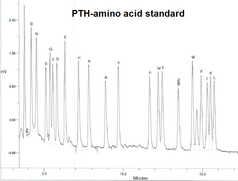
AGE-modified proteins are usually detected and analyzed using traditional approaches such as high-performance liquid chromatography (HPLC) and capillary electrophoresis (CE), and more recently with sensitive mass spectrometry-based methods.  Early studies of sugars and their derivatives by electron impact mass spectrometry were limited to volatile derivatives such as trimethylsilyl ethers, acetal derivatives, as well as acylated and methylated derivatives. However, with the development of so-called “soft” ionization techniques such as chemical ionization, field-desorption, fast-atom bombardment, and more recently electro-spray ionization and laser-desorption time-of-flight mass spectrometry, it became possible to study unmodified sugars as well as complex oligosaccharides, carbohydrates, nucleotides, peptides, proteins and related modified molecules. As a result, it is now possible to study glycoproteins as well as their oxidation products as found in AGEs.   

The mutarotation reaction of sugars is the key for the initial reaction step of the Maillard reaction.

Mutarotation of glucose in aqueous solution

Freshly prepared solutions of glucose in water gradually change in optical rotary power. The cause is a mutarotation reaction in which the dissolved glucose undergoes a transformation from one form to another. Figure 1 shows the mutarotation between the a-anomer and the b-anomer of glucose. 

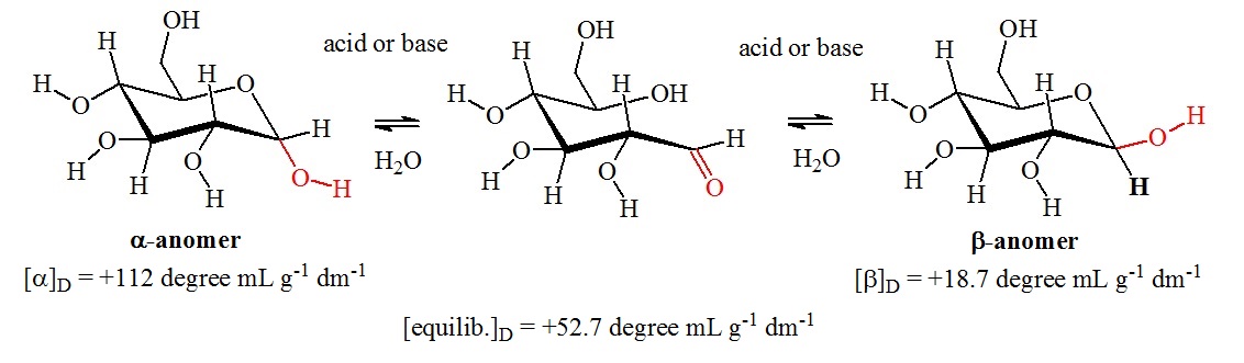
Figure 1: Mutarotation of glucose. Mutarotation is a characteristic of the cyclic hemiacetal forms of glucose. Aldehydes cannot undergo mutarotations. Mutarotation occurs by the opening of the pyranose ring to the free aldehyde form. This reaction is a reversal of a hemiacetal formation reaction. A rotation of 180° of the carbon-carbon bond to the carbonyl group allows reclosure of the hemiacetal ring via the reaction of the hydroxy group at the opposite site of the carbonyl carbon. In the glucose molecule, the two pyranose forms are interconverted. However, other carbohydrates can undergo more complex mutarotations. For example, D-fructose can mutarotate into pyranose and furanose forms.

Amadori Product Formation

Schiff base formation and Amadori rearrangement

Primary amines can react with aldehydes or ketones to form imines. This reaction is known as Schiff base formation.

Schiff base forming reaction:

R3-NH2 + R1HCO (or R1R2CO) -> R1HC=N-R3 (or R1R2C=N-R3)

The Amadori rearrangement occurs during cross-linking reactions often observed in collagen and protein glycosylation reactions. Chemically, the Amadori rearrangement refers to the conversion of N-glycosides of aldoses to N-glycosides of the corresponding ketoses. The reaction is catalyzed by acids or bases.


Steps of the Maillard reaction according to the Hodge Diagram.

1.    Initial reaction between a reducing sugar and an amino group forms
       an unstable Schiff base.

2.    The Schiff base slowly rearranges to form Amadori products.

3.    Amadori products degrade. 

4.    Formation of reactive carbonyl and dicarbonyl compounds.

5.    Production of Strecker aldehydes from amino acids and aminoketones.

6.    Production of aldol condensation products of furfurals, reductions, and
       aldehydes produced during steps 3, 4 and 5.

7.    Melanoidin formation: Furfurals, reductones, and aldehydes produced in
       steps 3, 4, and 5 react with amino compounds to form melanoidines.

8.    Free radicals can mediate the formation of carbonyl fission products
       resulting from reducing sugars.


Chemistry of the Maillard Reaction and Formation of Amadori Product


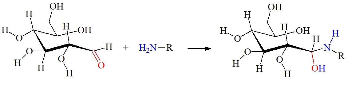
Reaction between an aldehyde group on a glucose molecule and a free amino group.


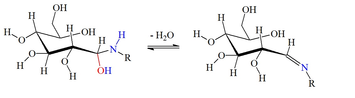
Dehydration reaction to form a Schiff base via β-elimination.


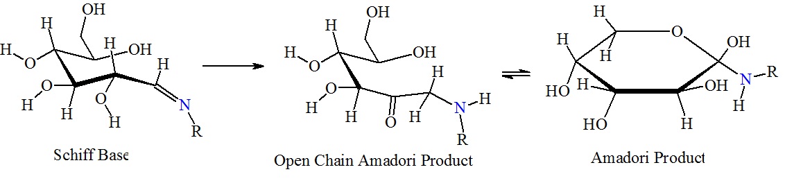
Formation of Amadori products. 

Figure 2:  Reaction between glucose and the amino group of amino acids, proteins or peptides. The nucleophilic attack by a free amino group on the aldehyde of glucose initially forms a carbinolamine. The carbinolamine subsequently dehydrates to a Schiff base. Next, the Schiff base undergoes a slow rearrangement to form the Amadori product. Only one Amadori product is shown here, however, due to the complexity of the Maillard reactions a mixture of several isoforms of Amadori products are generated during any Maillard reaction. Next, oxidative decomposition of Amadori products can lead to the formation of a wide range of reactive carbonyl and dicarbonyl compounds.  


The Schiff base, or imine, formation is catalyzed by acids, and the dehydration of the carbiolamine is the rate-limiting step of imine formation. Imine formation is a sequence of two reactions, namely, carbonyl addition followed by β-elimination.

According to Hodge et al., browned flavors generated by the Maillard reaction are essential for the recognition and taste of many processed foods.

Browned flavors include:

(1)    Food aromas that are described as toasted, baked, nutty, or roasted.

(2)    Corny and amine-like aromas from cooked grains and meals. This includes
         desirable and undesirable burnt aromas, bitter tastes, roasted malt, nuts,
         coffee, chicory, cocoa, meats, fruits, and vegetables.

Flavor compounds isolated from browning reactions allowed correlation of aromas to chemical structures. It was found that many of these flavor compounds were formed through sugar-amines condensations followed by Amadori rearrangement at lower temperatures. The Amadori compounds 1-amino-1-deoxy-2-ketoses are important nonvolatile precursor molecules originating from Maillard reactions.


Figure 3:  Maillard reaction and flvor formation in foods.

Maillard reaction products can have positive and negative effects on health. Maillard reaction products can act as antioxidants, bactericidal compounds, as antiallergic and antibrowning molecules, as prooxidants, and even carcinogens. The type of food processing appears to determine which properties are produced. It has been observed that acrylamides are formed in many foods via the Maillard reaction at high temperature.  

Reference

http://sphx.col.ynu.edu.cn/myfoodweb4/foodchemistry/maillard.html

Ames, J.M.; Dietary Maillard reaction products: Implications for human health and disease. Czech J. Food Sci. 2009, (27)  S66-S69. http://www.agriculturejournals.cz/publicFiles/07583.pdf

Hodge, J. E. (1953). "Dehydrated Foods, Chemistry of Browning Reactions in Model Systems". Journal of Agricultural and Food Chemistry. 1 (15): 928–43. doi:10.1021/jf60015a004.


Hodge, J.E., Mills, F.D., and Fisher, B.E.; Compounds of browned flavor derived from sugar-amine reactions. Cereal Science Today, 1972, vol. 17, No. 2, 34-40. https://naldc.nal.usda.gov/download/31078/PDF.

Loudon, Marc: Organic Chemistry. 5th edition. Roberts and Company Publishers. 2009.

http://www.macmillanlearning.com/Catalog/product/organicchemistry-sixthedition-loudon

Maillard LC. Action of amino acids on sugars. Formation of melanoidins in a methodical way. Compt 

Rend 1912; 154:66–68.

Tamanna, N., and Mahmood, N.; Food processing and Maillard reaction products: Effect on human health and nutrition. International Journal of Food Science. 2015. 1-6. https://www.hindawi.com/journals/ijfs/2015/526762/.

Zhang, Q., Ames, J.M., Smith, R. D., Baynes, J.W., and Metz, T. O.; A perspective on the Maillard reaction and the analysis of protein glycation by mass spectrometry: probing the pathogesis of chronic disease. J Proteome Res. 2009, 8(2): 754-769. https://www.ncbi.nlm.nih.gov/pmc/articles/PMC2642649/


What are Signature Peptides

$
0
0

What are Signature Peptides?

Signature peptides are unique tags or biomarkers, detected as molecular markers or as unique sequence tags. Signature peptides are useful tools for biomarker discovery and measurements.

Proteomic research involves the large-scale study of proteins in living organism. One important area of proteomics is the quantitative determinations of the protein content at a certain developmental or disease stage of an organism, including the human proteome. For this, absolute quantification is needed. Recent advancements made in mass spectrometry-based technologies has now enabled targeted protein quantification. However, many proteomic studies report only relative quantification and many methods for relative quantification now exist.

Absolute quantification is needed for biomarker analysis and system biology research. Typically quantitative proteomic approaches involve mass spectrometric determination of signature peptides which are usually enzymatically derived together with their isotope-labeled analogs. In general, tryptic peptides of target proteins are used. Unique peptide sequences are important for protein identification and selected signature peptides can be used as peptide or protein biomarkers.

A web tool called Unimap allows in-silico searching for signature peptides to find

(i)    a given molecular mass that is a unique molecular mass present or found in
       one human protein,

(ii)  a given peptide sequence or sequences found exclusively in one human protein,
       and 

(iii)  a specific protein for which unique masses or peptide sequences exist.

Already many novel protein candidates associated with various diseases have been identified. But because of the complexity of biological systems, the heterogeneity of human samples, and the lack of universal standardized quantitative technologies, biomarker validations have been challenging.

The human genome sequencing project has transformed biomedical research in the last decade. Also, a draft map of the human proteome was published in 2004 (Kim et al.). Proteomic profiling of 30 histological normal human samples resulted in the identification of 30,057 proteins encoded by 17,294 genes. A large number of peptides sequences were identified. These genes accounted for approximately 84% of the total annotated protein-coding genes in humans. The resulting peptide data is available as an interactive web-based resource. This data set is thought to complement human genome and transcriptome research which hopefully accelerates biomedical research in health and disease possibly leading to new and better therapeutic approaches.

For the experimental identification of signature peptides, data-dependent mass spectrometry experiments are performed. Different mass spectrometry platforms or workflows can be used. A typical setup consists of an on-line nanoLC chromatography system coupled to a mass spectrometer. The following platforms are examples: micro- or nano-LC systems coupled to Orbitrap type mass spectrometers (Thermo), to QTOF mass spectrometers (Agilent and Waters), to ion-trap mass spectrometers (Bruker), to TripleTOF mass spectrometers (Sciex), or to  MALDI-TOF/TOFs (ABI) or similar MALDI-MS instruments.

An example of this approach is the empirical peptide selection work flow for robust protein quantification reported by Fu et al. in 2015 (on-line publication). The research group compared the relative SRM signal intensity of 12 uromodulin-derived peptides between tryptic digests of 9 urine samples. Absolute quantification was performed using stable isotope–labeled peptides as internal standards. A standard curve needed to be prepared from a tryptic digest of purified uromodulin. The research group showed that the comparison of the peptide abundance of several peptides derived from the same target protein allows selection of signature peptides to detect and quantify proteins in biological samples, in this case, uromodulin. Also, the research group showed that one cannot take shortcuts in peptide selection if the development of a robust assay is desired.  

Uromodulin, UMOD or Tamm-Horsfall glycoprotein, was selected because it is the most abundant protein in healthy human urine. The uromodulin protein is encoded by the UMOD gene. Under physiological conditions, uromodulin is the most abundant protein in the mammalian urine. Uromodulin is thought to act as an inhibitor of calcium crystallization in renal fluids and its excretion in urine provides defense against urinary tract infections caused by uropathogenic bacteria. Gene defects of the UMOD gene are associated with the renal disorders medullary cystic kidney disease-2 (MCKD2), glomerulocystic kidney disease with hyperuricemia and isosthenuria (GCKDHI), and familial juvenile hyperuricemic nephropathy (FJHN). The gene is alternatively spliced. 

Fu et al. argue that exact quantification of urinary uromodulin can act as a biomarker for susceptibility to chronic kidney disease and hypertension. Uromodulin signature peptides can be potentially used as future diagnostic biomarkers for monitoring blood pressure-lowering treatments.


Uromodulin signature peptides selected by Fu et al. (2016) as biomarker peptides.


Peptide

Sequence

M/z mono

M/z average

DWVSV

DWVSVVTPAR

[M]    1,128.59280

[M+H]+  1,129.60008

[M]    1,129.28105

[M+H]+ 1,130.28832

YFIIQ

YFIIQDR

[M]   953.49711

[M+H]+  954.50439

[M]  954.09353

[M+H]+  955.10080

TLDEY

TLDEYWR

[M]   981.45564

[M+H]+  982.46291

[M]  982.06056

[M+H]+ 983.06784

FVGQG

FVGQGGAR

[M]   790.40863

[M+H]+  791.41591

[M]   790.87720

[M+H]+  791.88448


M/z values were calculated with the fragment ion calculator from the proteomicsToolkit
However, a researcher should always check experimentally how these peptides are detected in each individual mass spectrometer system used for the analysis. Methionine containing peptides where excluded because different levels of oxidation were observed during the study. According to Fu et al. purification of the digested peptides on an HLB microplate gave the best recoveries as validated with stable isotope-labeled peptides. The “Oasis HLB” resin from Waters consists of a strongly hydrophilic, water-wettable polymer with a unique hydrophilic-lipophilic balance. A typical workflow for the identification of signature biomarker peptides is shown below.

A Typical Peptide Selection Workflow

Theoretical = “in-silico”

In silico digestion.

Select peptides with 6 to 21 amino acids.

Identify constrained peptides for PTM and isoforms.

Eliminate peptides with methionines and cysteines.

Empirical and Experimental

Optimize trypsin digestion and peptide cleanup.

Assay 10 to 20 peptides by SRM in 10 to 20 biological samples.

Correlate (r2) peak areas for all pairs of peptides.

Select peptides with high correlation, strong signals, high signal to noise ratio, and sequences unique to the protein of interest.

Quantitative Assay

Synthesize or purchase 15N-labled internal standard peptides.

Optimize LC and SRM parameters.

Determine LLDQ and ULOQ with purified recombinant proteins.

Determine reproducibility.

Evaluate recovery.

Abbreviations: SRM,  selected reaction monitoring; MS, mass spectrometry; ARIC, Atherosclerosis Risk in Communities; SIL, stable isotope–labeled; LLOQ, lower limit of quantification.


Reference

Anastasia Alexandridou, George Th. Tsangaris, Konstantinos Vougas, Konstantina Nikita, George Spyrou; UniMaP: finding unique mass and peptide signatures in the human proteome. Bioinformatics 2009; 25 (22): 3035-3037. doi: 10.1093/bioinformatics/btp516.

Fu, Qin, Grote, Eric, Zhu, Jie, Jelinek, Christine, Köttgen, Anna, Coresh, Josef, Van Eyk, Jennifer E.; An Empirical Approach to Signature Peptide Choice for Selected Reaction Monitoring: Quantification of Uromodulin in Urine. Clinical Chemistry 2016, 62, 1, 198-207. http://clinchem.aaccjnls.org/content/62/1/198.abstract

Geng M, Ji J, Regnier FE.; Signature-peptide approach to detecting proteins in complex mixtures. J Chromatogr A. 2000 Feb 18;870(1-2):295-313.

Grant RP, Hoofnagle AN. From lost in translation to paradise found: Enabling protein biomarker method transfer by mass spectrometry. Clin Chem 2014;60:941-4.

Kim MS, Pinto SM, Getnet D et al., A draft map of the human proteome. Nature. 2014 May 29;509(7502):575-81. doi: 10.1038/nature13302.

Lee JW, Devanarayan V, Barrett YC, Weiner R, Allinson J, Fountain S, et al. Fit-for-purpose method development and validation for successful biomarker measurement. Pharmaceutical research 2006;23:312-28.

MacLean B, Tomazela DM, Shulman N, Chambers M, Finney GL, Frewen B, et al. Skyline: An open source document editor for creating and analyzing targeted proteomics experiments. Bioinformatics 2010;26:966-8.

Sheng Pan, Ruedi Aebersold, Ru Chen, John Rush, David R. Goodlett, Martin W. McIntosh, Jing Zhang, and Teresa A. Brentnall; Mass spectrometry based targeted protein quantification: methods and applications.  J Proteome Res. 2009 February ; 8(2): 787–797. doi:10.1021/pr800538n

Uromodulin gene info: https://www.ncbi.nlm.nih.gov/gene/7369.

Can single messenger RNAs (mRNAs) be tracked inside live cells?

$
0
0

Can single messenger RNAs (mRNA) be tracked inside live cells?

The answer is yes!

In recent years, single-cell biology has revealed that each cell is unique. However, single cells can vary significantly in their gene expression. Life is a dynamic process, and metabolic processes in cells are tightly regulated. The dynamics of RNA molecules including mRNAs can now be studied by tracking single RNA molecules. Recent advancements in fluorescence microscopy as well as in the synthesis of molecular probes now enable the study of cellular RNA dynamics. In 2006, Moon et al. reviewed the current state-of-the-art technology for tagging, delivery, and imaging useful for the tracking of single mRNA molecules in live cells.

Why is imaging of RNAs in living cells useful?

Imaging RNAs in cells allow studying the following events inside a living cell:

Transcription of RNA

Transcription of RNA is the initial step in gene expression and a regulation point for timing and production of gene products.

Imaging of RNA Lifetime

Investigation of each gene expression step, starting from transcription to translation. Imaging RNA allows the study of gene expression from transcription to translation. However, imaging of proteins only provides information of the location of the end product of the expressed gene.

Non-coding RNAs

Imaging and tracking RNA molecules also allow studying RNA molecules not translated into proteins. For example, the function of non-coding RNAs can be studied using labeled RNA.

RNA counting

Single molecule analysis of RNA may enable counting numbers of mRNA molecules inside single cells. This should allow measuring gene expression levels in a quantitative manner.

Ribonucleoprotein complexes

Single RNA imaging enables detection of subpopulations or transient states of messenger ribonucleoprotein (mRNP) complexes or particles.

mRNPs

Tracking single mRNP molecules in real time may allow studying the sequence of events or RNA processing and transport.


Having the ability to observe the organization and dynamics of RNA at the single molecule resolution in living cells will surely transform life science research in the near future.

How many molecules are there in a cell?

According to Moon et al. (2016), in chicken embryonic fibroblast cells there are approximately 2500 mRNA molecules present per cell, whereas there are approximately 108β-actin molecules present per cell. Therefore it is easier to count the number of RNA molecules in a single cell to study gene expression levels quantitatively than protein molecule numbers.

Labeling of mRNA

For tracking single particles organic dyes and fluorescent proteins are usually used for labeling mRNAs. However, hybridization probes or RNA motifs that bind to fluorescent molecules can also be used.

Oligodeoxynucleotide (ODN) probes

Target RNAs can be labeled via hybridization using short single-stranded DNA probes consisting of approximately 10 to 50 nucleotides. ODN probes are designed to be complementary to a target RNA sequence and are usually labeled with one or more fluorophore. To minimize background noise originating from free non-bound ODN probes, various strategies have been developed to only switch on fluorescence of the probes when bound to the target. This improves the signal to background ratio. To increase binding affinities to target sequences oligonucleotide mimics containing modified nucleotides are designed. Bridged Nucleotides (BNAs) can be used to enhance the stability and affinity of the probes.

Two main approaches are used for switching on the fluorescence signal: Förster resonance energy transfer (FRET) and static quenching.

Förster resonance energy transfer (FRET)    

FRET refers to the radiationless transmission of energy from a donor molecule to an acceptor molecule. FRET occurs when two fluorophores are in proximity, approximately between 2 to 10 nm, and when the emission spectrum of the donor overlaps with the excitation spectrum of the acceptor. FRET can be used for sensitive detection of molecular interactions.

For FRET to work, two ODN probes are designed to hybridize to target RNA side-by-side.  During hybridization, the donor and acceptor pair is brought together in the presence of the target (figure 1). 


Figure 1: RNA ODN probes for FRET. Two designed ODN probes are hybridized to target RNA side-by-side. The fluorescence signal is switched on.

Static Quenching

Static quenching is a process in which fluorescence is decreased when the distance between the fluorophore and the quencher is less than 2 nm.

For static quenching ODN probes, one probe is labeled with a fluorophore and a second complementary ODN is labeled with a quencher molecule. The two labeled ODN probes are annealed together. In this configuration, the fluorophore and the quencher are close to each other resulting in no fluorescence. If the target RNA is present, the ODN is hybridized to the target with a higher affinity than to the second oligo. This reaction restores the fluorescence of the ODN labeled with the fluorophore. The observed fluorescence signal indicates that the target RNA is present. Hence, the presence of the target RNA results in a fluorescent signal originating from the ODN probe labeled with the fluorophore (figure 2).

Figure 2: Static quenching– Annealed ODN probes labeled with fluorophore and quencher.

Other probe types

Other probe designs are also possible. For example, fluorescently labeled oligonucleotide mimic probes can be designed using BNAs to enhance their hybridization affinity and stability.

A brief list is shown below:

BNA modified probes

Exiton-controlled hybridization-sensitive fluorescent oligonucleotide (ECHO) probes

Forced Intercalation (FIT) probes

Peptide nucleic acid and nano-graphene oxide (PANGO) probes

Sticky flare probes.


BNA probes
contain nucleotide analogs that have a bridged structure in the sugar moiety. Optimal designed BNA probes increase base-discrimination, the stability of duplex or triplex formation, and show minimal cytotoxicity. These multi-functional synthetic RNA analogs can be spiked with DNA or RNA to modify structural formation of oligonucleotides. Because of their increased affinity to targets BNA based oligonucleotides enable detection of small or highly similar DNA or RNA targets.

Exiton-controlled hybridization-sensitive fluorescent oligonucleotide (ECHO) probes are designed to have thiazole orange (TO) dyes on a modified thymidine base. An exiton is defined as a bound state of an electron and an electron hole (electron-hole pair) that are attracted to each other via electrostatic Coulomb’s interaction. In other words, an exiton is an excited state formed by the recombination of an electron and an electron hole. During relaxation, the exiton gives off light and heat. Exitons are the main mechanism of light emission in semiconductors. Energy transfer processes occurring in exitons are radiative transfer, Förster transfer, and Dexter transfer. [https://en.wikipedia.org/wiki/Exciton].

FIT probes. Another probe type is a fluorescent probe designed as an oligonucleotide mimics containing dyes that replace one oligonucleotide in the middle of the probe. This type of probe is called a “forced intercalation probe” (FIT-probe). Oligonucleotide mimics labeled with asymmetric cyanine dyes, such as thiazole orange (TO), are forced intercalation probes that emit low background noise in the single-stranded state. When the probe is hybridized to the RNA target, the result is the intercalation of the TO dye within the duplex. The result is a strong fluorescence signal. A probe designed using peptide nucleic acid with a TO dye in the middle of the probe is an example for this. However, bridged nucleic acids (BNAs) can also be used for the design of these probes.

Sticky flare probes are made off 13nm gold particles functionalized with densely packed oligonucleotides. The gold core quenches the fluorescence of the target RNA. When the target RNA is recognized, sticky-flare transfers the fluorophore-conjugated ODNs to the RNA (figure 3). Sticky-flare probes can enter live cells by endocytosis. No transfection is needed. See also https://blog-biosyn.com/2013/08/29/what-are-nano-flares/.


Figure 3: Sticky-flare or nano-flare based detection of mRNA.

Molecular Beacons

Molecular beacons are a type of ODN probes designed with a hairpin structure that forms a loop and a stem via self-complementary 5’ and 3’ arms. A fluorophore is attached to the end of one arm, and a quencher is attached to the end of the other arm (figure 4). Base pairing of the two arms keeps the fluorophore and the quencher in proximity which quenches the fluorescence. When the beacon encounters the target molecule containing a sequence complementary to the loop structure a probe-target hybrid is formed. This hybrid is energetically more stable than the self-complementary hairpin structure. After hybridization, the conformation of the beacon is changed, and fluorescence is restored. Since the beacon has no background signal in the absence of target molecules, higher signal-to-noise ratios are achieved in comparison to other ODNs.  

Because of the advantages molecular beacons provide, they have already been used for the tracking and imaging of mRNAs of β-actin in fibroblasts, oskar in fruit fly oocytes, influenza virus mRNA in canine kidney epithelial cells, bovine respiratory syncytial virus RNA in bovine turbinate cells, and respiratory syncytial virus RNA in Vero cells.  

Figure 4: Molecular Beacon based assay.

Furthermore, molecular beacons have a high level of specificity for target RNAs than linear ODNs, and they can be designed to allow to distinguish single nucleotide polymorphisms (SNP) in live cells. However, the melting temperature of the matching hybrids need to be above 37 °C, and the melting temperature of single nucleotide mismatch hybrids need to be below 37 °C.

Please review: 

http://www.biosyn.com/moelcular-beacons.aspx,
http://www.biosyn.com/tew/molecular-beacon.aspx
http://www.biosyn.com/tew/Design-rules-for-Molecular-Beacons.aspx


Tentacle Molecular Beacons

To achieve an even higher specificity tentacle molecular beacons with increased kinetics and affinity have also been developed. [https://www.ncbi.nlm.nih.gov/pmc/articles/PMC1904288/]. Tentacle probes are similar to molecular beacons but the presence of a capture region allows for enhanced specificity. Tentacle molecular beacons contain a hairpin structure similar to molecular beacons but are modified by the addition of a capture probe. Tentacle molecular beacons have increased kinetics and affinities. Usually, kinetic rate constants are up to 200-fold faster than that for molecular beacons with corresponding stem strengths.

Multi-color Molecular Beacons and Wavelength-Shifting Molecular Beacons

To allow simultaneous detection of multiple RNAs, multi-color molecular beacons and wavelength-shifting molecular beacons have also been developed. In the absence of targets, the probes do not fluoresce, however, when the targets are encountered the probes usually fluoresce in the emission range of the emitter fluorophore. Wavelength-shifting molecular beacons are brighter than conventional molecular beacons.  [http://www.nature.com/nbt/journal/v18/n11/full/nbt1100_1191.html]

Dual FRET Molecular Beacons

To overcome false-positive signals of conventional molecular beacons, dual FRET molecular beacons have been designed. Dual FRET probes can achieve higher signal-to-noise ratios than can single molecular beacons.

Light Induced Molecular Beacons

Light-induced molecular beacons can hybridize to their target only when activated with UV light. This approach permits the fine control of timing and location of RNA labeling. Molecular beacons can be synthesized with caged nucleobases in the loop region. The resulting constructs remain non-fluorescent in the presence of the target RNA. The exposure to light (366 or 405 nm) in vitro or in cells fully activates the beacons. Molecular beacons synthesized with the caged nucleobases dANPE, dCNPE, or sGNPP cannot form normal Watson-Crick base pairs. However, after irradiation with light, the photo-labile caging groups are removed, and the unmodified nucleobases are regenerated. This restores the ability for base pairing of the molecular beacon resulting in a fluorescent signal of the probe-target hybrid.

[http://www.biosyn.com/tew/Light-sensitive-nucleotides.aspxhttp://pubs.rsc.org/is/content/articlehtml/2012/cc/c2cc16654b,  

http://www.biosyn.com/tew/Chemical-structures-of-caged-nucleobases.aspx]

Avoiding sequestering of molecular beacons into the nucleus

Sometimes molecular beacons tend to sequester into the nucleus, which can cause a nonspecific fluorescent signal. To prevent this, large proteins or nanoparticles have been attached to molecular beacons to prevent the passing of the beacon through the nuclear pores.

RNA Stem-Loop system

In this approach, mRNA is labeled in living cells using RNA stem-loop motifs. The MS2 bacteriophage coat protein (MCP) is known to exhibit strong affinity for the unique RNA stem-loop sequence MS2 binding site (MBS).[https://www.ncbi.nlm.nih.gov/pmc/articles/PMC413242/]. The MBS stem-loop sequence is a short oligonucleotide sequence, containing approximately 20 nucleotides. Because of this, multiple MBS stem-loops can be used for the tagging of the mRNA of interest. The signal of a single mRNA can be amplified by increasing the number of MBS stem-loops. Each MBS stem-loop can bind a dimer of MCP fused to a fluorescent protein (FB). With this approach, a single copy of mRNA can be labeled with many FBs. Other similar stem-loops such as the PP7 bacteriophage system and the λ-phage N system can also be used.

Figure 5: Structural model of the phage MS2 RNA hairpin-loop binding site.


Figure 6:  Structural models derived from the cocrystal structure of the PP7 bacteriophage coat protein in complex with its translational operator (Chao et al. 2008). The structure illustrated the molecular basis of the PP7 coat protein’s selective binding to the cognate RNA. The conserved beta-sheet surface recognizes the RNA hairpin. Different depictions are used for the image. 

Aptamer-fluorogenic System

An aptamer called “Spinach” was developed to bind and activate the fluorogen, 3,5-difluoro-4-hydroxy-benzylidend imidazolinone (DFHBI). DFHBI is a derivative of the green fluorescence protein’s (GFP) fluorphore 4-hydroxybenzlidine imidazolinone (HBI). This RNA aptamer induces fluorescence of a GFP-like chromophore.

Figure 7: Structural models of RNA Aptamer Spinach.

When Spinach binds to DFHBI a Spinach-DFHBI complex is formed which emits fluorescence. Molecular modules based on the Spinach sequence can be designed for the detection of other cellular molecules. New aptamers are constantly developed to enable investigation of a variety of molecules found in cells. However, because of some thermal instabilities and misfolding tendencies of aptamers when expressed or injected into living cells, aptamers with enhanced folding properties will need to be designed. Advance protocols using Systematic Evolution of Ligands by Exponential Enrichment (SELEX) can be coupled with fluorescence-activated cell sorting (FACS) for the development of brighter RNA aptamer-fluorogenic systems.

Glossary

Å                       Ångström: 1 Å = 0.1 nm

Broccoli            Newer aptamer

DFHBI             3,5-difluoro-4-hydroxy-benzylidene

ECHO              Exciton-controlled hybridization-sensitive oligonucleotide

FIT                   Forced intercalation

FRET              Förster resonance energy transfer

GFP                 Green Fluorescence Protein

HBI                 4-hydroxybenzlidene imidazolinone

MBS                MS2 binding protein

MCP                MS2 bacteriophage coat protein

mRNA             Messenger RNA

mRNP             Messenger riponucleoprotein

ODN               Oligodeoxynucleotide

PANGO          Peptide nucleic acid nano-graphene oxide

TO                   Thiazole ornage

SNR                Signal-to-noise ratio

Spinach           RNA aptamer specifically binding to DFHBI  

Sticky-flare     Functionalized gold particle with densely packed oligonucleotides


Appendix

Table 1:  Average Bond Lenghts

Bond   Bond length (Å)         Bond   Bond length (Å)

C-C     1.54                             N-N     1.47

C=C    1.34                             N=N    1.24

C≡C    1.20                             N≡N    1.10

C-N     1.43                             N-O     1.36

C=N    1.38                             N=O    1.22

C≡N    1.16                            

C-O     1.43                             O-O     1.48

C=O    1.23                             O=O    1.21

C≡O    1.13


Reference

Chao JA Patskovsky Y Almo SC Singer RH; Structural basis for the coevolution of a viral RNA-protein complex. Nat.Struct.Mol.Biol. (2008) 15 p.103.

Hyungseok C Moon, Byung Hun Lee, Kiseong Lim, Jae Seok Son, Minho S Song and Hye Yoon Park; TOPICAL REVIEW- Tracking single mRNA molecules in live cells. Journal of Physics D: Applied PhysicsVolume 49, (2016) Number 23.

Khashti Ballabh Joshi, Andreas Vlachos, Vera Mikat, Thomas Deller and Alexander Heckel; Light-activatable molecular beacons with a caged loop sequence. DOI: 10.1039/C2CC16654B (Communication) Chem. Commun., 2012, 48, 2746-2748.

Santangelo PJ, Nix B, Tsourkas A, Bao G.; Dual FRET molecular beacons for mRNA detection in living cells. Nucleic Acids Res. 2004 Apr 14;32(6):e57.

Brent C. SatterfieldJay A.A. West, and Michael R. Caplan; Tentacle probes: eliminating false positives without sacrificing sensitivity. Nucleic Acids Res. 2007 May; 35(10): e76.  PMCID: PMC1904288.

Smith JS, Nikonowicz EP.; Phosphorothioate substitution can substantially alter RNA conformation. Biochemistry. 2000 May 16;39(19):5642-52. RNA hairpin containing the binding sitwe for bacteriophage MS2 capsid protein.

Katherine Deigan Warner, Michael C. Chen, Wenjiao Song, Rita L. Strack, Andrea Thorn, Samie R. Jaffrey, and Adrian R. Ferré-D’Amaré; Structural basis for activity of highly efficient RNA mimics of green fluorescent protein. Nat Struct Mol Biol. 2014 Aug; 21(8): 658–663. https://www.ncbi.nlm.nih.gov/pmc/articles/PMC4143336/.

--...--


TRF Analysis of Telomeres using BNA Probes

$
0
0

In aging humans, the length of telomeres declines in dividing cells. Each time cells divide, telomeres can get shorter. When the telomeres are too short, the cells can no longer divide. The cells become inactive, or “senescent,” or die. The shortening process is associated with aging, cancer, and a high risk for death. Telomeres play a central role in cell fate and aging. Telomere repeats cap most chromosomes if not all to avoid activation of DNA repair pathways. Short telomeres are implicated in a variety of disorders. Telomeres shorten with physiological aging. However, during cancer immortalization telomeres undergo significant restoration. Determination of telomere lengths suggests that an age-based reference can be established for telomere studies. Therefore the availability and development of accurate and sensitive techniques and methods allowing measuring the lengths of telomeres in cells or cell tissue are needed.

Researchers at the UTSW Medical Center, Dallas, Texas have recently developed a method to measure telomere length.

Figure 1:  Structural models of homeobox telomere-binding protein 1 (HOT 1), a mammalian direct telomere repeat-binding protein. HOT1 is a positive regulator of telomere length that supports telomerase-depending telomere elongation.
 

A universal priming probe was used for TRFanalysis


5’-(Phos)GACTCTCAACTATC+T+A-3’


+N represents the location of BNAs.

Universal BNA Priming Probe
-.-

BNA Probes and Oligonucleotides can be ordered from Bio-Synthesis Inc. 

-.-


BNA-digoxigenin-probes for enhanced telomere length analysis


BNA-oligonucleotide probes were designed to specifically bind to telomere repeats. For this, the researchers designed a non-radioactive labeling method that uses 3’ fill-in combined with lambda exonuclease digestion for the incorporation of one or more digoxigenin molecules into bridged nucleic acid (BNA)-containing oligonucleotides. Using this method, the researchers generated probes for the detection of both C- and G-rich telomeric DNA strands. The use of this type of probes enhanced the sensitivity of telomere length measurements significantly.

In humans, telomere lengths have been associated with cancer and age-related diseases. Telomeres are located at the ends of chromosomes and are composed of tandem 5’-TTAGGG-3’ repeats. Shelterin proteins associate with telomeres and play essential roles in telomere protection, telomerase regulation and the prevention of chromosome degradation. As humans age, telomeres gradually shorten in all dividing cells. The shortening triggers DNA damage responses and cellular senescence that can lead to genomic instability and cancer progression, especially if oncogenic changes in cells occur.

Telomere-specific probes used for Southern blotting in combination with a technique called “Terminal Restriction Fragment Analysis” enables the direct detection of different sizes of telomeres.

Terminal restriction fragment (TRF) pattern analysis, also known as “Terminal Restriction Fragment Length Polymorphisms (T-RFLP) analysis, is a recently developed PCR-based method. This technique also allows studying microbial community structure and dynamics. 

How does the technique work?

1.  T-RFLP analysis measures the size polymorphism of terminal restriction
      fragments from PCR amplified markers. 

2.  Primers needed for this technique are designed with the help of comparative
      genomics. 

3.  Primers are designed against the amplification product or amplicon. 

4.  PCR amplifies the signal from a high background of unreleated markers. 

5.  Subsequent digestion with correctly selected restriction endonucleases
      produce terminal fragments.

6.  Fragments are separated on high resolution sequencing gels.

7.  A digital output is generated if separation is done in a capillary electrophoresis
      system.

8.  The use of fluorescently tagged or labeled primers limits the analysis to only
      the terminal fragments of the digestion.

9.  Using internal size markers with a different fluorophore makes the sizing
      very accurate.


Outline of TRF analysis protocol


According to Lai et al. 2016.

Step 1

a.     Prepare template DNA for DIG-labeled telomere C-rich (TC) or
   G-rich (TG) probe synthesis.

b.    Anneal G-rich or C-rich template oligonucleotides to a universal
  priming oligonucleotide.


Templates


The 5’-phosphorylated template oligonucleotide begins with seven telomeric repeats followed by a short non-telomeric sequence.

G-rich template

5’-(Phos)CCC TAA CCC TAA CCC TAA CCC TAA CCC TAA CCC TAA CCC TAF ATA GTT GAG AGT C-3’

C-rich template

5’-(Phos)GGG TTA GGGTTA GGGTTA GGGTTA GGGTTA GGGTTA GGGTTA GAT AGT TGA GAG TC-3’


Universal priming


The universal priming oligonucleotide is phosphorylated at the 5’-end and contains a sequence complementary to the non-telomeric sequence in the template oligonucleotide with additional thymine (T) and adenine (A) at the 3’-end to ensure that it anneals at the correct spot.

Oligonucleotides modified with BNAs are used to increase resistance to nuclease digestion and the affinity for the target DNA or RNA.

Universal primer

5’(Phos)GAC TCT CAA CTA TC+T+A-3’; +N = BNAs


Use Exo- Klenow Fragment together with a dNTP mix containing DIG-11-dUTP (Roche Applied Sciences, Mannheim, Germany) for 3’ fill-in reactions.


Remove additional nucleotides from 3’-end

Step 2

Apply T4 DNA polymerase to remove additional nucleotides at the 3’ end from the template DNA generated by 3’ fill-in reactions. This increases the specificity of DIG-labeled telomere probes.

Step 3

Use λ exonuclease to digest the 5’-phosphorylated template oligonucleotide and non-telomeric sequence in the priming oligonucleotide (5’->3’ direction). Note: λ exonuclease is unable to degrade BNA-containing telomeric-specific single-stranded DNA.

Step 4 A

At this step a dot blot on a nylon membrane can be performed to check that the experiment worked. [See DIG Application Manual for filter hybridization.

Step 4B

Perform Southern blot analysis.


Southern Blot Analysis

Step 1

Digest DNA and DIG-labled molecular weight marker II.

Step 2

Separate on a 7% agarose gel.

Step 3

Depurinate, denature and neutralize the gel.

Step 4

Transfer DNA fragments onto a positive  charged nylon membrane using a vacuum blotting system.

Step 5

Fix the DNA fragments on the membrane by UV-crosslinking.

Step 6

Pre-hybridize.

Step 7

Hybridize with DIG Easy Hyb solution containing one of the DIG-labeled telomeric probes over night.

Step 8

Wash membrane.

Step 9

Detect chemiluminescence signals.

Step 10

Analyze.


See Supplement “Material and methods” from Lai et al. for more details.


Example of DIG-probe synthesis


Start with templates and universal BNA probe(s):


                 Tandem 5’-TTAGGG-3’ repeat

                        GGGTTAGGGTTAGGGTTAGGGTTA...

5’(Phos)GACTCTCAACTATCTA-3’

     3’-CTGAGAGTTGATAGATCCCAATCCCAATCCCAATCCCAAT...N-PHOS


->Anneal -> 3’fill-in (Exo- KF)  ->  DNA blunting (T4 DNA polymerase)  ->  5 ->3’ digestion (λ exonuclease)  -> DIG probes.


The use of fluorescently labeled oligonucleotide probes instead of 32P-labeled oligonucleotides makes this technique more convenient and less hazardous since many waste disposal and safety issues are associated with radioactivity.


Reference

DIG RNA Labeling Kit (SP6/T7): http://www.sigmaaldrich.com/catalog/product/roche/11175025910?lang=en&region=US

Kyung H. Choi, Amy S. Farrell, Amanda S. Lakamp, Michel M. Ouellette; Characterization of the DNA binding specificity of Shelterin complexes. Nucleic Acids Res. 2011 Nov; 39(21): 9206–9223. Published online 2011 Aug 18. doi: 10.1093/nar/gkr665, PMCID:  PMC3241663.

Raffaella Diotti, Diego Loayza; Shelterin complex and associated factors at human telomeres. Nucleus. 2011 Mar-Apr; 2(2): 119–135. doi: 10.4161/nucl.2.2.15135, PMCID:  PMC3127094.

Kong PL, Looi LM, Lau TP, Cheah PL.; Assessment of Telomere Length in Archived Formalin-Fixed, Paraffinized Human Tissue Is Confounded by Chronological Age and Storage Duration. PLoS One. 2016 Sep 6;11(9):e0161720. doi: 10.1371/journal.pone.0161720. eCollection 2016.

Aubert G, Lansdorp PM.; Telomeres and aging. Physiol Rev. 2008 Apr;88(2):557-79. doi: 10.1152/physrev.00026.2007.

Lai, Tsung-Po, Wright, Woodring E., and Shay, Jerry W. ; 2016. Generation of digoxigenin-incorporated probes to enhance DNA detection sensitivity. BioTechniques 60:306-309 (June 2016).

Marsh TL; Terminal restriction fragment length polymorphism (T-RFLP): an emerging method for characterizing diversity among homologous populations of amplification products. Curr Opin Microbiol. 1999 Jun;2(3):323-7.

Dennis Kappei, Falk Butter, Christian Benda, Marion Scheibe, Irena Draškovi?, Michelle Stevense, Clara Lopes Novo, Claire Basquin, Masatake Araki, Kimi Araki, Dragomir Blazhev Krastev, Ralf Kittler, Rolf Jessberger, J Arturo Londoño?Vallejo, Matthias Mann, Frank Buchholz; HOT1 is a mammalian direct telomere repeat?binding protein contributing to telomerase recruitment. The EMBO Journal (2013) 32, 1681-1701.

-.-

BNAs as Tools for DNA or RNA Targeting

$
0
0

Bridged Nucleic Acids (BNAs) can be used as tools for DNA or RNA targeting!


Synthetic oligonucleotides have emerged as imported and established tools in the life sciences enabling many applications in biology, genetics, diagnostics, molecular biology and molecular medicine, as well as in other scientific fields. Natural oligodeoxynucleotides can form DNA:DNA and DNA:RNA duplexes but are often unstable and labile to nucleases. As a result, different nucleic acid analogs have been designed and synthesized for the enhancement of high-affinity recognition of DNA and RNA targets, duplex stability and to assist with cellular uptake. Bridged Nucleic Acids (BNAs) are a good example for these.


Bridged Nucleic Acids (BNAs) contain either a five-membered or six-membered bridged structure. BNAs contain a synthetically incorporated bridge at the 2’, 4’-position of the ribose to afford a 2’, 4’-BNA monomer. Monomers can be incorporated into oligonucleotide polymer structures using standard phosphoramidite chemistry.


BNAs oligonucleotide mimics exhibit 

(i)      Equal or higher binding affinities against RNA complements as well as
           excellent single-mismatch discriminating power, 

(ii)     Much better RNA selective binding, 

(iii)    Stronger and more sequence selective triplex-forming characters, and 

(iv)    A pronounced higher nuclease resistance, even higher than  
          Sp-phosphorthioate analogs, or than regular DNA, or RNA
          oligonucleotides.


 2',4-BNANC [NMe] 


BNA 2’, 4’-BNANC [NMe] has a six-membered bridged ring structure with a unique structural feature, an N-O bond, which is a hydrophilic amino-oxy moiety, wihin the sugar moiety. A methyl group is attached to the nitrogen atom located in the bridge. Oligonucleotides modified with this BNA have a very high affinity to RNA and are resistant to endonucleolytic cleavage by nucleases. Furthermore, 2’, 4’-BNANC [NMe]s are less toxic to hepatic cells than other bridged nucleic acids, for example LNAs. Bridged nucleic acids (BNAs) are very useful tools for DNA and RNA targeting, both in vivo and in vitro. Base modifications can be used for the tuning of base pairing. For example the targeting of RNA in vivo can be achieved using antisense and/or siRNA oligonucleotides. To enhance hybridization affinity, BNAs can be incorporated at strategic locations with the oligonucleotide sequence of oligonucleotide probes. 

Table 1:  Tools for targeting DNA and RNA


Natural

Artificial

Nuclease resistance

Poor

(Natural phosphoester linkage)

Good

(non-natural backbone)

Sequence recognition

Fixed

(Four bases)

Tunable

(modified base = BNA)

Recognition of mismatched base pair

Normal

Good

(flexibility of backbone)

Toxicity

Low

Usually low, but depends on the modification used.


Table 2:  BNANC [Me] as tools for targeting DNA and RNA


Natural

N-methyl-BNA

(BNANC[Me])

RNA selectivity

Normal

Very Good

Nuclease resistance

Poor

(Natural phosphoester linkage)

Very Good

(non-natural backbone)

Binding Affinity

Normal

Strong

Reduction of off-target effects

Normal

Good

Transfection

Good

Very Good

Compatibility with enzymes

Good

Good

Probes

Very suitable

Very suitable

Antisense Oligos

Usable

Very suitable

siRNA oligos

Usable

Very suitable

Sequence recognition

Fixed

(Four bases)

Tunable

(number of modified bases)

Recognition of mismatched base pair

Normal

Good

(flexibility of backbone)

Toxicity

Low

Very low


Backbone modifications

The bridging of the 2’-oxygen of the ribose with the 4’-carbon in bridged nucleic acids (BNAs) results in a 3’endo (N-type) conformation. This bridging locks the ribose sugar into the N-type conformation. When the bridge contains a methylene linkage between the 2’-oxygen and the 4’-carbon on the ribose the bridged nucleic acids is known as LNA.

Sugar rings are building blocks of oligonucleotides located between the nucleic acid nucleobase and phosphate backbone where they act as flexible links. Ribose and 2’-deoxyribose are the basic subunits that differentiate RNA and DNA. Ribose sugar rings can adopt different conformations and thereby influence the global structure of nucleic acids. The five-membered sugar ring in oligonucleotides is inherently nonpolar. The ribose ring structure can interconvert into different conformations by “pseudorotation.” The conformation with the lowest energy is usually a ring structure with one atom out of plane and four atoms in plane. The process of pseudorotation results in a ring pucker motion. Nonbonding interactions between substituents at the four ring carbon atoms are the cause for puckering resulting in nonplanar ring structures. The pseudorotation cycle describes the interconversion into different puckering modes as they have been observed in nucleic acid structures (see Saenger in Principles of Nucleic Acid Structure, page 57).  

The assumption based on structural analysis is that the ribose ring exists in a two-state conformational equilibrium between one N-type and one S-type conformation. If the ribose ring is fused to a second ring the conformation can be fixed.  

Figure 1:  Ribose sugar puckering modes in RNA and DNA.N- and S-type sugar puckering. The N-type conformation (C3-endo or A-form) exists predominantly in A-RNA and the S-type conformation (C2-endo or B-form) in duplexes with B-DNA helical structure.

Figure 2: Chemical structure for LNA.  A: Typical structure used in most reviews.  B: Structure illustrating the N-type conformation. 

Figure 3: Chemical structures for RNA and various bridged nucleic acids. The numbering nomenclator is shown in the upper panel and the models are shown in the lower panel. The stick models were created using Pymol in the builder and sculpting mode. RNA = ribonucleic acids; LNA = locked nucleic acid; ENA = ethylene nucleic acid; BNA = bridged nucleic acid. 

BNA Applications

·        Duplex Formation through Hybridization;

·        RNA targeting;

·        Gene silencing;

·        Antisense;

·        siRNA;

·        Aptamer capping;

·        Probes for real-time qPCR;

·        BNAClampTM PCR;

·        Probes for in-situ hybridization (ISH or FISH)

·        Triplex-Forming Oligonucleotides (TFO), thermodynamically favored;

·        RNase H Activation;

·        Enhanced Nuclease Resistance and Serum Stability;

·        Cells Delivery;

·        Low In Vivo Toxicity;

·        Development of Therapeutic and Antisense Agents;

·        BNAzymes.

·        Tools for Antigene Strategies, targeting of long noncoding RNAs (lncRNAs);

·        Design of BNA aptamers;

·        Design of novel reagents and probes for diagnostics;

·        Single Nucleotide (SNP) Polymorphism (SNP) Detection;

·        RNA Capture Probes, miRNA Detection.


Therapeutic Applications


Antisense

Antigene


Antisense Oligonucleotides

Triplex Forming Oligonucleotides (TFO)

siBNAs

Aptamer based approaches

BNAzyme

Transcription Elongation Inhibitors

Targeting non-coding RNAs

Strand Invasion

Gapmers


Reference

Ming Huang, Timothy J. Giese, Tai-Sung Lee, and Darrin M. York; Improvement of DNA and RNA Sugar Pucker Profiles from Semiempirical Quantum Methods. J Chem Theory Comput. 2014 Apr 8; 10(4): 1538–1545. Published online 2014 Mar 3. doi:  10.1021/ct401013s PMCID: PMC3985690. https://www.ncbi.nlm.nih.gov/pmc/articles/PMC3985690/]

Mayer, G.; The Chemical Biology of Nucleic Acids. 2010. WILEY. ISBN: 978-0-470-51974-5.

---...---

CRISPR Screening, Myomixer, Skeletal Muscle, and single-guide RNAs

$
0
0

Myomixer, a micropeptide, controls formation of skeletal muscle.


Recently the fusogenic micropeptide called myomixer was found to control the formation of mammalian skeletal muscles.

Researchers at the UTSW Medical Center in Dallas, Texas, used a genome-wide CRISPR genetic loss-of-function screen to identify genes required for myoblast fusion and myogenesis. The researchers identified the new regulator of myogenesis using 130,209 single-guide RNAs (sgRNA) in C2C12 myoblasts of a mouse muscle cell line.

First, the scientists reported that the gene Gm7325 is required for myoblast fusion. This gene is expressed in embryonic stem (ES) cells and germ cells. However, its function was not known until now.  

Secondly, using the CRISPR screen, the scientists discovered an 84-amino acid muscle-specific peptide, named “myomixer.”

This peptide was previously predicted from an RNAseq intron as an uncharacterized precursor. Because of its expression coinciding with myoblast differentiation and its essential role in the fusion of skeletal muscle as well as for the formation of skeletal muscle during embryogenesis the scientists named it “myomixer.” In addition, the researchers reported that myomixer is localized in the plasma membrane. Myomixer promotes myoblast fusion and associates with myomaker, a fusogenic membrane protein.

According to Millay et al. (2016), Myomaker is the only identified muscle-specific protein required for myoblast fusion. CRISPR/Cas9 mutagenesis screening was used for structure-function analysis to understand the fusion process of skeletal muscle cells. 

Amino acid sequence of myomixer as determined by Bi et al. (2017).

 >NP_001302423.1 uncharacterized LOC101929726 precursor  [Homo sapiens]

MPTPLLPLLLRLLLSCLLLPAARLARQYLLPLLRRLARRLGSQDMREALLGCLLFILSQRHSPDAGEASR


VDRLERRERLGPQK


>NP_001170939.1 uncharacterized LOC101929726 homolog isoform 1  [Mus musculus]

MPVPLLPMVLRSLLSRLLLPVARLARQHLLPLLRRLARRLSSQDMREALLSCLLFVLSQQQPPDSGEASR VDHSQRKERLGPQK  

Alignment using BioEdit:


Skeletal muscle is the largest tissue in humans. Skeletal muscle accounts for approximately close to 40 % or more of human body mass.  

Myogenesis refers to the formation of muscular tissue, usually during embryonic development. In general muscle fibers form from the fusion of myoblasts to form multi-nucleated fibers. 

Muscle fibers are called myotubes. The formation of skeletal muscle occurs through fusion of embryotic muscle cells (myoblasts) forming multinucleated muscle fibers (myofibers). Skeletal muscles are made up of strings or bundles of muscle fibers. These are large cells which are approximately 50 μm in diameter and up to several centimeters long. These skeletal muscle bundles are formed by fusion of many individual muscle cells during development. 

The fusion of myoblasts needs cell recognition, migration, adhesion, signaling, and the joining of different muscle cells. Extracellular calcium and changes in cell membrane topography and cytoskeletal organization are required for myoblast fusion. Several cell-surface and intracellular proteins are now known to mediate myoblast fusion. Also, myoblast fusion appears to be also regulated by activation of specific cell-signaling pathways. The activation of these pathways is essential for the fusion process and cytoskeletal rearrangement.

Formation of skeletal muscle begins with speciation of muscle cells induced by the myogenic transcription factors Pax7 and MyoD, followed by the expression of a large number of genes that establish muscle structure and function. 

The Pax7 gene is a transcription factor and a member of the paired box (PAX) family. This gene family typically contain a paired box domain, and octapeptide, and a paired-type homeodomain. PAX family genes play critical roles during fetal development as well as in cancer growth.

The MyoD protein has a major role in regulating muscle cell differentiation. The expression of MyoD is necessary for the expression of muscle-related genes. MYOD1 encodes a nuclear protein belonging to the helix-loop-helix family of transcription factors. MYOD1 regulates muscle cell differentiation by inducing cell cycle arrest. It is also involved in muscle regeneration, and it activates its own transcription. 

The fusion of mononucleated myoblasts, embryonic muscle cells containing one nucleus, forms multinucleated myofibers or fused muscle cells containing multiple nuclei.


When muscle cells are injured, healthy human muscle cells respond by activating pro-genitor cells, or muscle stem cells that can only develop into muscle fiber cells, present within adult muscles that now fuse to generate new myofibers or bundled muscle cells. However, components and the molecular basis of fusion of muscle cells are not yet fully defined or understood.  


Reference

PENGPENG BI, ANDRES RAMIREZ-MARTINEZ, HUI LI, JESSICA CANNAVINO, JOHN R. MCANALLY, JOHN M. SHELTON, EFRAIN SÁNCHEZ-ORTIZ, RHONDA BASSEL-DUBY, ERIC N. OLSON; Control of muscle formation by the fusogenic micropeptide myomixer. Science 2017, 323-327. DOI: 10.1126/science.aam9361.

Millay DP, Sutherland LB, Bassel-Duby R, Olson EN. Myomaker is essential for muscle regeneration. Genes & Development. 2014;28(15):1641-1646. doi:10.1101/gad.247205.114.

Millay DP, Gamage DG, Quinn ME, et al. Structure–function analysis of myomaker domains required for myoblast fusion. Proceedings of the National Academy of Sciences of the United States of America. 2016;113(8):2116-2121. doi:10.1073/pnas.1600101113.} 


The End   
-.-

Web resources for the CRISPR Cas System

$
0
0

Drosophila CRISPR Web Resources

Name

Link

Description

OxfCRISPR

(Liu Lab)

http://www.oxfcrispr.org

Oxford Fly CRISPR Resources

CRISPRflydesign (Bullock Lab)

http://www.crisprflydesign.org/

Offers Cas9 transgenic stocks

DRSC CRISPR finder (Perrimon Lab)

http://www.flyrnai.org/crispr/

A web tool to identify CRISPRs for fly study

FlyCas9

(Ueda Lab)

http://www.shigen.nig.ac.jp/fly/nigfly/cas9/index.jsp

Provides reagents, protocols and online tools for genome engineering by the designer nuclease Cas9 in Drosophila

flyCRISPR(O’Connor-Giles Lab)

http://flycrispr.molbio.wisc.edu/

Fly CRISPR resources

flyCRISPR discussion group


https://groups.google.com/forum/#!forum/flycrispr-discussion-group

A forum for sharing developments, insights, ideas and asking questions related to fly CRISPRs

Fly target sites

(Zhang lab)

http://www.genome-engineering.org/crispr/?page_id=41

For application of Cas9 for site-specific genome editing in eukaryotic cells and organisms

General CRISPR Resources

Name

Link

Description

Addgene CRISPR plasmids

 http://www.addgene.org/CRISPR/

A collection of CRISPR plasmids and reagents

Crass: The CRISPR Assembler

http://ctskennerton.github.io/crass/

A program that searches through raw metagenomic reads for CRISPRs

CRISPI

http://crispi.genouest.org/

A web interface with graphical tools and functions allows users to find CRISPR in personal sequences.

CRISPR Discussion Forum

https://groups.google.com/forum/#!forum/crispr

A forum to discuss Genome Engineering using CRISPR/Cas Systems

CRISPRmap

http://rna.informatik.unifreiburg.de/CRISPRmap


Web server provides an automated assignment of newly sequenced CRISPRs to standard classification system

CRISPRs web server


http://crispr.u-psud.fr/

A gateway to publicly accessible CRISPRs database and software, including CRISPRFinder, CRISPRdb and CRISPRcompar

CRISPRTarget

http://bioanalysis.otago.ac.nz/CRISPRTarget


Predicts the most likely targets of CRISPR RNAs

E-CRISP

http://www.e-crisp.org

A software tool to design and evaluate CRISPR target sites

Goldstein lab CRISPR

http://wormcas9hr.weebly.com/

A genome engineering resource for the C. elegans research community

Joung lab CRISPR

http://www.crispr-cas.org/

A genome engineering resource for zebrafish research community

Zhang lab Genome Engineering

http://www.genomeengineering.org/


CRISPR genome engineering resources website

ZiFiT target design tool

http://zifit.partners.org/ZiFiT/


Identifies potential target sites in DNA sequences

Contact us for all the synthetic RNA oligonucleotides you need!

Long RNA oligos are available at Biosynthesis Inc.

Please inquire by calling 1-800-227-0627

or by clicking   www.biosyn.com

Structural Models of the CRISPR Cas System

$
0
0

As of December 2013 90 solved structures were available in the PubMed structural database related to the CRISPR Cas System!

A few these models are shown below!

A PubMed search for “CRISPR Cas” showed that close to 90 solved structures related to this system were available by the end of December in 2013. The model of the CRISPR associated Cse3 protein, in complex with RNA published by Gesner et al. in 2011 is shown below.

cse5-rna-complex

Figure 1:  Model of the structure of Thermus Thermophilus Cse3 bound to RNA.

(Note: To generate the models PDB files were retrieved from the PubMed structure database and rendered using the modelling software packages Cn3D 4.3 and Pymol)

 -.-

Jore et al. in 2011 reported the structure of CRISPR RNA (crRNA) which contains a 5’handle connected to a spacer sequence and a 3’handle. As can be seen from the figure below, the 3’handle is thought to form a hairpin structure.  This type of loop is an unpaired loop of RNA that is created when an RNA strand folds and forms base pairs with another section of the same strand. The resulting structure is called a hairpin structure and looks like a loop or a U-shape.

Figure 2:  Structure of crRNA. (As proposed by Jore et al. in 2011).

Jinek et al. in 2012 reported models of the naturally occurring and engineered RNA-guided nuclease systems. The model for the naturally occurring RNA-guided nuclease system is shown below.

Figure 3:  Schematic model of the naturally occurring RNA-guided nuclease systems.

The naturally occurring dual RNA-guided Cas9 nuclease is illustrated. crRNA interacts with the complementary strand of the DNA target site harboring an adjacent PAM sequence, shown as green and red text. TracrRNA base pairs with the crRNA, and the overall complex is recognized and cleaved by Cas9 nuclease shown in light blue color. Folding of the crRNA and tracrRNA molecules is predicted by the program Mfoldand the association of the crRNA to the tracrRNA is partially based on the model proposed by Jinek et al. (2012).

Spilman et al. in 2013 reported that they have solved the structure of an RNA Silencing Complex of the CRISPR Cas Immune System using cryoelectron microscopy. The research group reconstructed a functional Cmr complex bound with a target RNA at a resolution of approximately 12 A°. They showed that pairs of the Cmr4 and Cmr5 proteins form a helical core that is asymmetrically capped on each end by distinct pairs of the four remaining subunits: Cmr2 and Cmr3 at the conserved 50 crRNA tag sequence and Cmr1 and Cmr6 near the 30 end of the crRNA. The structure revealed that the shape and organization of the RNA targeting Cmr complex is strikingly similar to the DNA-targeting Cascade complex. In addition these results revealed a remarkably conserved architecture among very distantly related CRISPR Cas complexes.

The next figure shows the model of the functional Cmr complex bound to a target RNA.


olo-cmr-complex-mode


Figure 4:  Overview of the P. furiosus CRISPR/Cas Locus and the Cmr Complex Structure

(adapted from Spilman et al. 2013). The structure was determined by cryoelectron microscopy.

(A)  The genes encoding the Cmr complex protein subunits are color coded to match those used for structure images in subsequent figures. The CRISPR repeats are shown in black and spacers in various colors.

(B)  The mature 39 nt and 45 nt crRNAs contain a 50 repeat-derived 8 nt sequence (50 tag) and a 31 nt or 37 nt spacer-derived sequence (guide), respectively. The sequence of the target RNA used in RNA cleavage assays and assembly with the Cmr complex is shown in blue.

(C)  Color-coded EM density of the Cmr complex bound with the 45 nt crRNA and the target RNA is shown in two orientations. Cmr1, red; Cmr2, light blue; Cmr3, orange; Cmr4, three different shades of green; Cmr5, three different shades of yellow; and Cmr6, magenta.

Do you need long RNA oligonucleotides?

Modified or unmodified?

Biosynthesis Inc. can synthesize them for you!

Please inquire calling 1-800-227-0627

or by clicking   www.biosyn.com


Reference

Gesner EM, Schellenberg MJ, Garside EL, George MM, Macmillan AM; Structure of Thermus Thermophilus Cse3 Bound to an RNA Representing a Product Complex. Nat.Struct.Mol.Biol. (2011) 18 p.688. PDB ID: 3QRR]

Matthijs M Jore, Magnus Lundgren, Esther van Duijn, Jelle B Bultema, Edze R Westra, Sakharam P Waghmare, Blake Wiedenheft, Ümit Pul, Reinhild Wurm, Rolf Wagner, Marieke R Beijer, Arjan Barendregt, Kaihong Zhou, Ambrosius P L Snijders, Mark J Dickman, Jennifer A Doudna, Egbert J Boekema, Albert J R Heck, John van der Oost & Stan J J Brouns; Structural basis for CRISPR RNA-guided DNA recognition by Cascade. Nature Structural & Molecular Biology 18, 529–536 (2011). doi:10.1038/nsmb.2019.

Jinek M, et al. A programmable dual-RNA-guided DNA endonuclease in adaptive bacterial immunity. Science. 2012; 337:816–821. [PubMed]

Michael Spilman,Alexis Cocozaki,Caryn Hale,Yaming Shao,Nancy Ramia,Rebeca Terns,Michael Terns,Hong Li,and Scott Stagg; Structure of an RNA Silencing Complex of the CRISPR-Cas Immune System. Molecular Cell 52, 146–152, October 10, 2013.


BNA Gapmers revert splicing and reduce RNA foci

$
0
0
BNANCgapmers revert splicing defects in myotonic dystrophy type 1 (DM1) cells. DM1 is a multisystemic disease caused by an expanded CTG repeat in the 3’-untranslated region (UTR) of the dystrophia myotonica protein kinase (DMPK) gene. Gapmers targeting within the repetitive region of DMPK preferentially degrades the mutant allele. 


Myotonic dystrophy is the most common type of muscular dystrophy. In DM1 the expanded CUG repeat RNA (CUGexp RNA) is retained in the nucleus where it forms RNA foci which lead to defects in regulated alternative splicing events during development. Therefore if the development of CUG expanded repeat RNA foci could be prevented, the disorder will not materialize.

To test this, Manning et al. used the antisense BNA gapmer strategy to degrade CUG expanded repeat (CUGexp) RNA in immortalized human TeloMyoD fibroblast cell lines expressing telomerase and containing a tetracycline-inducible MyoD to promote the myogenic program in response to growth. A FISH probe targeting the repeat RNA was used for visualization of the RNA foci in the untreated and treated cells.

BNANC gapmers targeting within the repetitive region of DMPK preferentially degrade the mutant allele thereby decreasing RNA foci. Manning et al. were able to show that antisense BNANC gapmers could be used to potentially revert splicing defects in myotonic dystrophy type 1 (DM1) cells.

RNA foci are a result of expanding RNA repeats that are retained in the nucleus, and adopt unusual secondary structures, sequester various RNA binding proteins, and can become toxic to the cell. RNA-RNA binding protein complexes or aggregates form insoluble nuclear foci causing cellular defects. The abnormal expansion of nucleotide repeats leads to numerous effects on genes such as the inhibition of transcription and the loss-of-function of proteins, leading to the disease.


Reference

Kassie S. ManningAshish N. RaoMiguel Castro, and Thomas A. Cooper; BNANC Gapmers Revert Splicing and Reduce RNA Foci with Low Toxicity in Myotonic Dystrophy Cells. ACS Chem. Biol., Article ASAP. DOI: 10.1021/acschembio.7b00416. 

---...---

BNA Application Overview

$
0
0

The Bridged Nucleic Acid 2',4'-BNANC (2'-O,4'-aminoethylene bridged nucleic acid) is a molecule that contains a six-membered bridged structure with an N-O linkage. This novel nucleic acid analog can be synthesized and easily incorporated into oligonucleotides.

When compared to the earlier generations of bridged nucleic acids (e.g., LNA, ENA), BNA was found to possess:

  • Higher binding affinity to an RNA complement.

  • Excellent single-mismatch discrimination.

  • Enhanced binding selectivity to RNA.

  • Stronger and more sequence selective triplex-forming characteristics.

  • Stronger nuclease resistance to endo and exo-nucleases, even higher than S(p)-phosphorothioate analogs.

Based on the above observations, BNA has shown great promise for applications in antisense and antigene technologies.

BNAs, or bridged nucleic acids, are well suited for the following applications:

Also, BNAs are useful tools for hybridization assays that require high specificity and/or reproducibility.

The BNA modification is perfectly suited for SNP detection.

  • Reduction in probe size maximises the impact of mismatch in the stability of the probe/target duplex.

  • BNA modifications increase the specificity of the probe and also its power of discrimination.


Advantages

Affinity

  • BNA increases the thermal stability of duplexes due to its RNA-like structure. 

  • BNA-BNA duplex formation creates a very stable Watson-Crick base pairing system

Tm modulation

  • Depending on their position along the sequence, BNA bases allow reaching the desired Tm level without losing specificity.

  • The introduction of BNA allows for shorter probes while maintaining the same Tm.

Ease of use

  • BNA enhances hybridization performance relative to native DNA, RNA or phosphorothioate.

  • BNA lowers experimental error rates due to better mismatch discrimination.  

  • BNA improves the signal-to-noise ratio.

Enzyme compatibility

  • BNA exhibits increased resistance to certain exo- and endonucleases with a high biostability.

  • DNA-BNA gapmers readily activate RNAse H.

  • BNA acts as a substrate for standard molecular biology enzymes: T4 PNK, T4 DNA ligase, DNA polymerases.

Simplicity

  • BNA behaves like DNA. Therefore it is easily transferable to DNA-based assays.

  • BNA is highly soluble in water.

  • BNA can be used in oligonucleotide synthesis and analysis methods (QC, purification, etc.).

  • BNA exhibits the same salt dependence as DNA and RNA.

---...---

Analysis of Proteins and Peptides

$
0
0

The following contains a list of techniques for the analysis of proteins and peptides with protein identification or protein sequence analysis as the final step. Most of these methods have been used and tested for minute sample quantities (> 50 picomoles) in the past decades in many laboratories and by many researchers.

Depending on the solubility of the proteins studied, an extraction and possibly concentration step will be needed before the use of gel electrophoresis, liquid chromatography, ion-chromatography, reversed phase (RP) HPLC, or hydrophobic interaction liquid chromatography (HILIC).


Over the years classical protein biochemistry has evolved into proteomics. Historically, the characterization of unknown proteins began with the extraction and solubilization of selected proteins from tissue samples using an appropriate method or combination of methods. The nature of the protein source determines which extraction method will need to be selected. As many researchers have found out, a complete biochemical characterization of a protein can be quite complex and tricky. Downstream methods used for purification of the protein, generally include affinity chromatography, amino acid analysis, electrofocusing, gel electrophoresis, ion exchange chromatography, mass spectrometry, protein sequencing, as well as size-exclusion or gel filtration chromatography, including spectrophotometry.

Many biochemical methods and assays for the analysis of proteins, peptides, and protein-based products have been developed in the past. To determine the N-terminal end of unknown proteins, Edman based N-terminal protein sequencing is still the method of choice. On the other hand, the availability of more sensitive and highly specific mass spectrometry instruments now allow protein identification at exceedingly lower levels.

For protein identification or proteomic approaches, proteins can be identified and analyzed from:

#

Sample type

Technique or method used

A

Protein(s)
in solution,
or in LC fractions

Protein extracts in mixtures are separated using 1D or 2D PAGE, reversed phase HPLC, or in combination with ion-exchange followed by reversed phase HPLC. Fractions containing the protein(s) are collected and enzymatic (e.g. tryptic) in-solution digest is performed. The resulting peptides are analyzed using LC-MS/MS followed by Data Base Searches using a search engine such as Mascot.

Result:  -> Internal peptide identification -> List of identified peptides -> Possibly whole sequence coverage of a protein.

B

Gel bands

Protein bands are cut out from gels and enzymatic in-gel digest is performed, peptides are extracted and analyzed via LC-MS/MS. Data Searches are perfume using the peptide mass fingerprint patter, for example using the search engine Mascot or similar.

Result -> Internal peptide identification -> List of identified peptides.

 When modern methods and instrumentation is used, analysis results can cover the complete sequence of the identified protein(s) including some post-translational modifications.

C

PVDF pieces

Protein mixtures are separated using 1D or 2D PAGE, followed by electro-blotting onto a membrane such as PVDF, PVDF piece(s) containing the protein of interest are cut out, and chemical sequencing using Edman Protein Sequencing is performed.

Result: The N-terminal sequence of the protein is observed and reported.

D

PVDF pieces

Protein mixtures are separated using 1D or 2D PAGE, followed by electro-blotting onto a membrane such as PVDF,  the membrane piece containing the protein is cut out and an enzymatic digest is performed, followed by extracting peptides, the analysis via LC-MS/MS, and Data Base Searches, for example using the search engine Mascot.

 Result:  -> Internal peptide identification -> List of identified peptides.

E

Lyophylized Proteins

Proteins are dissolved in a buffer suitable for downstream analysis. Analysis can be performed using various chromatographic methods as well as gelelectrophoresis as described in A to C.


Examples for protein bands in a gel (left) and on a PVDF membrane (right) are shown in figure 1 below. Bands that are as darkly stained may contain between 25 to 50 pm for 50 to 100 kDa proteins. However, bands that only show up as faint bands may contain femtomole amounts of protein. The use of nano-spray LC-MS/MS methods may enable the identification of faint bands as well.

Examples for protein bands in a gel (left) and on a PVDF membrane (right) are shown in figure 1 below. Bands that are as darkly stained may contain between 25 to 50 pm for 50 to 100 kDa proteins. However, bands that only show up as faint bands may contain femtomole amounts of protein. The use of nano-spray LC-MS/MS methods may enable the identification of faint bands as well. 


We recommend performing top-down mass spectrometry on protein or peptide samples after the primary sequence is determined. This type of analysis will confirm the sequence assignment, or, reveal covalent modifications. For example, an observed HexNAc ion (m/z = 204; as observed as a delta mass) may indicate the presence of a glycopeptide (Carr, S.A., Huddleston, M.J., & Bean, M.F. 1993).

A comparison of the observed sequence with the known consensus sequences may help to determine types of modifications present in the protein or peptide (see A. Aitken's Identification of Protein Consensus Sequences). The use of peptide digestions in combination with LC-MS(MS) approaches for assigning modified peptides may also be useful. However, results can be misleading if an unexpected or incomplete digestion occurs, or if the modification is partial or heterogeneous.

Use orthogonal methods during isolation, purification and enrichment steps. For example, we recommend not to use electro-elution twice, rather use electro-blotting or HPLC as the last purification step.

For protein sequencing using Edman chemistry, the higher the protein amount per volume, or per mm2 in the case of a membrane blot, the better the recovery of the protein will be.


Abbreviations and nomenclature

AAA

Amino acid analysis

Blotting or Electroblotting

Electroblotting of proteins and peptides onto polymeric membranes of the PVDF-type

CE

Capillary electrophoresis

DTT

Dithiothreitol

EC

Exclusion chromatography

HexNAc

Hexose N-acetyl

HIC

Hydrophilic ion chromatography

HPFA

Heptafluoroacetone

HPFP

Heptafluoropropanol

HPLC

High performance liquid chromatography

LC

Liquid chromatography

LC-MS

Liquid chromatography-mass spectrometry

LC-MS/MS      

Liquid chromatography-tandem mass spectrometry

Peptide mapping

Creating a map of peptide fragments ('finger print') for a protein using chemical or enzymatic digesting methods followed by microseparation methods using either SDS-PAGE, microbore or nanobore  HPLC, or capillary or nano-spray LC-MS/MS.

PVDF

Polyvinylidene difluoride

RPC

Reversed phase chromatography

SDS-PAGE

Sodium dodecyl sulfate-polyacrylamide gel electrophoresis

SEC

Size exclusion chromatography

TFA

Trifluoroacetic acid

UV Spectrophotometer

Ultra violet spectrophotometer

XLA

Analytical ultracentrifuge


Since properties of proteins are different and often protein specific different approaches may need to be designed or developed to achieve success.

Classical methods of protein characterization are generally used for the following type of samples:

  • Crude extracts, e.g. from tissues or microbial cells.
  • Separation and purification of individual proteins.
  • Protein or peptide characterization by determining the molecular mass, the amino acid composition, the protein sequence, as well as potential post-translational modifications.

In general, purification techniques selected can be based on the molecular size of the selected protein. Techniques like dialysis, ultracentrifugation, or size-exclusion chromatography maybe used. Aslo, the solubility of the proteins or peptides of interest may determine which methods to select. Examples are isoelectric point precipitation or salting out methods. If the electric charge of a protein or peptide is known, ion-exchange chromatography or electrophoresis based method maybe selected.

The following flow charts illustrate the use of instrumentation and methodologies for sample preparations of protein and peptides in small or minute amounts. However if higher amounts of protein are available, these protocols can be up scaled as well.


Please note:  Microsequencing can be done using the classical approach via Edman based chemical sequencing or by LC-MS/MS based protein or peptide identification methods (proteomics) via mass finger print pattern matching using database searches of genomic data.

A brief description of techniques used for protein analysis, characterization, or proteomics.


Technique

Result

Brief description


HPLC

Detection of purity of protein as a single peak. A chromatogram is usually reported.

Analytical HPLC is used for determination of the protein/peptide purity and to estimate amounts of protein present in a sample. However, the measurement of exact concentrations is not possible unless a control protein with the exact sequence is available for establishing a calibration curve. In addition, HPLC/UV or HPLC/DAD allows the analysis of small organic compounds, peptides, oligonucleotides, antibodies, enzymes, proteins, as well as their conjugates. A variety of reversed-phase columns, including C4, C8, C18, and phenyl-modified columns can be used.

SDS-PAGE

An image of a 1D gradient gel is usually reported with apparent molecular weights.

This method removes buffers, lipids, PBS and sugars from the protein.

Electroblotting

An image of the stained membrane containing the protein band(s) is usually reported.

Needed for N-terminal Sequencing.

N-terminal sequencing

A typical report will contain the observed sequence plus the chromatogram for each cycles of PTH-amino acids released.

This method will provide sequence information starting from the N-terminal end of the protein. Often up to 40 or 45 cycles may be observed. However, when the technique was young sequences up to 70 or 80 cycles have been reported.

UV absorbance at 280 nm

The absorbance of the protein at 280 nm is measured and reported. A BSA standard curve is often used to estimate the specific absorbance.

Unless the specific absorbance for the protein is known this will only result in an estimate of the protein concentration.

MALDI-MS

This will result in a mass measurement of the protein.

The mass spectra showing the observed peaks are reported.

This analysis is more accurate than molecular weight determination via SDS-PAGE. Impurities originating from peptides and protein fragments will be observed as well. Newer instrument types together with accurate calibration can result in highly accurate mass data.

LC-MS/MS

Proteomics. Protein sequence identification is done usually from gel pieces containing the protein of interest via enzymatic (tryptic) digest, followed by LC-MS/MS analysis and database searches.

Protein identification via mass pattern matching. The N-terminal and C-terminal end may be present in the data. However it is possible that the N- and C-terminal peptides are missing.

AAA

Determination of protein concentration in a sample. Recovered amino acids are usually reported in picomoles or nanomoles, and nanograms, micrograms, or mgs.

Amino acid analysis is still considered to be the most accurate method for determination of protein content. However, matrix effects due to interfering compounds in the formulation buffer are possible. Sometimes the method only works accurately if the protein is highly purified prior to the analysis.

 
Books to review

Aitken's Identification of Protein Consensus Sequences.  https://www.ncbi.nlm.nih.gov/nlmcatalog/9103409

Ghon, a. S.; Protein/Peptide Sequence Analysis: Current Methodologies. 1988. CDC Press.

Bollag, D. M., and Edelstein, S. J.; Protein Methods. 1991. Wiley-Liss.

Chen, G.; Characterization of protein therapeutics using mass spectrometry. Springer Science & business Media.

Cutler, P.; Protein Purification Protocols. 2nd edition. Methods in Molecular Biology. Vol 244. Humana Press.

Elzinga, M.; Methods in Protein Sequence Analysis. Humana Press.

Marshak, D., Lin, S-H, Brennan, W.E., Knurth, M., Burgess, R. R.; Strategies for protein purification and characterization. Bookbarn International.

Smith, B. J.; Protein Sequencing Protocols. Methods in Molecular Biology. Vol 211. Humana Press.

Wittman-Liebold, B., Salnikow, J., and Erdman, V. A.; Advanced methods in protein microsequence analysis. Springer Verlag. 1986.

References

Abersold, R.H., Teplow, D.B, Hood, L.E., andKent, S.B.H (1986) J. Bio. Chem. 261:4229-4238.

Aebersold, R.H., Teplow, D.B., Hood, L.E. and Kent St.B.H., (1986);  Electroblotting onto activated glass: High efficiency preparation of proteins from analytical SDS-polyacrylamide gels for direct sequence analysis. J. Biol. Chem.  261, (9) 4229-4238. 

Abersol, R.H., Leavitt, J., Saavedra, R.A., Hood, L.E. and Kent, S.B.H., (1987) Proc. Natl. Acad. Sci. USA 84:6970-6974.

Aebersold, R.H., Leavitt, J., Hood, L.E. and Kent, S.B.H., (1987);  Sequence analysis of proteins from whole cell lysates after separation  in analytical two-dimensional gels, p. 277-294; In K. Walsh (Ed.), Methods in Protein Sequence Analysis. Humana Press,NJ.

Bauw, G., De Loose, M., Inzé, D., Van Montagu, M., and Vanderkerckhove, J. (1987) Proc. Natl. Acad. Scie. USA 84:4806-4910.

Bonaventura, C., Bonaventura, J., Stevens, R. and Millington, D. 1994, Anal. Biochem. 222, 44-48. Acrylamide in polyacrylamide gels can modify proteins during electrophoresis.

Carr, S.A., Huddleston, M.J., & Bean, M.F. (1993)  Selective identification and differentiation of N- and O-linked oligosaccharides in glycoproteins by liquid chromatography-mass spectrometry.  Protein Sci. 2,  183-196. https://www.ncbi.nlm.nih.gov/pubmed/24225996

Hunkapillar and Lujan, in "Methods of Protein Characterization"  (JE Shively, ed.) p. 89. Humana Press, Clifton, N.J. (1986).

Kamp, R.M., 1986, High performance liquid chromatography of proteins in Adv. Meth. in Prot. Microseq. Anal. Ed. Wittman-Liebold, Springer 1986; pp. 63-73.

Laemmli, U.K.; Cleavage of Structural Proteins during the Assembly of the Head of Bacteriophage T4 (1970) Nature (London) 227, 680-685.https://www.ncbi.nlm.nih.gov/pubmed/5432063

Matsudaira. P.;  Sequence from picomole quantities of proteins electroblotted onto polyvinylidene difluoride membranes. J. Biol. Chem. 1987 262:10035-8. http://www.jbc.org/content/262/21/10035.full.pdf+html

M Moos, Jr, N Y Nguyen, and T Y Liu; Reproducible high yield sequencing of proteins electrophoretically separated and transferred to an inert support. J. Biol. Chem. 263:6005 (1988). http://www.jbc.org/content/263/13/6005.full.pdf

Perides, G., Plagens, U., and Traub, P., (1986) Anal. Biochem. Protein transfer from fixed, stained, and dried polyacrylamide gels and immunoblot with protein A-gold.https://www.ncbi.nlm.nih.gov/pubmed/2420231

Renart, J. , Reiser, J., and Stark, G.R., (1979)  PNAS 76, 3116-3120. Transfer of Proteins from gels to diazobenzyloxymethyl-paper and detection with antisera: A method for studying antibody specificity and antigen structure. https://www.ncbi.nlm.nih.gov/pmc/articles/PMC383774/

Towbin, H., and Gordon, J., (1984)  Journ. Immuno. Meth. 72, 313-340 Immunoblotting and Dot Immunobinding - Current Status and Outlock. https://www.ncbi.nlm.nih.gov/pubmed/?term=Towbin%2C+H.%2C+and+Gordon%2C+J.%2C+(1984)++Journ.+Immuno.+Meth.+72%2C+313-340+Immunoblotting+and+Dot+Immunobinding+-+Current+Status+and+Outlock.

Towbin, H., Staehlin, Th., and Gordon, J., (1979)  PNAS 76, 4350-4354.  Electrophoretic transfer of proteins from polyacrylamide gels to nitrocellulose sheets: Procedure and some applications. https://www.ncbi.nlm.nih.gov/pubmed/388439

Vandekerckhove, J., Bauw, G., Puype, M.,Van Damme, J. & Van Montagu, M. (1985) Protein-blotting on polybrene-coated glass-fiber sheet. Eur: J. Biochem. 152, 9-19. https://www.ncbi.nlm.nih.gov/pubmed/3899644

Wessel and Fluegge, 1984;  A method for the quantitative recovery of protein in dilute solution in the presence of detergents and lipids. Anal. Biochem. 138,141-143.https://www.ncbi.nlm.nih.gov/pubmed/6731838

Wilson, K. J., 1988; Purification of protein/peptides for structural studies in Protein / Peptide Sequence Analysis: Current Methodologies  pp. 1-33.

--...--- 


How to minimize sample losses

$
0
0

Low concentrations of biological samples is one of the largest challenges encountered when attempting to structurally characterize biomolecules such as lipids, oligonucleotides, peptides and proteins. Therefore, any loss of these important biological molecules during isolation and analysis should be avoided. To this end, modification in sample handling protocols may be necessary.

Follow the KISS principle (keep it simple).

The following summarizes steps necessary to minimize sample losses:

  • Attempt to eliminate or reduce the number of steps involving drying, lyophilization, and/or dialysis (It may be wise to totally eliminate dialysis as part of a purification procedure).

  • When performing a series of chromatographic steps, reduce organic or salt concentrations by dilution before loading on to the next column.

  • Arrange chromatographic separations intelligently, e.g., IEC, HIC, and/or SEC carried out prior to RPC.

  • When necessary, utilize a cold acetone, methanol, ethanol or a chloroform/methanol/water precipitation method for optimal sample recovery.

  • Keep all the solutions including the ones containing the waste (discarded material) until your experiment is over and you are satisfied with your data.

  • Minimize sample exposure to new surfaces (pipette tips, vial etc.). Do not transfer concentrated samples from one vial to another.

  • Avoid reducing HPLC fractions to complete dryness. For micro sequencing: Freeze fractions of interest using liquid nitrogen immediately after collection and store at -70° C until just prior to analysis.

  • If buffer changes are necessary for other studies, attempt using volatile buffers such as ammonium formate or bicarbonate, and, if possible, avoid dialysis.

  • Volatile buffers can be easily evaporated without compromising the sample. The sample will be dissolved in aqueous phase.

  • Concentrated buffer stocks can be added to the sample for pH or buffer adjustments.

  • Freeze sample-rich fractions at -70º C until analyzed and avoid multiple freeze/thaw cycles.


---...---


KRAS alleles and Cancer

$
0
0

The RAS gene family is one of the most studied and best characterized cancer-related genes (Arrington et al. 2012). KRAS is one of the three RAS isoforms that is the most frequently altered or mutated gene. KRAS mutations occur in 17 to 25% of all cancers. The Kristen Rat Sarcoma Virus and Murine Sarcoma Virus were co-discovered in 1982 by Chang and coworkers, and Der and Cooper (Jančík et al. 2010). The Harvey and Kirsten murine sarcoma viruses each encode a structurally and functionally related 21-kilodalton protein called p21. Ultimately the p21 protein was found to be the transforming protein of each virus. KRAS belongs to a group of small GTP-binding proteins also known as the RAS superfamily of RAS-like GTPases.

Figure 1:  Molecular model of H-ras p21:  The crystal structure for the H-ras oncogene protein 21 in complex with a slowly hydrolyzing GTP analog GppNp was determined at a resolution of 1.35 Å in 1990 (Pai et al. 1990). The binding site of the nucleotide and the magnesium could be revealed in high detail. This high resolution model allowed Pai et al. to propose a mechanism for GTP hydrolysis.


The KRAS gene, or Ki-ras2 Kirsten rat sarcoma viral oncogene homolog, is an oncogene encoding a small guanosine triphosphatase (GTPase) transducer protein called KRAS. The normal form of human c-Ras has also been called KRAS or KRAS2 or “Kristen Rat Sarcoma Viral oncogene homolog” or “Kristen Murine Sarcoma Virus 2 homolog.” The mammalian KRAS gene belongs to the ras gene family found on chromosomes 12p12.1 encoding a small GTPase. A single amino acid substitution or mutation results in a transforming protein.

In its natural, non-mutated or unchanged form the KRAS gene encodes for a protein called KRAS. KRAS is involved in cell signaling pathways that control cell growth, cell maturation, and cell death or apoptosis. The mutated or changed versions of the KRAS gene have been found in some types of cancer, including non-small cell lung cancer (NSCLC), colorectal cancer, and pancreatic cancer.

If a patient knows that his tumor has a wild-type or mutated KRAS gene, an optimal cancer treatment can be devised.

KRAS signaling

KRAS is one of the front-line sensors that initiate activation of an array of signaling molecules transmitting transducting signals from the cell surface to the nucleus. Transduction signaling pathways affect cell differentiation, growth, chemotaxis, and apoptosis. Signal transduction occurs in a cell when a molecule such as a hormone binds to a receptor on the cell membrane and initiates a set of chemical reactions inside the cell. 

Many human tumors contain Ras mutations characteristically mutated at codons 12 or 61 but more rarely at 13. Most Gly12/Gln61 mutations are the chemical reason for the oncogenicity (the capability) of inducing tumor formation. The mutation prevents the RAS form of the protein from being switched off. The oncogenic Ras mutants remain constitutively activated and contribute to the abnormal growth of tissue of tumor cells. Missense mutations in RAS proteins alter the cells balance of GDP and GTP binding and thereby the active state of the protein. This can either occur by reducing GTP hydrolysis or by increasing the rate of GTP loading. 

KRAS is frequently mutated in human cancers. The KRAS gene encodes for two distinct protein forms KRAS4A and KRAS4B through alternative splicing. The common mutations of KRAS produce mutant forms of KRAS4A and KRAS4B.

Mutations in the KRAS codon 12 are present in approximately 90% of ductal adenocarcinomas and in undifferentiated carcinomas of the pancreas. In colorectal cancer, KRAS gene mutations are correlated with increased proliferation and spontaneous apoptosis. Approximately 30 to 50 % of colorectal tumors have mutated KRAS genes. 

Mutations in the KRAS oncogene are typically heterozygous. The ratio of the mutant allele to wild-type allele can be balanced or unbalanced. Sometimes, but relatively rare, the mutant allele can become dominant. This can occur either through deletion of the wild-type allele or copy number gain of the mutant allele. A mutation distribution observed in patients with KRAS-mutated pancreatic adenocarcinomas and undifferentiated pancreatic carcinomas is shown in table 1.

When the mutant becomes dominant, it is called “mutant allele-specific imbalance” or MASI. Cancers that have KRAS MASI appear to behave aggressively as is the case in lung and colon adenocarcinomas (cancers).

Table 1: Observed mutation distribution in patients with KRAS-mutated pancreatic adenocarcinomas and undifferentiated pancreatic carcinomas. (Source: Krasinskas et al. 2013.; Uniprot: http://www.uniprot.org/uniprot/P01116).

Glossary

Cancer

Description

Link


Adenocarcinoma

A malignant tumor formed from glandular structures in epithelial tissue.

https://www.webmd.com/colorectal-cancer/what-is-adenocarcinoma#1


Ductal adenocarcinoma

A malignant tumor present in the duct cells within a gland.

https://www.cancer.gov/publications/dictionaries/cancer-terms?cdrid=45099

https://www.mayoclinic.org/diseases-conditions/dcis/basics/definition/con-20031842


Heterozygous

A pair of genes in which one is dominant and the other recessive.

https://www.vocabulary.com/dictionary/heterozygous


Malignant

Very dangerous. Uncontrolled growth that can become harmful such as in a disease or cancer.

https://www.cancer.gov/publications/dictionaries/cancer-terms?cdrid=45771


Oncogene

A gene that can transform a cell into a tumor cell. This gene confers the potential to cause cancer to a cell.

https://en.wikipedia.org/wiki/Oncogene


Recessive Gene

A gene that can be masked by a dominant gene.

https://www.vocabulary.com/dictionary/recessive


Reference

Arrington AK, Heinrich EL, Lee W, et al. Prognostic and Predictive Roles ofKRAS Mutation in Colorectal Cancer. International Journal of Molecular Sciences. 2012;13(10):12153-12168. doi:10.3390/ijms131012153. https://www.ncbi.nlm.nih.gov/pmc/articles/PMC3497263/

Ahmadian MR, Zor T, Vogt D, et al. Guanosine triphosphatase stimulation of oncogenic Ras mutants. Proceedings of the National Academy of Sciences of the United States of America. 1999;96(12):7065-7070.

Chang EH, Gonda MA, Ellis RW, Scolnick EM, Lowy DR. Human genome contains four genes homologous to transforming genes of Harvey and Kirsten murine sarcoma viruses. Proceedings of the National Academy of Sciences of the United States of America. 1982;79(16):4848-4852.
https://www.ncbi.nlm.nih.gov/pmc/articles/PMC346782/.

Der, C. J., & Cooper, G. M. (1983). Altered gene products are associated with activation of cellular rasK genes in human lung and colon carcinomas. Cell, 32(1), 201-208. DOI: 10.1016/0092-8674(83)90510-X, http://www.sciencedirect.com/science/article/pii/009286748390510X

Hagis, K.M.; KRAS Alleles: The Devil Is in the Detail. Trends in Cancer, October 2017, 3, 10, 686-697. http://www.cell.com/trends/cancer/pdf/S2405-8033(17)30163-2.pdf

Krasinskas, Alyssa M, Moser, A James, Saka, Burcu, Adsay, N Volkan, Chiosea, Simion I; KRAS mutant allele-specific imbalance is associated with worse prognosis in pancreatic cancer and progression to undifferentiated carcinoma of the pancreas. Mod. Pathol. 2013/10/ 26 / 10 /1346 – 1354. 
http://dx.doi.org/10.1038/modpathol.2013.71

Kauke MJ, Traxlmayr MW, Parker JA, et al. An engineered protein antagonist of K-Ras/B-Raf interaction. Scientific Reports. 2017;7:5831. doi:10.1038/s41598-017-05889-7.

Liu X, Jakubowski M, Hunt JL.; KRAS gene mutation in colorectal cancer is correlated with increased proliferation and spontaneous apoptosis. Am J Clin Pathol. 2011 Feb;135(2):245-52. doi: 10.1309/AJCP7FO2VAXIVSTP.

Pai EF, Krengel U, Petsko GA, Goody RS, Kabsch W, Wittinghofer A. Refined crystal structure of the triphosphate conformation of H-ras p21 at 1.35 A resolution: implications for the mechanism of GTP hydrolysis. The EMBO Journal. 1990;9(8):2351-2359. https://www.ncbi.nlm.nih.gov/pubmed/2196171

Custom Aptamer Synthesis

$
0
0

Aptamers are nucleic acid sequences or single-stranded oligonucleotides with selected sequences that specifically fold and bind to selected molecules or targets. The single strand of an aptamer folds into a well-defined three-dimensional (3D) structure.

Aptamers (named by Ellington and Szostak in 1990) are single-stranded DNA or RNA oligonucleotides (ssDNA and ssRNA) usually 20 to 80 nucleotides long with molecular weights ranging from 6 to 30 kDa that fold into unique 3D conformations.

If the oligonucleotide sequence of an aptamer is known, chemical solid phase oligonucleotide synthesis can be used for its production. However, aptamer synthesis can also be achieved using DNA, RNA or modified nucleic acids. Both, chemical as well as enzymatic aptamer synthesis is possible. Sometimes chemical and enzymatic synthesis are combined for the production of specific aptamers.

Aptamers bind to their targets with high affinity via van der Waals forces, hydrogen bonding, electrostatic interactions, stacking of flat moieties, and shape complementarities. Dissociation constants (Kd) can range from pico- to nanomolar.

Target molecules recognized by aptamers can be small molecules such as cocaine as well as proteins and peptides or others. Aptamers allow for the design of reagents with high affinity for desired compounds or molecules. The unique features of aptamers make them suitable for clinical applications or diagnostics.

For example, Sassanfar and Szostak in 1993 described an in vitro selection procedure to find an RNA motif that binds to ATP. A few years later, in 1996, Dieckman and others reported the structure of an ATP-binding RNA aptamer with a novel fold.  Figure 1 illustrates the aptamer fold in the structural model as determined by NMR.

Figure 1: Different models of an ATP-binding RNA aptamer as determined by NMR. Cn3D and PyMOL was used for the creation of the images using the structural pdb data form 1RAW.   


Specific detection by aptamers depends on specific interactions with the analyte and base pairing between different parts of the aptamer. The specificity of aptamers is often as high as that obtained with antibodies. Specifically designed oligonucleotide sequences can be used for the detection of a variety of different molecules.

The sequence of an aptamer can be determined using the SELEX process. The SELEX approach or “systematic evolution of ligands by exponential enrichment” allows for the evolution of aptamers (defined by Tuerk and Gold in 1990).

Random sequences are used as the input for the SELEX process that produces functional sequences. The process includes multiple rounds of exponential amplification and enrichment, allowing for the evolution of aptamers with high target-specific affinity from random oligonucleotide pools.

Several steps are needed for the generation of the final aptamer.

1

Preparation of the initial oligonucleotide pool of approximately 1014 to 1015 random sequences, 30 to 50 nucleotides in length between two primer binding sites.

2

Incubation in which random sequences in the initial pool fold into different secondary and tertiary structures and form aptamer-target complexes when optimal conditions occur.

3

Partitioning in which unbound sequences are separated from target-bound sequences using methods such as membrane filtration, affinity columns, magnetic beads, or capillary electrophoresis.

4

Amplification in which target-bound sequences are amplified by PCR, in the case of DNA aptamers, or RT-PCR, in the case of RNA aptamers. Reaction products are used as a new aptamer sub-pool for the next round of selection.

5

Sequencing of enriched aptamer sequences using Sanger sequencing or newer high-throughput sequencing methods.


Several negative-target selections are often added to the process that eliminates non-specific sequences. Often specific aptamers can be obtained after 8 to 20 rounds of selection. The whole selection process can take weeks to months.  

References

Dieckmann T, Suzuki E, Nakamura GK, Feigon J. Solution structure of an ATP-binding RNA aptamer reveals a novel fold. RNA. 1996;2(7):628-640.

Ellington, Andrew D., Szostak, Jack W.; In vitro selection of RNA molecules that bind specific ligands. Nature 346, 818-882, 1990. doi:10.1038/346818a0. 

Sassanfar, Mandana, Szostak, Jack W.; An RNA motif that binds ATP. Nature 364, 550-553. 

Milan N. Stojanovic, Paloma de Prada, andDonald W. Landry; Aptamer-Based Folding Fluorescent Sensor for Cocaine.  J. Am. Chem. Soc., 2001, 123 (21), pp 4928–4931. DOI: 10.1021/ja0038171. http://pubs.acs.org/doi/abs/10.1021/ja0038171

Sassanfar, Mandana, Szostak, Jack W.; An RNA motif that binds ATP. Nature 364, 550-553.

Hongguang Sun and Youli Zu; A Highlight of Recent Advances in Aptamer Technology and Its Application. Molecules 2015, 20(7), 11959-11980; doi:10.3390/molecules200711959. https://www.ncbi.nlm.nih.gov/pubmed/26133761

Tuerk C, Gold L.; Systematic evolution of ligands by exponential enrichment: RNA ligands to bacteriophage T4 DNA polymerase. Science. 1990 Aug 3;249(4968):505-10. https://www.ncbi.nlm.nih.gov/pubmed/2200121

Wang RE, Zhang Y, Cai J, Cai W, Gao T. Aptamer-Based Fluorescent Biosensors. Current medicinal chemistry. 2011;18(27):4175-4184.


---...---

PEDF and Stem Cell Fate

$
0
0

Recently, in 2015, a research group reported that PEDF is a determinant of stem cell fate.  The research group published an overview of our present understanding how PEDF and PEDF peptides regulate stem cells. The review also covered potential clinical applications of PEDF proteins or peptides.

In the same year, a second research group reported that the cytoprotective effects of pigment epithelium-derived factor (PEDF) requires interactions between a region with a distinct ectodomain on the PEDF receptor (PEDF-R). A peptide scanning approach employing custom made synthetic peptides and peptide binding assays was used for studying the interaction of PEDF with its receptor, PEDF-R. It was found that the sequence region composed of positions 98 to 114 of PEDF contains critical residues for PEDF-R interaction that mediates survival effects.

These findings added new functions to the already known multiple functions pigment epithelium-derived factor is known to have in mammalian cells.

What is PEDF?

Pigment epithelium-derived factor (PEDF) is a secreted glycoprotein widely expressed in multiple organs with broad biological activities. PEDF exhibits multiple functions including antiangiogenic (inhibits the process of new blood vessel formation), antitumor (inhibits the formation or growth of a tumor), anti-inflammatory (reduces inflammation), neurotrophic (regulates growth, differentiation, and survival or neurons) properties, cytoprotection and inhibiting oxygen-glucose deprivation (OGD)-induced cardiomyocytes apoptosis, as well as several other functions.

PEDF is a member of the serpin superfamily with non-inhibitory functions. PEDF exhibits neurotrophic, neuro-protective and antiangiogenic properties and is widely expressed in the developing and adult nervous systems. Furthermore, PEDF is important for bone development as well as for the modulation of resident stem cell populations in the brain, muscle, and eye. PEDF also promotes stem cell renewal and is important as a regulator of bone development.

The crystal structure of glycosylated human PEDF was reported in 2001 at a resolution of 2.85 Å.

Figure 1: Structure of human PEDF. The model illustrating the secondary structure is on the left, and the model showing the surface of the protein is on the right. PEDF is an anti-angiogenic and neurite growth-promoting factor.  PDB ID: 1IMV. Mw: ~ 50 kDa; but 44,277 dalton, based on the sequence, pI is estimated at 5.8.
 

Known functions of PEDF and PEDF peptides

  • Determines stem cell fate.
  • Potent inhibitor of angiogenesis in the mammalian ocular compartment (eye).
  • Potent proliferation inhibitor in various cell types.
  • Anti-tumor effect.
  • Increases bone mass.
  • Improves bone plasticity.

  • Regulates wound healing.

  • Acts against a broad range of angiogenic/proliferative inducer molecules possibly through an apoptotic mechanism.
  • Induces a neuronal phenotype in both cultured human retinoblastoma Y79 and Weri cells when added at nanomolar concentrations.

  • Promotes neuronal survival of the cerebellar granule.

  • Promotes both survival and differentiation of developing spinal motor neurons.

  • Prevents death of cerebellar granule neurons.

  • Prevents death of spinal motor neurons.

  • Prevents death of developing primary hippocampal neurons caused by glutamate cytotoxicity.

  • Prevents hydrogen peroxide-induced apoptosis of retinal neurons.

  • Delays death of photoreceptors in the mouse model of retinitis pigmentosa.

  • Supports both normal development of the photoreceptor neurons and opsin expression after removal of the retinal pigment epithelium.

  • Inhibits microglial growth.

  • Modulates different apoptotic pathways.

  • Protects against hypoxia-induced cell death.

  • PEDF production decreases with age and in some diseases, such as nephropathy.

  • Others

Functional epitopes

Functional epitopes have been identified. A PEDF-derived short peptide (PSP) is known to induce satellite cell proliferation and promotes muscle regeneration. Whereas a 44-amino acid peptide fragment of PEDF binds receptors on the surfaces of different neuron types were it determines neurotrophic activity and neuronal differentiation. So far four isoforms of secreted human and bovine PEDF have been observed. The PEDF protein contains 418 amino acids. The first 35 residues at the N-terminus are not structured, and the anti-angiogenic properties and neurotrophic activities are localized in the N-terminal region. The C-terminal region interacts with the membrane receptor. The functional domains of PEDF are listed in Table 1.

Table 1:Functional domains of PEDF (Source: Belkacemi et al. 2016).

Function

Peptide sites

Anti-angiogenesis

34-mer peptide region (residues 24–57).

SPPEEGSPDPDSTGALVEEEDPFFKVPVNKLAAA

Collagen binding

(anti-angiogenesis)

Asp256,Asp258, Asp300 (negatively charged), Arg149, Lys166, Lys167 (positively charged).

Asp255, Asp257 and Asp299 are critical to collagen-I-binding.

Cell differentiation

44-mer peptide region (residues 58–101)

VSNFGYDLYRVRSSTSPTTNVLLSPLSVATALSALSLGAEQRTE


Heparin binding

Arg145, Lys146 and Arg148

Hyaluronan

Lys189, Lys191, Arg194 and Lys197 form a motif that is critical for hyaluronan binding.

Laminin binding

34-mer peptide region (residues 44–77).

DPFFKVPVNKLAAAVSNFGYDLYRVRSSTSPTTN

Phosphorylation

Ser24, Ser114, Ser227

Neurotrophy

44-mer peptide region (residues in humans 78–121)

VLLSPLSVATALSALSLGAEQRTESIIHRALYYDLINNPDIHGT,

ILLSPLSVATALSALSLGAEQRTESVIHRALYYDLINNPDIHST

Tumor cell apoptosis

34-mer peptide region (residues 24–57)

SPPEEGSPDPDSTGALVEEEDPFFKVPVNKLAAA


Figure 2: Schematic diagram of PEDF functions. The N-terminal end contains anti-angiogenesis effects. This region has been reported to inhibit Wnt receptor, LRP6, in differentiated cells. The 44-mer and a 20-mer peptide stretch within this region appear to function in neuronal differentiation and muscle progenitor proliferation. The full-length PEDF protein induces mesenchymal stem cells (MSC) to the osteoblast linesage and affect pluripotency of embryonic stem cells (ESC) and apoptosis of inducible pluripotent stem cells.


Figure 2: Functional domains of PEDF. Locations for the 34mer peptide and the 44mer peptide are shown in yellow within the structure of human PEDF. The carbohydrate moiety of N-acetyl-D-glucosamine is shown in blue. For the 44mer peptide, the structure is rotated to allow for a better view of the peptide domain.


PEDF-PEDF-R Interactions 

In 2015 Kenealey et al. used a peptide scanning approach for studying the interaction of PEDF with its receptor PEDF-R. The goal was to elucidate the mechanism how PEDF exerts cytoprotection function.  Earlier molecular docking studies suggested that the ligand binding site of PEDF-R interacts with the neurotrophic region of PEDF (44-mer, positions 78–121). The use of binding assays demonstrated that PEDF-R binds to the 44-mer peptide.  The peptide P1from the PEDF-Rectodomain was demonstrated to have affinity for the 44-mer and a shorter fragment within it, a 17-mer peptide (positions 98–114). Alanine scanning using small peptide fragments (17-mers) of PEDF revealed key interacting residues responsible for binding to PEDF-R. The 17-mer contains a novel PEDF-R binding region important for retino-protection. Kenealey suggested that altered PEDF peptides could be exploited pharmacologically to improve protection of photoreceptors from degeneration.

Table 2:PEDF peptides used for studying receptor PEDF binding

             (Source: Kenealey et al. 2015).

Name

Sequence

P1

TSIQFNLRNLYRLSKALFPPEPLVLREMCKQGYRDGLRFL

34-mer

FFVPVNKLAAVSNFGYDLYRVRSSMSPTTN

44-mer

VLLSPLSVATALSALSLGADQRTESIIHRALYYDLISSPDIHGT

17-mer

QRTESIIHRALYYDLIS

This peptide still retains affinity to PEDF-R.

Q98A

ARTESIIHRALYYDLIS

R99A

QATESIIHRALYYDLIS

T100A

QRAESIIHRALYYDLIS

E101A

QRTASIIHRALYYDLIS

S102A

QRTEAIIHRALYYDLIS

I103A

QRTESAIHRALYYDLIS

I104A

QRTESIAHRALYYDLIS

H105A

QRTESIIARALYYDLIS

R106A

QRTESIIHAALYYDLIS

L108A

QRTESIIHRAAYYDLIS

Y109A

QRTESIIHRALAYDLIS

Y110A

QRTESIIHRALYADLIS

D111A

QRTESIIHRALYYALIS

L112A

QRTESIIHRALYYDAIS

I113A

QRTESIIHRALYYDLAS

S114A

QRTESIIHRALYYDLIA


PEDF Protein Sequence

>1IMV_A Chain A, 2.85 A Crystal Structure Of Pedf

NPASPPEEGSPDPDSTGALVEEEDPFFKVPVNKLAAAVSNFGYDLYRVRSSMSPTTNVLLSPLSVATALS

ALSLGADERTESIIHRALYYDLISSPDIHGTYKELLDTVTAPQKNLKSASRIVFEKKLRIKSSFVAPLEK

SYGTRPRVLTGNPRLDLQEINNWVQAQMKGKLARSTKEIPDEISILLLGVAHFKGQWVTKFDSRKTSLED

FYLDEERTVRVPMMSDPKAVLRYGLDSDLSCKIAQLPLTGSMSIIFFLPLKVTQNLTLIEESLTSEFIHD

IDRELKTVQAVLTVPKLKLSYEGEVTKSLQEMKLQSLFDSPDFSKITGKPIKLTQVEHRAGFEWNEDGAG

TTPSPGLQPAHLTFPLDYHLNQPFIFVLRDTDTGALLFIGKILDPRGP


Osteodystrophy

Osteodystrophy, the defective development of bone, appears to be caused by defects in PEDF expression. Recently, it was shown that null mutations in PEDF, the protein product of the SERPINF1 gene, are the cause of osteogenesis imperfecta (OI) type VI. A PEDF-knockout (KO) mouse exhibited elements similar to the human disease. The result of missing PEDF is diminished bone mineralization and the propensity to bone fracture.

PEDF and mesenchymal stem cell

PEDF directs human mesenchymal stem cell (hMSC) commitment to the osteoblast lineage and modulates Wnt/β-catenin signaling. Wnt/β-catenin is a major regulator of bone development.

PEDF peptides inhibit Wnt/β-catenin signaling and increase mineralization

Belinsky et al. showed that PEDF peptides inhibit Wnt/β-catenin signaling. A 34-mer fragment of PEDF (44–77 aa) and its mutated versions were used for the study (sequence: DPFFKVPVNKLAAAVSNFGYDLYRVRSSTSPTTN, N-acetylation, and C-amidation). The research group could show that short-term exposure to PEDF peptides or DKK1 antagonizes Wnt signaling in hMSCs. Adding the native PEDF 34-mer and a k→a PEDF 34-mer significantly increased mineralization when added during the last 7 days of the differentiation protocol.

Reference

Belkacemi L, Zhang SX. Anti-tumor effects of pigment epithelium-derived factor (PEDF): implication for cancer therapy. A mini-review. Journal of Experimental & Clinical Cancer Research : CR. 2016;35:4. doi:10.1186/s13046-015-0278-7.


Belinsky GS, Sreekumar B, Andrejecsk JW, et al. Pigment epithelium–derived factor restoration increases bone mass and improves bone plasticity in a model of osteogenesis imperfecta type VI via Wnt3a blockade. The FASEB Journal.
2016;30(8):2837-2848. doi:10.1096/fj.201500027R.

Ho T-C, Chiang Y-P, Chuang C-K, et al. PEDF-derived peptide promotes skeletal muscle regeneration through its mitogenic effect on muscle progenitor cells. American Journal of Physiology - Cell Physiology. 2015;309(3):C159-C168. doi:10.1152/ajpcell.00344.2014.

Kenealey J, Subramanian P, Comitato A, et al. Small Retinoprotective Peptides Reveal a Receptor-binding Region on Pigment Epithelium-derived Factor. The Journal of Biological Chemistry. 2015;290(42):25241-25253. doi:10.1074/jbc.M115.645846.

Sagheer U, Gong J, Chung C. Pigment Epithelium-Derived Factor (PEDF) is a Determinant of Stem Cell Fate: Lessons from an Ultra-Rare Disease. Journal of developmental biology. 2015;3(4):112-128. doi:10.3390/jdb3040112.

Simonovic M, Gettins PGW, Volz K. Crystal structure of human PEDF, a potent anti-angiogenic and neurite growth-promoting factor. Proceedings of the National Academy of Sciences of the United States of America. 2001;98(20):11131-11135. doi:10.1073/pnas.211268598.

---...---


Specific labeling of RNA

$
0
0

Structured small RNAs natural, labeled or modified with nonstandard nucleotides can all be custom synthesized. These types of RNA are very useful and of high interest for structural and functional investigations.

Using modern chemical and biochemical methods, a diverse collection of RNA molecules, short, medium, and long can be prepared using solid-phase chemical synthesis or enzymatic synthesis or a combination of both synthesis methods.

Tracking RNA with fluorophores in vivo allows studying their complex cellular functions. The design of specific labels and labeling methods allows RNA painting as well as the study of various RNA species such as messenger RNA (mRNA), ribosomal RNA (rRNA), transfer RNA (tRNA), interference RNA (RNAi), small interfering RNA (siRNA), small nucleolar RNA (snoRNA), long-non-coding RNA (lncRNA), and others.


Splint ligation method


Longer RNA sequence fragments or blocks can be synthesized using ligation methods in combination with solid phase or enzymatic synthesis methods. The splint ligation approach allows assembly of smaller RNAs into larger RNA molecules. RNA fragments either synthesized via chemical solid phase synthesis or in-vitro transcription (IVT) are starting materials. This method allows precise labeling of long RNAs with modified groups or stable and radioactive isotopes. Ligation is possible using chemical synthesis methods or enzymatic methods using DNA and RNA ligases. A molecular “splint” is used to bring the two substrates together. The use of a splint enhances the ligation efficiency. 

DNA ligase-based ligation


Ligation with T4 DNA ligase appears to be the most widely used method. T4 DNA ligase is largely sequence independent, tolerates modifications, and has minimal ligase activities with RNAs that are not hybridized to the DNA splint. In this approach, segments are joined using T4 DNA ligase-mediated splinted ligation, of two or more RNA fragments. RNA segments to be ligated are hybridized to a complementary DNA strand. This DNA strand is called the “splint.”  Hybridization creates a nicked duplex in which the 3’-hydroxyl group of one RNA substrate is placed proximal to the 5’-monophosphate group of the other substrate.


Figure 1: T4 DNA or T4 RNA ligase ligation.

Sets of RNA fragments containing modified nucleotides can also be used. RNA fragments hybridize with the help of a complementary DNA splint forming a ternary ligation-competent-complex (LCC). This complex is turned over by the DNA ligase. 

For highly structured RNAs, long splints are used to overcome the low propensity for hybridization and to significantly improve ligation efficiencies.

RNA ligase-based ligation


RNA ligases can also be used with similar strategies. However, T4 RNA ligase prefers ligation of single-stranded residues. In addition, the efficiency differs greatly between different RNA systems and efficiencies can vary between 40 to 80%.  

Figure 2: Structural model of a nicked 5’-adenylated nucleic acid duplex containing a 2’-deoxyribonucleotide at the nick. The figure was created using the structural data from the T4 RNA ligase 2 nicked nucleic acid duplex, PDB 2HVS. 

Chemical ligation


Chemical ligation is also possible. The phosphodiester bond linkage between two RNA substrates can be formed either by activating the phosphomonoester group using a reactive imidazolide or by using a condensing reagent such as cyanogen bromide. A disadvantage of chemical ligation is that it can also result in the creation of a 2’-5’ phosphodiester linkage, together with the desired 3’-5’ phosphodiester linkages. A purification step may be needed to produce a clean product.

Labeling RNA by hybridization


Labeled oligonucleotides are useful for the visualization of RNA using fluorescence resonance energy transfer (FRET). In this approach long target RNA recognize labeled oligonucleotides through Watson-Crick base-pairing. For FRET to work, two short labeled oligonucleotides are hybridized to two regions of a target RNA. The two modified oligonucleotides contain donor and acceptor fluorophores. The hybridization sites should be located such that the fluorophores are in the range of efficient energy transfer. To probe RNA tertiary structures the two hybridization sites can be located distantly in the primary sequence but should be located such that FRET can occur in the final tertiary structure. Also, oligonucleotides labeled with biotin are useful for single-molecule FRET. Synthetic labeled short DNA or RNA probes can be custom designed thereby allowing for a flexible and simple approach. It is important to design the probes such that they only anneal with non-functional regions of the target RNA to avoid steric hindrance during hybridization. To avoid false positive signals, correct experimental conditions need to be selected to avoid dissociation of non-covalent probes.


Figure 3: Hybridization based RNA labeling.

Aptamer based labeling method

Aptamers are oligonucleotides with sequences optimized for specific binding to a molecular target of interest. Aptamers have been made to specifically bind to fluorogenic dyes such as malachite green, Hoechst dye, fluorescein, and others. Also, RNA-binding proteins (RBPs) fused to probes, such as a green fluorescing protein, can be used to introduce probes into RNA molecules. For example, the phage MS2 coat protein that bind to hairpins in the 3’-untranslated region can be used to track the localization and dynamics of RNA. Unfortunately, GFP-RBP conjugates also emit fluorescence in the unbound form. This can interfere with the detection of target RNAs.

Figure 4: Aptamer Spinach. Spinach is an in vitro-selected RNA aptamer that binds a green fluorescein protein (GFP)-like ligand to activate its green fluorescence. Spinach is an RNA analog of GFP. Spinach binds the phenolate form of 3,5-difluoro-4-hydroxy-benzylidene imidazolinone (DFHBI) and selectively activates its fluorescence.

End-Labeling of Oligonucleotides


Several strategies for labeling the ends of oligonucleotides have been developed.

5’-end labeling using T4 PNK


This strategy makes use of the ability of bacteriophage T4 polynucleotide kinase (T4 PNK) to transfer a phosphate to the 5’-end of RNA or DNA oligonucleotides. If ATP is substituted with the ATP analog adenosine 5’-[γ-thio]triphosphate, the product of the transfer reaction is a phosphorylated oligonucleotide with a reactive sulfur at the 5’-end (1st reaction step). Incubation of the oligonucleotide with a haloacetamide derivative allows the addition of a chemical tag (2nd reaction step).

For example, 5-(iodoacetamido)fluorescein (5-IAF) can be used for conjugation of the fluorophore to the 5’-end of DNA and RNA oligonucleotides. Other compounds with different chemical properties can be used as well. However, for each reagent set used, optimization of the reaction will be necessary.

Figure 5: Attachment of a phosphorothioate to the 5’-terminal nucleotide. 


Figure 6: Reaction of 5-IAF with phosphorothioates. An S-alkylated thiophosphate diester is formed.

3’-end labeling using sodium periodate


This strategy makes also use of a two step reaction. First, sodium periodate oxidizes the 3’-terminal end of the ribose sugar. A reactive aldehyde is formed. Second, the oxidized sugar is conjugated to an aldehyde-reactive chemical tag. Often fluorescein 5-thio-semicarbazide (FITSC) is used. The periodate oxidation reaction requires the presence of vicinal hydroxyl groups. Therefore the reaction is specific for RNA and only modifies the 3’-terminal ribose.

Step 1: Formation of reactive aldehydes on 3’-end.


Figure 7: Oxidation of the diols on the 3’-terminal sugar. Sodium periodate is used for the oxidation to form reactive aldehydes. (For mechanism see Loudon M.; Organic chemistry. 5th edition. 2009, 506-507).

Step 2: Formation of a semicarbazone linkage between a fluorophore and an oligonucleotide.

Figure 8: Formation of fluorescein 5-thiosemicarbazone. Fluorescein 5-thiosemicarbazide is incubated with the oxidized 3’-terminal sugar.


A variety of aldehyde-reactive fluorescent compounds are now commercially available for selective labeling of 3’-end RNA. 
Also, oligonucleotides can be conjugated to biotin using (+)-biotinamidohexanoic acid hydrazide (BACH), or to a solid support using adipic-acid dihydrazide-agarose.

Reference

Eastberg JH, Pelletier J, Stoddard BL. Recognition of DNA substrates by T4 bacteriophage polynucleotide kinase. Nucleic Acids Research. 2004;32(2):653-660. doi:10.1093/nar/gkh212.https://www.ncbi.nlm.nih.gov/pmc/articles/PMC373337/

Huang H, Suslov NB, Li N-S, et al. A G-Quadruplex-Containing RNA Activates Fluorescence in a GFP-Like Fluorophore. Nature chemical biology. 2014;10(8):686-691. doi:10.1038/nchembio.1561.

KURSCHAT WC, MÜLLER J, WOMBACHER R, HELM M. Optimizing splinted ligation of highly structured small RNAs. RNA. 2005;11(12):1909-1914. doi:10.1261/rna.2170705.

Liu, Y., Sousa, R., and Wang, Y.-X.; Specific labeling: An effective tool to explore the RNA world. Bioessays 38: 192-200.

Maroney, Patricia A;  Chamnongpol, Sangpen; Souret, Frédéric; Nilsen, Timothy W.; Direct detection of small RNAs using splinted ligation. Nature Protocols 3, 279 – 287 (2008). http://dx.doi.org/10.1038/nprot.2007.530

Nandakumar, Jayakrishnan et al.; RNA Ligase Structures Reveal the Basis for RNA Specificity and Conformational Changes that Drive Ligation Forward. Cell , Volume 127 , Issue 1 , 71 – 84. 

---...---

Carbamylation in aging and disease

$
0
0

Carbamylation or homocitrullination is a post-translational modification (PTM) known for many years in the context of uremia, a metabolic disorder caused by the accumulation of waste products in the blood normally excreted in urine.

Carbamylation involves the non-enzymatic reaction of urea-derived cyanate with free NH2- groups on lysine (K, Lys) residues in proteins to yield homocitrulline (Hcit). Protein N-termini can also be carbamylated. Carbamylation can also be artificially introduced during sample preparation using urea.


Figure 1: Carbamylation of amino groups on lysine residues. (1) Urea decomposition occurs in aqueous solutions. (2) The released cyanate can now react with free amino groups in proteins, peptides or lysine to carbamylate the molecules. (3) The relationship between ammonium ion, cyanate concentrations and protein carbamylation in fixed urea solution concentration and temperature is illustrated as proposed by Sun et al. in 2014.


Carbamylation adds an NHCO group to lysine to convert it to homocitrulline. Carbamylated proteins and peptides can be detected by a mass increase of the modified peptide by a delta mass of 43 mass units when detected in a mass spectrometer. If the ratio of homocitrulline is high in proteins or peptides, amino acid analysis may be used for its analysis. However, for the detection of low amounts of homocitrulline in tissue or protein mixtures, selective enrichment of the modified peptides followed by analysis using nano-spray liquid chromatography tandem mass spectrometry (LC-MS/MS) will be the analysis of choice.

Isocyanic acid (CNOH, ΔM 43) is derived from spontaneously dissociating urea forming cyanate and ammonia in aqueous solutions. Temperature, incubation time and pH are factors that affect the rate of urea dissociation and the degree of protein/peptide carbamylation.

Urea is commonly used for denaturing proteins in aqueous solutions. In these solutions, urea is in equilibrium with ammonium and isocyanate. Under alkaline conditions isocyanate can react with primary amines of free N-terminal group and ε-amino groups of lysines in proteins and peptides to form carbamylates. Prolonged incubation of proteins in urea buffers can introduce unwanted carbamylation
by-products and thereby interfere with modern proteomics workflows including stable isotope-labeling methods.

A detailed analysis of urea stability in different solutions by Panyachariwat and Steckel showed that urea is more stable at a pH range of pH 4 to 8. However, the stability of urea decreased with increased temperature for all pH values. The lowest urea degradation occurred in lactate buffer at pH 6.0. Furthermore, urea decomposition rates in solution and pharmaceutical preparations depend on the initial urea concentration. At a higher initial concentration of urea, the degradation rate is lower.


Table 1: Residue and Delta Masses

Amino Acid

Residue Composition

Residue
Monoisotopic Mass

Delta Mass

Lysine

C6H12N2O

128.09496

0

Carbamyl Lysine

C7H13N3O2

171.10078

43.00582

Carbamylation

* NHCO

43.00582

-

*Note: A proton is lost from the amino group on the protein during carbamylation. The change in composition is NHCO. {Source: Ionsource}


Gorisse et al. in 2016 could show that carbamylation is associated with aging and life expectancy in mammalian species including humans. Carbamylation promotes molecular aging through alteration of protein functions such as of long-lived extracellular matrix proteins. Accumulation of carbamylated proteins in tissue is now considered as a general hallmark of aging.

Carbamylation in the skin

Carbamylation-derived products (CDPs) have been found to accumulate in the skin with age. Carbamylation occurs throughout the whole lifespan of mammals as well as in humans and leads to the accumulation of carbamylated proteins in tissues. Matrix proteins such as type I collagen and elastin are preferentially carbamylated. Homocitrulline accumulates more intensely than carboxymethyl-lysine. However, carboxymethyl-lysine is one of the major glycation end products. Hence, carbamylation may play a more prominent role in age-related tissue alterations then do glycoxidation reactions. Furthermore, it is known that carbamylated proteins are present in sera of patients with rheumatoid arthritis (RA) and can be used to predict joint damage. Shi et al. in 2011 reported that autoantibodies are present in sera of RA patients that recognized carbamylated proteins. The presence of IgG antibodies that recognize carbamylated antigens were found in 45% of RA patients.

Protein carbamylation rates

Protein carbamylation rates can be detected and analyzed in skin extracts by quantification of homocitrulline (HCit) or homocitrulline residues. The study by Gorisse et al. revealed that during aging, HCit concentrations significantly increase with age in all species studied. For example, HCit increased from 0.08 mmol/mol Lys in 1-d-old mice up to 2.2 mmol/mol Lys in 2-y-old mice, representing a 29-fold increase. Type I collagen carbamylation increased in all species as well, and human elastin also showed an age-dependent increase in HCit content. HCit content did not exceed 2.5 mmol/mol Glu in younger subjects (<4 y old), but it reached 11.7 mmol/mol Lys in older subjects (>70 y old), representing a 4.7-fold increase.


Urea in skin creams


Urea is also used in many skin creams to keep the creams and skin moist. However, stability studies of urea indicate that skin cream may need to be rather formulated in buffers containing ammonia or tertiary amines at pH 4 to 6 or above pH 7, if possible, to prevent carbamylation of skin proteins. Adding certain amino acid may also be beneficial.

Carbamylation and myeloperoxidase

In tissue, carbamylation can also be mediated in vivo by myeloperoxidase (MPO), the enzyme responsible for the inflammation-driven carbamylation of proteins via the MPO/H2O2/SCN- system. MPO has recently attracted attention as a potential trigger factor for atherogenesis and inflammation.

Apparently MPO has a negative impact on the extent of adaptive immune responses, but does not inhibit proinflammatory processes. 


Figure 2: Structure of Human Myeloperoxidase 5FIW.   Myeloperoxidase (MPO) is known as a front-line defender against phagocytosed microorganisms. MPO utilizes H2O2 to generate hypochlorous acid (HClO) and other reactive species to kill pathogens during infections but can also promote inflammation and causing tissue damage. Elevated levels of MPO have been observed in autoimmune diseases such as in the central nervous system (CNS) of multiple sclerosis (MS) and in the joints of rheumatoid arthritis (RA) patients.

Additionally, carbamylation of serum albumin has been implicated as a risk factor for mortality in patients with kidney failure (Berg et al. 2013).

In biochemistry and proteomics, urea solutions are the most widely used as denaturants. When proteins are digested in the presence of urea carbamylation can occur.

What is the result of carbamylation?

(1)    Carbamylation block N-terminal ends of proteins and peptides and react with
         the amino acid side chains of lysine and arginine residues. Hence, carbamylated
         amino groups are now no longer available for downstream labeling reactions.

(2)    Carbamylated amino groups prevent enzymatic digestions resulting in
         incompletely digested peptides.

(3)    Carbamylated proteins and peptides have different and sometimes unexpected
         retention times when studied using chromatography or SDS-PAGE as well as
         increased and unpredicted masses. This increases the complexity of samples.

(4)    Carbamylation affects peptide and protein identification as well as quantification.

(5)    In vitro-carbamylation affects the study of in-vivo carbamylation as being
         observed in metabolic diseases such as in uremia, and severe renal and
         cardiovascular disorders.

For the study of in-vivo carbamylated molecules, carbamylation reactions occurring during sample handling steps need to be prevented.

How can carbamylation of proteins and peptides be prevented?

Sun et al. recently showed that NH4HCO3 buffer is more effective in protecting proteins and peptides against carbamylation in urea solution than phosphate (PB) or Tris-HCl buffer. The research group found that the inhibition efficiency increased with increased NH4HCO3 concentrations and that a 1M NH4HCO3 buffer nearly completely prevented carbamylation on proteins and peptides during tryptic digestion in urea solution without inhibiting trypsin. Also, other ammonium or tertiary ammonium containing buffers also inhibited protein carbamylation during protein digestion in urea solution.

Reference

Berg, A. H., Drechsler, C., Wenger, J., Buccafusca, R., Hod, T., Kalim, S., … Karumanchi, S. A. (2013). Carbamylation of Serum Albumin as a Risk Factor for Mortality in Patients with Kidney Failure. Science Translational Medicine, 5(175), 175ra29. http://doi.org/10.1126/scitranslmed.3005218

Gorisse, L., Pietrement, C., Vuiblet, V., Schmelzer, C. E. H., Köhler, M., Duca, L., Gillery, P. (2016). Protein carbamylation is a hallmark of aging.Proceedings of the National Academy of Sciences of the United States of America, 113(5), 1191–1196.
http://doi.org/10.1073/pnas.1517096113.

https://pubchem.ncbi.nlm.nih.gov/compound/Isocyanic_acid#section=Top

http://pubs.acs.org/doi/abs/10.1021/ja01623a011

Kollipara LZahedi RP.; Protein carbamylation: in vivo modification or in vitro artefact? Proteomics. 2013 Mar;13(6):941-4. doi: 10.1002/pmic.201200452.

Panyachariwat N, Steckel H.; Stability of urea in solution and pharmaceutical preparations.J Cosmet Sci. 2014 May-Jun;65(3):187-95.

William H. R. Shaw, John J. Bordeaux; The Decomposition of Urea in Aqueous Media. J. Am. Chem. Soc., 1955, 77 (18), pp 4729–4733. DOI: 10.1021/ja01623a011. https://www.ncbi.nlm.nih.gov/pubmed/25043489

Jing Shi, Rachel Knevel, Parawee Suwannalai, Michael P. van der Linden, George M. C. Janssen, Peter A. van Veelen, Nivine E. W. Levarht, Annette H. M. van der Helm-van Mil, Anthony Cerami, Tom W. J. Huizinga, Rene E. M. Toes, and Leendert A. Trouw; Autoantibodies recognizing carbamylated proteins are present in sera of patients with rheumatoid arthritis and predict joint damage PNAS 2011 108 (42) 17372-17377; published ahead of print October 10, 2011, doi:10.1073/pnas.1114465108.

Strzepa, A., Pritchard, K. A., & Dittel, B. N. (2017). Myeloperoxidase: A new player in autoimmunity. Cellular Immunology317, 1–8. http://doi.org/10.1016/j.cellimm.2017.05.002https://www.ncbi.nlm.nih.gov/pmc/articles/PMC5665680/

Sun, S., Zhou, J.-Y., Yang, W., & Zhang, H. (2014). Inhibition of Protein Carbamylation in Urea Solution Using Ammonium Containing Buffers.Analytical Biochemistry, 446, 76–81. http://doi.org/10.1016/j.ab.2013.10.024. https://www.ncbi.nlm.nih.gov/pmc/articles/PMC4072244/


---...---

Minor Groove Binders or MGBs

$
0
0

Minor Groove Binders or MGBs are crescent-shaped molecules that selectively bind non-covalently to the minor groove of DNA, a shallow furrow in the DNA helix. Binding to DNA with specific sequences usually takes place by a combination of directed hydrogen bonding to base pair edges.

Natural Minor Groove Binder Molecules

Duocarmycin A and its analogs are naturally occurring antitumor agents. These molecules belong to a group of DNA minor groove binding molecules that exhibit AT-sequence selective adenine-N3-alkylations. Duocarmycins have been isolated from Streptomyces bacteria, and a number of these compounds have already been used during clinical trials to test them for their potential as anticancer drugs.


Figure 1: Structure and molecular model of duocarmycin A (Source: PDB 107D). Duocarmycins are known to derive their potent antitumor activity through sequence-selective minor groove alkylation of N3 adenine in double-stranded DNA. Duocarmycins belong to a new class of antitumor antibiotics containing a unique  spiro-cyclo-propyl-hexadienone moiety responsible for DNA alkylation. The water-soluble derivative, KW-2189, has broad-spectrum antitumor activity in a series of experimental tumor models and is presently in Phase II clinical trials. 


Figure 2:  Different renderings of molecular models of a duocarmycin A-DNA duplex complex (Source: PDB 107D).  The model is based on the solved solution structure using a combined NMR-molecular dynamics study including NOE based intensity refinement by Lin and Patel in 1995. The structure revealed that the antitumor antibiotic duocarmycin A binds covalently to the minor groove N-3 position of adenine with sequence specificity for the 3'-adenine in a d(A-A-A-A) tract in duplex DNA. The adenine ring is protonated during duocarmycin adduct formation resulting in charge delocalization over the purine ring system. The model shows a minimally perturbed right-handed duplex in the B-form helix and duocarmycin A is positioned within the walls of the minor groove.

Hoechst dyes are Minor Groove Binders 

Hoechst dyes are a family of blue fluorescent dyes useful for the staining of DNA. These bis-benzimide dyes were developed by the Hoechst AG. The three dyes Hoechst 33342, 333580, and 333258 are the most commonly used.  

The bisbenzimide Hoechst 33258 and its derivatives are starting molecules for the design of minor groove binders. These molecules are widely used for staining DNA in cells and belong to a class of small molecules that preferably bind to AT-rich sequences. The dye Hoechst 33258 is known to induce apoptosis and enhance over expression of transgenes. 


Figure 3:  Chemical structures of the dyes Hoechst 33258 and 33342.

Figure 4:  Molecular model of the dye Hoechst 33342.

Figure 5:  Molecular structure of d(CGC[e6G]AATTCGCG in complex with Hoechst 33342 containing O6-ethyl-G-C base pairs (Source: PDB 129D).  O6-ethyl-G (e6G) is an important DNA lesion caused by the exposure of cells to alkylating agents such as N-ethyl-N-nitrosourea.

---...---

Minor Groove Binder Oligonucleotides

$
0
0

MGB oligonucleotide probes

Oligonucleotides conjugated to MGB molecules are known to form stable duplexes with single-stranded DNA targets. The addition of MGBs to DNA probes allows the design of shorter hybridization probes useful for molecular diagnostics, for example for their use in quantitative PCR based assays. Several designs are possible, including Taqman MGB probes. MGB oligonucleotides form stable hybrid complexes with complementary DNA and RNA target sequences. Several solved 3D structures of MGBs with DNA oligonucleotides as well as a structure of a MGB-oligonucleotide conjugate hybrid provided insight of molecular interactions between minor groove binding moieties and DNA.

What are Minor Groove Binder Molecule?

Minor Groove Binder (MGB) molecules are crescent-shaped molecules that selectively bind non-covalently to the minor groove of DNA, a shallow furrow in the DNA helix. Binding to DNA with specific sequences usually takes place by the combination of directed hydrogen bonding to base pair edges. Tri-imidazole polyamide and the tripeptide of dihydropyrroloindole-carboxylate are good examples of MGBs.


Tri-imidazole polyamides recognize the minor groove of DNA


The design of molecules that can recognize base pairs or mismatched base pairs is desirable for studying mutations leading to mismatched base pairs. The T:G mismatch has been shown to be responsible for most common mutations in human ras oncogenes. Spontaneous deamination of 5-methylcytosine or errors in replication can introduce a T:G mismatch. The combination of pyrrole (Py), hydroxypyrrole (Hp) and imidazole (Im) units of polyamides can confer specific recognition properties for all four different base pairs, A:T, T:A, G:C, and C:G.


Yang et al. in 1999 proposed that an imidazole-imidazole pair containing polyamide may contain a good molecular motif for selective recognition of T:G/G:T base pairs. The researchers reasoned, since the N2 amino group of the G base in a T:G mismatch is not involved in base pairing with T, the free amino group could form two hydrogen bonds, each with one imidazole nitrogen atom of the polyamide. Studying the binding of two polyamides, AR-1-144 and Im-Py-Im, to DNA sequences using NMR, showed that an imidazole-imidazole pair is a good motif for the recognition of a T:G/G:T base pair.

The structures for AR-1-144 and Im-Py-Im are illustrated in figure 1 and the structure of AR-1-144 in complex with a CCGG containing DNA duplex solved by NMR is shown in figure 2.

Figure 1: Chemical structure of AR-1-144 and Im-Py-Im polyamides. The molecular model of AR-1-144 is shown as well (Yang et al. 1999).

Figure 2: NMR structure of AR-1-144 in complex with a CCGG containing DNA duplex. The tri-imidazole AR-1-144 {N-[2-(dimethylamino)ethyl]-1-methyl-4-[1-methyl-4-[4-formamido-1-methylimidazole-2-carboxamido]imidazole-2-carboxamido]-imidazole-2-carboxamide} is a minor groove binder. AR-1-144 favors the CCGG sequence (Yang et al. 1999).

A dihydropyrroloindole tripeptide CDPI3 is a minor groove binder

Another minor groove binder (MGB) derivative, a dihydropyrroloindole tripeptide (N-3-carbamoyl-1,2-dihydro-3H-pyrrolo[3,2-e]indole-7-carboxylate tripeptide or CDPI3), was shown to arrest sequence-specific primer extension on single-stranded DNA when linked to oligodesoxyribonucleotides.

Afonina et al. in 1996 showed that CDPI3 conjugated to DNA oligonucleotides increased both the specificity and the strength of hybridization using absorption thermal denaturation and slot-blot hybridization studies. Furthermore, a complementary 16-mer conjugated to 5'-CDPI3 when hybridized to phage DNA blocked primer extension by a modified form of phage T7 DNA polymerase (Sequenase).

Kutyavin et al. in 1997 showed that CDPI3 when conjugated to DNA oligonucleotides enhanced the stability of all duplexes studied. The observed stabilization was backbone and sequence-dependent and reached a maximum value of 40-49 °C for d(pT)8: d(pA)8. However, duplexes with a phosphorothioate DNA backbone were less stable than phosphodiesters analogs. Duplexes with a 2'-O-methyl RNA backbone were modestly stable, and the conjugated CDPI3 residue stabilized GC-rich DNA duplexes to a lesser extent than AT-rich duplexes of the same length.

In 1998, Kumar et al. reported the solution structure of a duplex consisting of the oligodeoxyribonucleotide 5'-TGATTATCTG-3' conjugated at the 5'-end to CDPI3 with its complementary strand using NMR. This hybrid duplex was shown to be very stable.


Figure 3: Solution structure of CDPI3-decamer conjugate duplex solved using NMR. (A) The overall shape of the duplex is that of a straight B-type helix and the CDPI3 moiety is bound snugly in the minor groove stabilized by van der Waal’s interactions. (B) Chemical structure of CDPI3. (C) Molecular stick model of CDPI3. The crescent-shape of the molecule is nicely demonstrated here.

In summary,

(i)    oligonucleotides conjugated to minor groove binders (MGBs) stabilize DNA
         hybrid duplexes,

(ii)   conjugation of MGBs to A/T rich oligonucleotides increase the melting
         temperature of DNA hybrids by as much as 44 °C,

(iii)  conjugates also form stabilized hybrids with complementary RNA with
         G/C-rich DNA,

(iv)  CDPI3 covalently linked to the 5’-end of oligonucleotides blocks primer
         extension by DNA polymerase,

(v)   CDPI3 conjugates as short as 8 to 10 mers can function as primers in PCR.


Reference

I Afonina, I Kutyavin, E Lukhtanov, R B Meyer, and H Gamper; Sequence-specific arrest of primer extension on single-stranded DNA by an oligonucleotide-minor groove binder conjugate. Proc Natl Acad Sci U S A. 1996 April 16; 93(8): 3199–3204.

Kumar, S., Reed, M. W., Gamper, H. B., Gorn, V. V., Lukhtanov, E. A., Foti, M., … Schweitzer, B. I. (1998). Solution structure of a highly stable DNA duplex conjugated to a minor groove binder. Nucleic Acids Research, 26(3), 831–838.

Kutyavin, I. V., Lukhtanov, E. A., Gamper, H. B., & Meyer, R. B. (1997). Oligonucleotides with conjugated dihydropyrroloindole tripeptides: base composition and backbone effects on hybridization. Nucleic Acids Research, 25(18), 3718–3723.

Yao Y, Nellåker C, Karlsson H.; Evaluation of minor groove binding probe and Taqman probe PCR assays: Influence of mismatches and template complexity on quantification. Mol Cell Probes. 2006 Oct;20(5):311-6. Epub 2006 Apr 21.

Yang XL, Kaenzig C, Lee M, Wang AH.; Binding of AR-1-144, a tri-imidazole DNA minor groove binder, to CCGG sequence analyzed by NMR spectroscopy. 1999 Aug;263(3):646-55.

Xiang-Lei Yang, Richard B. Hubbard, Moses Lee, Zhi-Fu Tao, Hiroshi Sugiyama, Andrew H.-J. Wang; Imidazole-imidazole pair as a minor groove recognition motif for T:G mismatched base pairs, Nucleic Acids Research, Volume 27, Issue 21, 1 November 1999, Pages 4183–4193.


---...---

Minor Groove Binder Phosphoramidites

$
0
0

Minor groove binder (MGB) phosphoramidites are new tools that allow adding the MGB moiety dihydropyrroloindole-carboxylate (CDPI3) to the 3’- or 5’-end of oligonucleotides.

MGB phosphoramidites can be used for the synthesis of modified oligonucleotides that contain the MGB moiety and have increased duplex stability useful in PCR, DNA array and antisense applications.


Adding bridged nucleic acids to selected positions within the oligonucleotide sequence will increase the stability of the final DNA duplex even more.

The structures of the two commercially CDPI3 MGB phosphoramiditesare shown below.

Figure 1: 5’-CDPI3, MGB Phosphoramidite. This phosporamidite allows adding the MGB moiety to the 5’-end of an oligonucleotide during solid phase oligonucleotide synthesis.  

Figure 2: CDPI3, MGB CPG. This phosporamidite allows adding the MGB moiety to the 3’-end of an oligonucleotide during solid phase oligonucleotide synthesis. 


Kutyavin et al. in 2000 designed and synthesized a fluorogenic DNA probes conjugated to a minor groove binder moiety. These new MGB probes were more specific than standard DNA probes. The probes stabilized A/T rich duplexes more than G/C duplexes.

Figure 3: DNA duplex formed with MGB-oligonucleotide probe.


If a MGB-oligonucleotide probe is designed using FRET pairs such as a reported dye, for example, fluorescein, on the 5’-end, and an internal quencher, for example, TAMRA, upstream of the MGB moiety, the probe can be used for real-time measurements in PCR.

The hybridized probe is cleaved during the primer extension step by the 5’-exonuclease activity of Taq polymerase which results in the release of a fluorescent signal. The fluorescent signal can be detected by any quantitative PCR instrument that combines thermal cycler functions with a fluorimeter.

Several types of MGB probes have been developed and tested in recent years. These are:

(1)        5’-F-3’-Q MGB TAQMAN probes

(2)        3’-F-5’ Q FRET MGB probes

(3)        3’-Q-5’-F FRET MGB probes

(4)        miRNA inhibitor probes

These new types of probes increase the repertoire of molecular tools useful for diagnostics and gene expression modulation.

We at Biosynthesis Inc. have extensive experience in DNA and RNA synthesis as well as in probe design.


Reference

MGB-probes: http://www.glenresearch.com/GlenReports/GR29-11.html

Kutyavin, I. V., Afonina, I. A., Mills, A., Gorn, V. V., Lukhtanov, E. A., Belousov, E. S., … Hedgpeth, J. (2000). 3′-Minor groove binder-DNA probes increase sequence specificity at PCR extension temperatures. Nucleic Acids Research, 28(2), 655–661. https://www.ncbi.nlm.nih.gov/pmc/articles/PMC102528/

Viewing all 572 articles
Browse latest View live


Latest Images