Quantcast
Channel: Latest Articles of Bio-Synthesis Inc.
Viewing all 572 articles
Browse latest View live

Can single messenger RNAs (mRNAs) be tracked inside live cells?

$
0
0

Can single messenger RNAs (mRNA) be tracked inside live cells?

The answer is yes!

In recent years, single-cell biology has revealed that each cell is unique. However, single cells can vary significantly in their gene expression. Life is a dynamic process, and metabolic processes in cells are tightly regulated. The dynamics of RNA molecules including mRNAs can now be studied by tracking single RNA molecules. Recent advancements in fluorescence microscopy as well as in the synthesis of molecular probes now enable the study of cellular RNA dynamics. In 2006, Moon et al. reviewed the current state-of-the-art technology for tagging, delivery, and imaging useful for the tracking of single mRNA molecules in live cells.

Why is imaging of RNAs in living cells useful?

Imaging RNAs in cells allow studying the following events inside a living cell:

Transcription of RNA

Transcription of RNA is the initial step in gene expression and a regulation point for timing and production of gene products.

Imaging of RNA Lifetime

Investigation of each gene expression step, starting from transcription to translation. Imaging RNA allows the study of gene expression from transcription to translation. However, imaging of proteins only provides information of the location of the end product of the expressed gene.

Non-coding RNAs

Imaging and tracking RNA molecules also allow studying RNA molecules not translated into proteins. For example, the function of non-coding RNAs can be studied using labeled RNA.

RNA counting

Single molecule analysis of RNA may enable counting numbers of mRNA molecules inside single cells. This should allow measuring gene expression levels in a quantitative manner.

Ribonucleoprotein complexes

Single RNA imaging enables detection of subpopulations or transient states of messenger ribonucleoprotein (mRNP) complexes or particles.

mRNPs

Tracking single mRNP molecules in real time may allow studying the sequence of events or RNA processing and transport.


Having the ability to observe the organization and dynamics of RNA at the single molecule resolution in living cells will surely transform life science research in the near future.

How many molecules are there in a cell?

According to Moon et al. (2016), in chicken embryonic fibroblast cells there are approximately 2500 mRNA molecules present per cell, whereas there are approximately 108β-actin molecules present per cell. Therefore it is easier to count the number of RNA molecules in a single cell to study gene expression levels quantitatively than protein molecule numbers.

Labeling of mRNA

For tracking single particles organic dyes and fluorescent proteins are usually used for labeling mRNAs. However, hybridization probes or RNA motifs that bind to fluorescent molecules can also be used.

Oligodeoxynucleotide (ODN) probes

Target RNAs can be labeled via hybridization using short single-stranded DNA probes consisting of approximately 10 to 50 nucleotides. ODN probes are designed to be complementary to a target RNA sequence and are usually labeled with one or more fluorophore. To minimize background noise originating from free non-bound ODN probes, various strategies have been developed to only switch on fluorescence of the probes when bound to the target. This improves the signal to background ratio. To increase binding affinities to target sequences oligonucleotide mimics containing modified nucleotides are designed. Bridged Nucleotides (BNAs) can be used to enhance the stability and affinity of the probes.

Two main approaches are used for switching on the fluorescence signal: Förster resonance energy transfer (FRET) and static quenching.

Förster resonance energy transfer (FRET)    

FRET refers to the radiationless transmission of energy from a donor molecule to an acceptor molecule. FRET occurs when two fluorophores are in proximity, approximately between 2 to 10 nm, and when the emission spectrum of the donor overlaps with the excitation spectrum of the acceptor. FRET can be used for sensitive detection of molecular interactions.

For FRET to work, two ODN probes are designed to hybridize to target RNA side-by-side.  During hybridization, the donor and acceptor pair is brought together in the presence of the target (figure 1). 


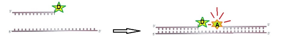
Figure 1: RNA ODN probes for FRET. Two designed ODN probes are hybridized to target RNA side-by-side. The fluorescence signal is switched on.

Static Quenching

Static quenching is a process in which fluorescence is decreased when the distance between the fluorophore and the quencher is less than 2 nm.

For static quenching ODN probes, one probe is labeled with a fluorophore and a second complementary ODN is labeled with a quencher molecule. The two labeled ODN probes are annealed together. In this configuration, the fluorophore and the quencher are close to each other resulting in no fluorescence. If the target RNA is present, the ODN is hybridized to the target with a higher affinity than to the second oligo. This reaction restores the fluorescence of the ODN labeled with the fluorophore. The observed fluorescence signal indicates that the target RNA is present. Hence, the presence of the target RNA results in a fluorescent signal originating from the ODN probe labeled with the fluorophore (figure 2).

Figure 2: Static quenching– Annealed ODN probes labeled with fluorophore and quencher.

Other probe types

Other probe designs are also possible. For example, fluorescently labeled oligonucleotide mimic probes can be designed using BNAs to enhance their hybridization affinity and stability.

A brief list is shown below:

BNA modified probes

Exiton-controlled hybridization-sensitive fluorescent oligonucleotide (ECHO) probes

Forced Intercalation (FIT) probes

Peptide nucleic acid and nano-graphene oxide (PANGO) probes

Sticky flare probes.


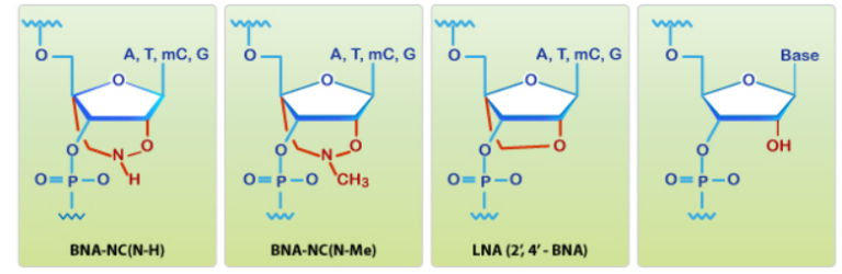
BNA probes
contain nucleotide analogs that have a bridged structure in the sugar moiety. Optimal designed BNA probes increase base-discrimination, the stability of duplex or triplex formation, and show minimal cytotoxicity. These multi-functional synthetic RNA analogs can be spiked with DNA or RNA to modify structural formation of oligonucleotides. Because of their increased affinity to targets BNA based oligonucleotides enable detection of small or highly similar DNA or RNA targets.

Exiton-controlled hybridization-sensitive fluorescent oligonucleotide (ECHO) probes are designed to have thiazole orange (TO) dyes on a modified thymidine base. An exiton is defined as a bound state of an electron and an electron hole (electron-hole pair) that are attracted to each other via electrostatic Coulomb’s interaction. In other words, an exiton is an excited state formed by the recombination of an electron and an electron hole. During relaxation, the exiton gives off light and heat. Exitons are the main mechanism of light emission in semiconductors. Energy transfer processes occurring in exitons are radiative transfer, Förster transfer, and Dexter transfer. [https://en.wikipedia.org/wiki/Exciton].

FIT probes. Another probe type is a fluorescent probe designed as an oligonucleotide mimics containing dyes that replace one oligonucleotide in the middle of the probe. This type of probe is called a “forced intercalation probe” (FIT-probe). Oligonucleotide mimics labeled with asymmetric cyanine dyes, such as thiazole orange (TO), are forced intercalation probes that emit low background noise in the single-stranded state. When the probe is hybridized to the RNA target, the result is the intercalation of the TO dye within the duplex. The result is a strong fluorescence signal. A probe designed using peptide nucleic acid with a TO dye in the middle of the probe is an example for this. However, bridged nucleic acids (BNAs) can also be used for the design of these probes.

Sticky flare probes are made off 13nm gold particles functionalized with densely packed oligonucleotides. The gold core quenches the fluorescence of the target RNA. When the target RNA is recognized, sticky-flare transfers the fluorophore-conjugated ODNs to the RNA (figure 3). Sticky-flare probes can enter live cells by endocytosis. No transfection is needed. See also https://blog-biosyn.com/2013/08/29/what-are-nano-flares/.


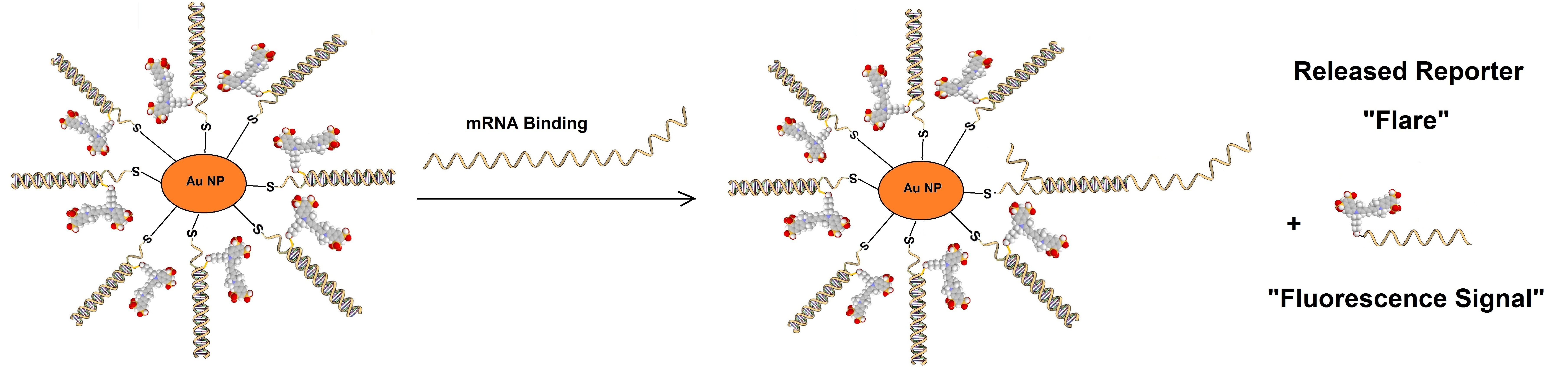
Figure 3: Sticky-flare or nano-flare based detection of mRNA.

Molecular Beacons

Molecular beacons are a type of ODN probes designed with a hairpin structure that forms a loop and a stem via self-complementary 5’ and 3’ arms. A fluorophore is attached to the end of one arm, and a quencher is attached to the end of the other arm (figure 4). Base pairing of the two arms keeps the fluorophore and the quencher in proximity which quenches the fluorescence. When the beacon encounters the target molecule containing a sequence complementary to the loop structure a probe-target hybrid is formed. This hybrid is energetically more stable than the self-complementary hairpin structure. After hybridization, the conformation of the beacon is changed, and fluorescence is restored. Since the beacon has no background signal in the absence of target molecules, higher signal-to-noise ratios are achieved in comparison to other ODNs.  

Because of the advantages molecular beacons provide, they have already been used for the tracking and imaging of mRNAs of β-actin in fibroblasts, oskar in fruit fly oocytes, influenza virus mRNA in canine kidney epithelial cells, bovine respiratory syncytial virus RNA in bovine turbinate cells, and respiratory syncytial virus RNA in Vero cells.  

Figure 4: Molecular Beacon based assay.

Furthermore, molecular beacons have a high level of specificity for target RNAs than linear ODNs, and they can be designed to allow to distinguish single nucleotide polymorphisms (SNP) in live cells. However, the melting temperature of the matching hybrids need to be above 37 °C, and the melting temperature of single nucleotide mismatch hybrids need to be below 37 °C.

Please review: 

http://www.biosyn.com/moelcular-beacons.aspx,
http://www.biosyn.com/tew/molecular-beacon.aspx
http://www.biosyn.com/tew/Design-rules-for-Molecular-Beacons.aspx


Tentacle Molecular Beacons

To achieve an even higher specificity tentacle molecular beacons with increased kinetics and affinity have also been developed. [https://www.ncbi.nlm.nih.gov/pmc/articles/PMC1904288/]. Tentacle probes are similar to molecular beacons but the presence of a capture region allows for enhanced specificity. Tentacle molecular beacons contain a hairpin structure similar to molecular beacons but are modified by the addition of a capture probe. Tentacle molecular beacons have increased kinetics and affinities. Usually, kinetic rate constants are up to 200-fold faster than that for molecular beacons with corresponding stem strengths.

Multi-color Molecular Beacons and Wavelength-Shifting Molecular Beacons

To allow simultaneous detection of multiple RNAs, multi-color molecular beacons and wavelength-shifting molecular beacons have also been developed. In the absence of targets, the probes do not fluoresce, however, when the targets are encountered the probes usually fluoresce in the emission range of the emitter fluorophore. Wavelength-shifting molecular beacons are brighter than conventional molecular beacons.  [http://www.nature.com/nbt/journal/v18/n11/full/nbt1100_1191.html]

Dual FRET Molecular Beacons

To overcome false-positive signals of conventional molecular beacons, dual FRET molecular beacons have been designed. Dual FRET probes can achieve higher signal-to-noise ratios than can single molecular beacons.

Light Induced Molecular Beacons

Light-induced molecular beacons can hybridize to their target only when activated with UV light. This approach permits the fine control of timing and location of RNA labeling. Molecular beacons can be synthesized with caged nucleobases in the loop region. The resulting constructs remain non-fluorescent in the presence of the target RNA. The exposure to light (366 or 405 nm) in vitro or in cells fully activates the beacons. Molecular beacons synthesized with the caged nucleobases dANPE, dCNPE, or sGNPP cannot form normal Watson-Crick base pairs. However, after irradiation with light, the photo-labile caging groups are removed, and the unmodified nucleobases are regenerated. This restores the ability for base pairing of the molecular beacon resulting in a fluorescent signal of the probe-target hybrid.

[http://www.biosyn.com/tew/Light-sensitive-nucleotides.aspxhttp://pubs.rsc.org/is/content/articlehtml/2012/cc/c2cc16654b,  

http://www.biosyn.com/tew/Chemical-structures-of-caged-nucleobases.aspx]

Avoiding sequestering of molecular beacons into the nucleus

Sometimes molecular beacons tend to sequester into the nucleus, which can cause a nonspecific fluorescent signal. To prevent this, large proteins or nanoparticles have been attached to molecular beacons to prevent the passing of the beacon through the nuclear pores.

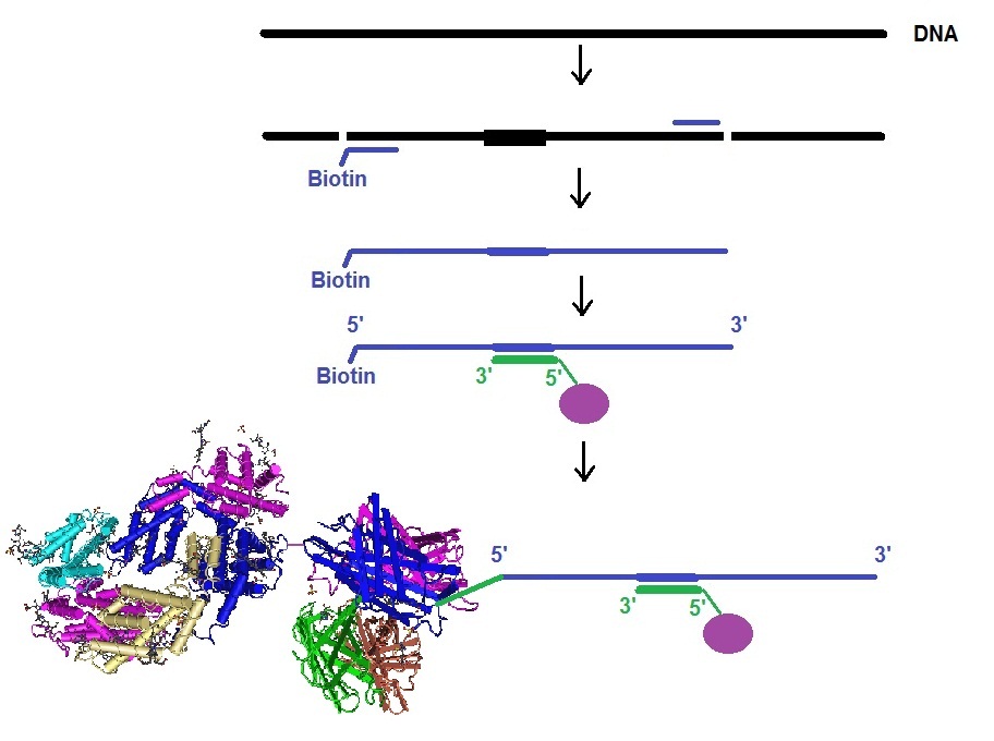
RNA Stem-Loop system

In this approach, mRNA is labeled in living cells using RNA stem-loop motifs. The MS2 bacteriophage coat protein (MCP) is known to exhibit strong affinity for the unique RNA stem-loop sequence MS2 binding site (MBS).[https://www.ncbi.nlm.nih.gov/pmc/articles/PMC413242/]. The MBS stem-loop sequence is a short oligonucleotide sequence, containing approximately 20 nucleotides. Because of this, multiple MBS stem-loops can be used for the tagging of the mRNA of interest. The signal of a single mRNA can be amplified by increasing the number of MBS stem-loops. Each MBS stem-loop can bind a dimer of MCP fused to a fluorescent protein (FB). With this approach, a single copy of mRNA can be labeled with many FBs. Other similar stem-loops such as the PP7 bacteriophage system and the λ-phage N system can also be used.

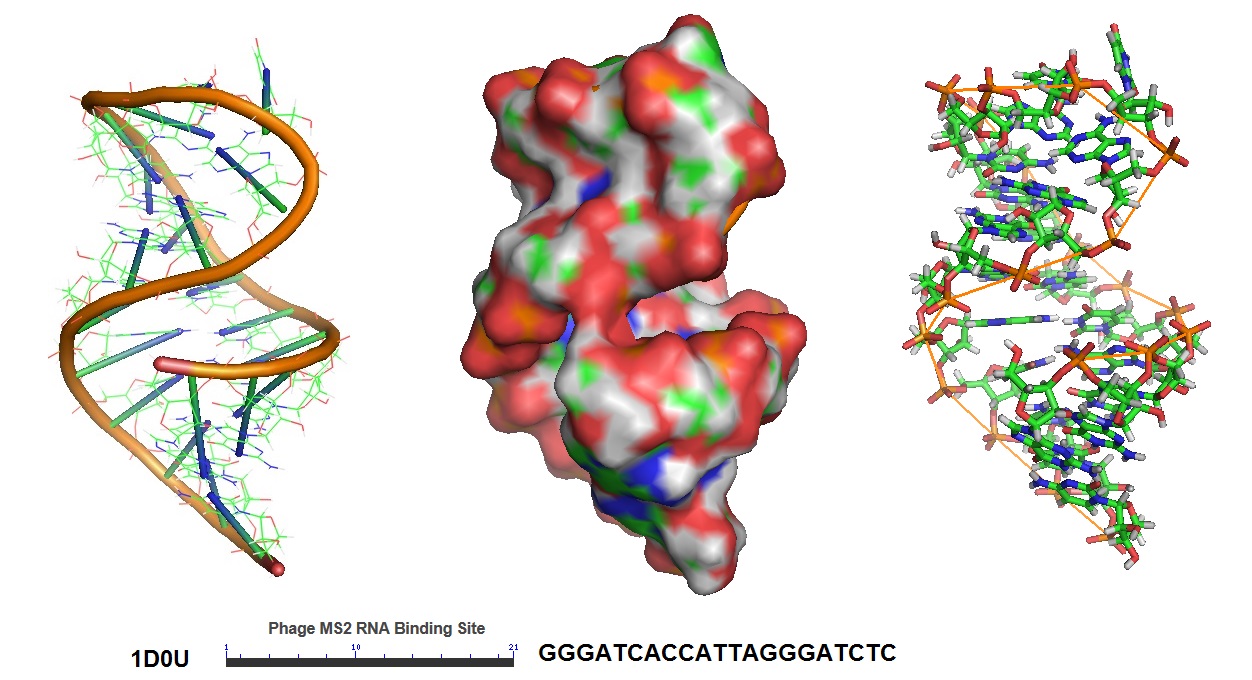
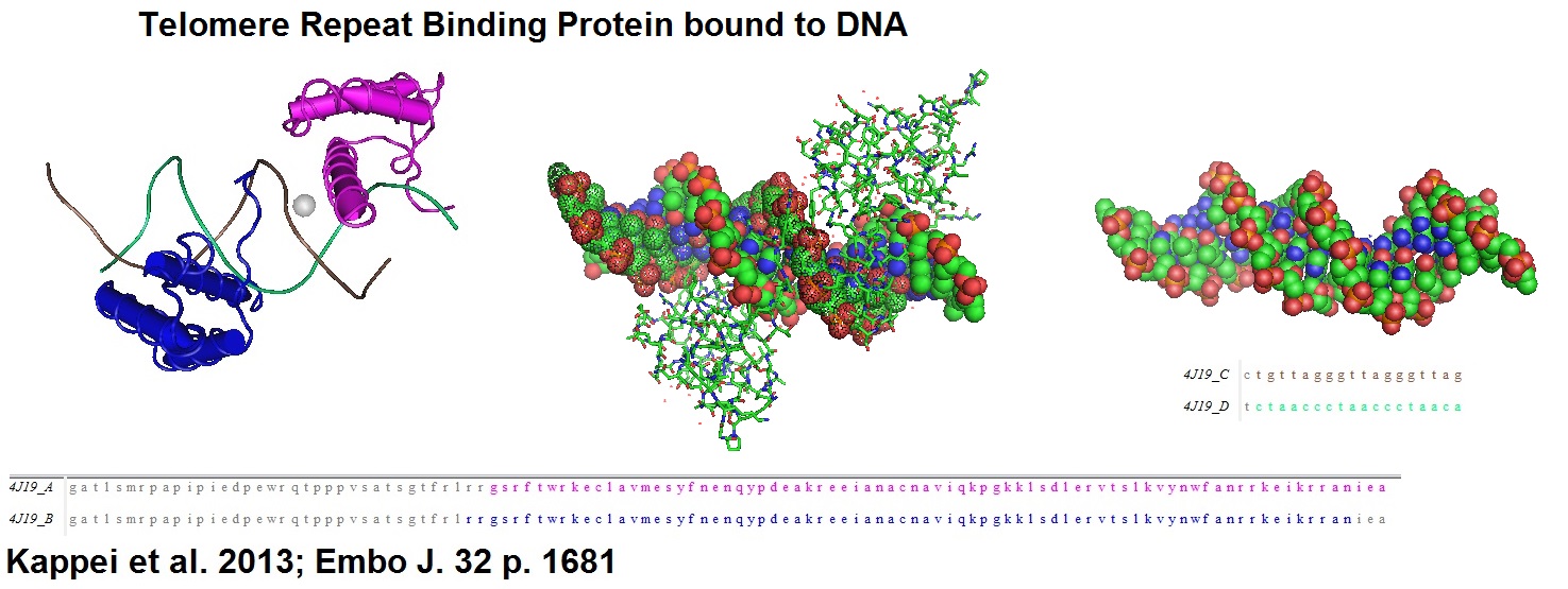
Figure 5: Structural model of the phage MS2 RNA hairpin-loop binding site.


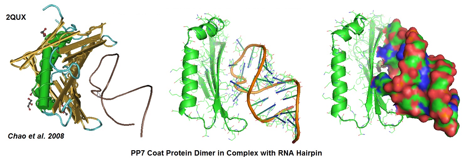
Figure 6:  Structural models derived from the cocrystal structure of the PP7 bacteriophage coat protein in complex with its translational operator (Chao et al. 2008). The structure illustrated the molecular basis of the PP7 coat protein’s selective binding to the cognate RNA. The conserved beta-sheet surface recognizes the RNA hairpin. Different depictions are used for the image. 

Aptamer-fluorogenic System

An aptamer called “Spinach” was developed to bind and activate the fluorogen, 3,5-difluoro-4-hydroxy-benzylidend imidazolinone (DFHBI). DFHBI is a derivative of the green fluorescence protein’s (GFP) fluorphore 4-hydroxybenzlidine imidazolinone (HBI). This RNA aptamer induces fluorescence of a GFP-like chromophore.

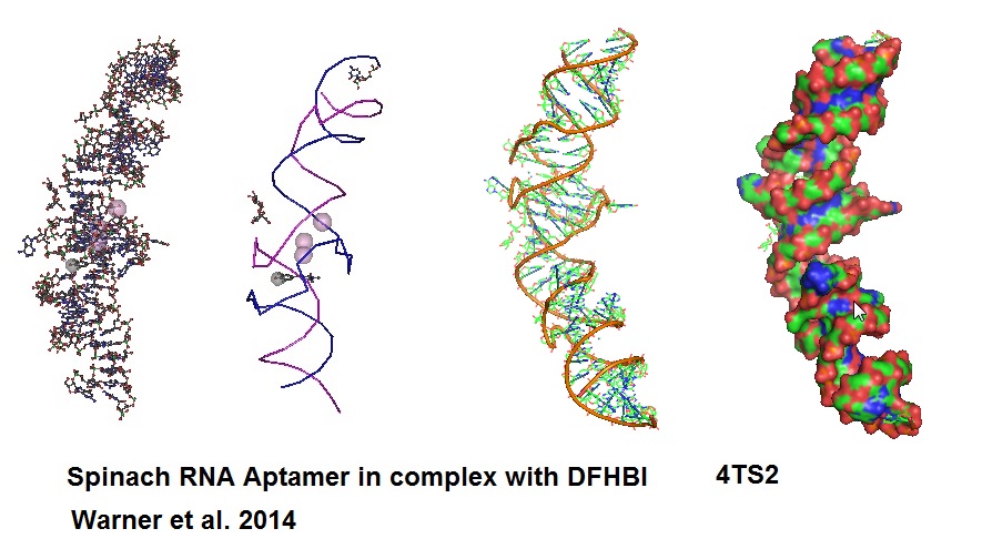
Figure 7: Structural models of RNA Aptamer Spinach.

When Spinach binds to DFHBI a Spinach-DFHBI complex is formed which emits fluorescence. Molecular modules based on the Spinach sequence can be designed for the detection of other cellular molecules. New aptamers are constantly developed to enable investigation of a variety of molecules found in cells. However, because of some thermal instabilities and misfolding tendencies of aptamers when expressed or injected into living cells, aptamers with enhanced folding properties will need to be designed. Advance protocols using Systematic Evolution of Ligands by Exponential Enrichment (SELEX) can be coupled with fluorescence-activated cell sorting (FACS) for the development of brighter RNA aptamer-fluorogenic systems.

Glossary

Å                       Ångström: 1 Å = 0.1 nm

Broccoli            Newer aptamer

DFHBI             3,5-difluoro-4-hydroxy-benzylidene

ECHO              Exciton-controlled hybridization-sensitive oligonucleotide

FIT                   Forced intercalation

FRET              Förster resonance energy transfer

GFP                 Green Fluorescence Protein

HBI                 4-hydroxybenzlidene imidazolinone

MBS                MS2 binding protein

MCP                MS2 bacteriophage coat protein

mRNA             Messenger RNA

mRNP             Messenger riponucleoprotein

ODN               Oligodeoxynucleotide

PANGO          Peptide nucleic acid nano-graphene oxide

TO                   Thiazole ornage

SNR                Signal-to-noise ratio

Spinach           RNA aptamer specifically binding to DFHBI  

Sticky-flare     Functionalized gold particle with densely packed oligonucleotides


Appendix

Table 1:  Average Bond Lenghts

Bond   Bond length (Å)         Bond   Bond length (Å)

C-C     1.54                             N-N     1.47

C=C    1.34                             N=N    1.24

C≡C    1.20                             N≡N    1.10

C-N     1.43                             N-O     1.36

C=N    1.38                             N=O    1.22

C≡N    1.16                            

C-O     1.43                             O-O     1.48

C=O    1.23                             O=O    1.21

C≡O    1.13


Reference

Chao JA Patskovsky Y Almo SC Singer RH; Structural basis for the coevolution of a viral RNA-protein complex. Nat.Struct.Mol.Biol. (2008) 15 p.103.

Hyungseok C Moon, Byung Hun Lee, Kiseong Lim, Jae Seok Son, Minho S Song and Hye Yoon Park; TOPICAL REVIEW- Tracking single mRNA molecules in live cells. Journal of Physics D: Applied PhysicsVolume 49, (2016) Number 23.

Khashti Ballabh Joshi, Andreas Vlachos, Vera Mikat, Thomas Deller and Alexander Heckel; Light-activatable molecular beacons with a caged loop sequence. DOI: 10.1039/C2CC16654B (Communication) Chem. Commun., 2012, 48, 2746-2748.

Santangelo PJ, Nix B, Tsourkas A, Bao G.; Dual FRET molecular beacons for mRNA detection in living cells. Nucleic Acids Res. 2004 Apr 14;32(6):e57.

Brent C. SatterfieldJay A.A. West, and Michael R. Caplan; Tentacle probes: eliminating false positives without sacrificing sensitivity. Nucleic Acids Res. 2007 May; 35(10): e76.  PMCID: PMC1904288.

Smith JS, Nikonowicz EP.; Phosphorothioate substitution can substantially alter RNA conformation. Biochemistry. 2000 May 16;39(19):5642-52. RNA hairpin containing the binding sitwe for bacteriophage MS2 capsid protein.

Katherine Deigan Warner, Michael C. Chen, Wenjiao Song, Rita L. Strack, Andrea Thorn, Samie R. Jaffrey, and Adrian R. Ferré-D’Amaré; Structural basis for activity of highly efficient RNA mimics of green fluorescent protein. Nat Struct Mol Biol. 2014 Aug; 21(8): 658–663. https://www.ncbi.nlm.nih.gov/pmc/articles/PMC4143336/.

--...--


Streptide, a newly discovered naturally occuring cyclic peptide

$
0
0

Streptide is a newly discovered macrocyclic naturally occurring streptococcal peptide.

Streptococcal bacteria are thought to use peptides as communication signals. Often these types of peptides contain unusual post-translational modifications. Many different biosynthetic strategies for the formation of cyclic peptides have now been observed in nature. Several cyclization reactions in which the N-terminal end is connected to the C-terminal end of the peptides have been well documented. Other biosynthetic cyclization types have also been observed, such as using isopeptide bonds, disulfide bonds, esters and thiolactones. However, the biosynthesis of the newly reported streptide peptide does not fall into any of these categories. Hence, a member of a new family of cyclic peptides has been found.


Figure 1 shows the chemical structure of streptide as deduced by Schramma et al. in 2015. The research group used several tools, such as HPLC-UV purification, NMR, mass spectrometry, solid-phase peptide synthesis as well as genetic and biochemical studies to determine the structure and conformation of streptide and its biochemical synthesis.
 

Figure 1: The chemical structure of streptide including the stereochemistry as deduced by Schramma et al. is shown in the left part of the panel. The middle and right part of the panel shows models of the peptides generated using a simple force field method. However, a NMR based model can be reviewed in the cited paper.

Streptide is produced by the pathogen Streptococcus thermophilus and was found to have a lysine-to-tryptophan (K-W) crosslink in its peptide backbone creating a cycle in the N-terminal part of the peptide. Streptococcus thermophilus or Streptococcus salivarius subspecies thermophilus is a gram-negative bacteria. This bacteria is found in fermented milk products. It is usually used for the production of yoghurt. It has been reported that yoghurt helps alleviate symptoms of mucositis which can be caused by chemotherapy in cancer patients. Mucositis is an inflammation of the mucous membranes in the digestive system which also can cause ulcers in the digestive tract. Streptococcus thermophilus is also classified as a lactic acid bacterium and is therefore considered to be a probiotic microorganism.

Reference

Kelsey R. Schramma, Leah B. Bushin and Mohammad R. Seyedsayamdost; Structure and biosynthesis of a macrocyclic peptide containing an unprecedented lysine-to-tryptophan crosslink. Nature Chemistry 2015, vol 7, 431-437.

Whitford, E. J.; Cummins, A. G.; Butler, R. N.; Prisciandaro, L. D.; Fauser, J. K.; Yazbeck, R; Lawrence, A; Cheah, K. Y.; Wright, T. H.; Lymn, K. A.; Howarth, G. S. (2009). “Effects of Streptococcus thermophilus TH-4 on intestinal mucositis induced by the chemotherapeutic agent, 5-Fluorouracil (5-FU)”. Cancer biology & therapy 8 (6): 505–11.


Teixobactin, a new antibiotic peptide that has the potential to avoid the development of resistance

$
0
0

Teixobactin is a newly discovered natural occurring antibiotic peptide.

Teixobactin is a newly discovered antibiotic peptide identified from soil samples using isolation chip (iChip, Ichip, or ichip) technology. In recent years, researchers developed the iChip technology to allow for the identification of antibiotic compounds from soil bacteria or other microbial species that are termed as “unculturable” by in-situ incubation in tiny diffusion chambers. In the early days of antibiotic discovery, most antibiotics were produced by screening soil microorganisms. Historically, molds and moldy bread are known to have been used to combat wound infections. The antibiotic penicillin is the prime example of an antibiotic compound isolated by Alexander Fleming from the mold Penicillium notatum.

Because of the heavy use of antibiotics since their introduction to human and veterinary medicine newly evolving bacteria are rapidly becoming resistant to the now commonly used antibiotics. Also, antibiotic resistance is now spreading faster than the development and introduction of new compounds that have antibiotic activities. Therefore, the quest to identify novel antibiotic compounds that avoid the development of resistance is of paramount importance. Teixobactin is reported to be one of these compounds.

Figure 1 shows the chemical structure of teixobactin as deduced by Ling et al. in 2015. 

Figure 1: Structural models of teixobactin.
Ling et al. used a multichannel device called iChip for the isolation and growth of uncultured bacteria found in soil. The iChip technology was used to screen 10,000 bacterial isolates for their antimicrobial activity against Staphyloccocus aureus. The research group succeeded in isolating an active compound with mass 1,242 Da. The use of NMR together with advanced Marfey’s analysis allowed to assign the sterochemical conformation of amino acid residues for the peptide termed teixobactin. The peptide is a depsipeptide that contains enduracididine, methylphenuylalanine, and four D-amino acids. The researchers also identified the gene cluster via a homology search using sequence data from genomic DNA from E. terrae. Furthermore, the research group was able to show that teixobactin expressed excellent activity against Gram-positive pathogen, including drug-resistant strains..

Reference

D. Nichols, N. Cahoon, E. M. Trakhtenberg, L. Pham, A. Mehta, A. Belanger, T. Kanigan, K. Lewis, and S. S. Epstein; Use of Ichip for High-Throughput In Situ Cultivation of “Uncultivable” Microbial Species. APPLIED AND ENVIRONMENTAL MICROBIOLOGY, Apr. 2010, Vol. 76, No. 8, p. 2445–2450. doi:10.1128/AEM.01754-09.

Ling, Losee L., Schneider, Tanja, Peoples, Aaron J., Spoering, Amy L., Engels, Ina, Conlon, Brian P., Mueller, Anna, Schaberle, Till F., Hughes, Dallas E., Epstein, Slava, Jones, Michael, Lazarides, Linos, Steadman, Victoria A., Cohen, Douglas R., Felix, Cintia R., Fetterman, K., Ashley, Millett, William P., Nitti, Anthony G., Zullo, Ashley M., Chen, Chao, Lewis, Kim; A new antibiotic kills pathogens without detectable resistance. Nature 2015, 517, 455-459.

Cancer genes and food that fight cancer

$
0
0

News from the Annual Meeting of the American Association for Cancer Research (AACR) 2015 held in Philadelphia April 18 – 22, 2015 at the Pennsylvania Convention Center, Philadelphia, Pennsylvania

http://www.aacr.org/Meetings/Page/MeetingDetail.aspx?EventItemID=25


Disclaimer: Due to the many posters and program items covered at this meeting and to my personal inability to attend them all I will only list highlights that are of personal interest to me.


How can cancer be prevented?

Reducing tobacco use, improving diet and physical activity, reducing obesity, and expanding the use of established screening methods is recommended to avoid suffering and death from cancer.

Furthermore, vaccination against human papillomavirus and hepatitis B virus together with minimizing extensive exposure to ultraviolet radiation is recommended to reduce major risk factors of cancer.

More and more evidence has accumulated in recent years indicating that the food a human eats affects her or his risk for developing cancer. Many scientists now estimate that between 30 to 60% of all cancers are caused by environmental factors. Since all of us have to eat the type of food we eat and what lifestyle we have may increase or decrease our risk of developing cancer. In addition, there are many indications that the majority of cancers may be preventable. Altering one’s lifestyle may achieve this goal. However, the range in percentage of these estimates varies depending on which review articles are consulted.

Two out of three humans are resistant to the development of cancer and never get it.

According to George Klein two out of three humans, never get cancer. Klein suggests that resistance genes act at the level of tissue organization in a fashion that this prevents the development of cancer in close to 60% of all humans. Many genetic and epigenetic changes appear to promote cancer development and progression. But there is epidemiological evidence that a certain proportion of the human population is very resistant to the development of cancer. However, there is also further evidence that another significant proportion is highly susceptible to cancer.

George Klein has identified five to possibly six mechanisms that have anticancer surveillance functions in mammals including humans.George Klein has identified five to possibly six mechanisms that have anticancer surveillance functions in mammals including humans.

Mechanisms of cancer resistance


MechanismNameDescription
1Immunological resistanceThis mechanism is acting mainly against virus-induced or virus-associated cancer.
2Genetic resistanceA large genetic set of DNA repair mechanisms appear to be present in the human genome.
3Epigenetic resistanceThe stringency of genetic imprinting appears to contribute to this type of resistance.
4Intracellular resistance The optimal triggering of apoptosis in cells with incorrectly activated oncogenes or extensive DNA damage may prevent the development of cancer
5Intercellular resistanceThis mechanism is also described as micro-environmental control. It is known that normal cells can inhibit the growth of neighboring polyoma-transformed cells. Oncogens, for example, myc, ras, scr and other genetic factors may be involved.
6HAMLET(a newly discovered possible surveillance mechanism): This is a protein-lipid complex formed by the interaction between compounds in human milk and a lipid cofactor at the pH in the stomach of breast-fed children. HAMLET induces p53-independent apoptosis in tumor cells. However, it leaves normally differentiated cell unaffected.

–.–

However, it may be too early to tell how exactly these mechanisms work in detail since most disease research including cancer research study non-functioning cell mechanisms. As George Klein points out, the genetics of cancer resistance has remained largely unexplored. However, it may well be that optimal food intake and lifestyle may help normalization these surveillance mechanisms in susceptible humans and thereby help to prevent cancer in these individuals. Furthermore, it seems natural to reason that foods that contain pesticides, insecticides, fungicides or antibiotics should be avoided when selecting a healthy lifestyle.

–.–

Genes that are involved in the development of cancer

GenesFunction
 OncogenesThe normal function of these genes is the regulation of cell division. Mutations in these genes can start and support aberrant growth.
 Tumor suppressor genesThese genes can suppress or block the development of cancer in mammals and humans. The stop the unwanted proliferation of cancer cells. Tumor suppressor genes regulate proteins that regulate cell division. A tumor suppressor gene no longer does its job when mutated. The result is uncontrolled cell growth which may contribute to the development of cancer.
 DNA replication and repair genesFor a cell to function normally DNA replication and repair must be accurate during its life time. Any chemical change in the DNA must be corrected to keep the encoded information uncorrupted. Mutations in these genes can lead to DNA processing and repair disorders which can ultimately lead to cancer.
Apoptosis genesApoptosis or “programmed cell death” is essential for normal development of a mammalian organism. Apoptosis is a genetically regulated process of cell self-destruction. It is a normal physiological process by which DNA-damaged or unwanted cells are removed. A functional apoptotic mechanism appears to help prevent the development of cancer. Mutations in apoptosis genes can lead to pathogenesis in mammals and humans, and the development of cancer.
  

The “American Institute for Cancer Research” (AICR) now suggests that most common cancers could be prevented if people ate a healthy diet, had at least 30 minutes of daily physical activity and maintained a healthy weight. Furthermore, to lower the risk for cancer AICR now recommends eating a mostly plant-based diet, limiting the intake of red meat and avoiding processed meat. Plant foods contain many kinds of substances that fight cancer such as vitamins, minerals, fiber and phytochemicals or natural occurring plant substances. Hence, organic food is the choice of food to eat since analytical studies suggest that this type of food has the lowest level of contaminations.

Phytochemicals that are present in the human diet are known to work together and help preventing cancer and other diseases.

Potentially, phytochemicals stimulate the immune system, slow down the growth of cancer cells and prevent DNA damage which may lead to cancer. However, no single phytochemical or food protects from cancer or any other disease. Therefore, eating a varied diet with lots of fruits, vegetables, beans and whole grains is highly recommended. Existing evidence indicates that this can offer the most protection against cancer and other diseases. For good health the United States Department of Agriculture (USDA) recommends to eat a healthy diet that includes fruits, vegetables, whole grains and legumes. The reported best health protection is gained when eating a wide variety of foods within each food group.

–.–

Phytochemicals in the diet

Phytochemicals  Plant SourcesPossible Benefits
Allium Compounds

Allicin
Alliin
Allyl sulfides

Chives, garlic, leeks, onions, scallions, shallotsSlow or stop the growth of tumors.

Foods in the allium family probably protect against stomach cancer.
Garlic probably decreases the risk of developing colorectal cancer.
Laboratory studies suggest that allium compounds might also protect against leukemia and
cancers of the prostate, bladder, skin and lung.

Hint: Cutting or crushing garlic at least 10 minutes before cooking makes more cancer fighters available.

Carotenoids

Alpha-carotene
Beta-carotene
Beta-cryptoxanthin
Lutein
Lycopene
Zeaxanthin

Red, orange, yellow and some dark green
Fruits:Apricots, cantaloupe, citrus fruits, nectarines,
papayas, peaches, watermelon
Vegetables:

Bok choy, broccoli, carrots, corn, greens (collards, kale, lettuce, spinach), pumpkin, red peppers, sweet potatoes, tomatoes and tomato products, winter squash

Act as an antioxidant
Inhibit cancer cell growth
Improve immune response 
Foods containing carotenoids probably protect against cancers of the mouth, pharynx and larynx.Carotenoids in dark leafy vegetables may inhibit the growth of cancers of the skin, lung, stomach and some types of breast cancer cells.
Flavonoids

(a group of Polyphenols)

Anthocyanidins (cyanidin, delphinidin, malvidin, pelargonidin, peonidin)
Flavan-3-ols (catechin, epicatechin, epicatechin gallate, epigallocatechin,
epigallocatechin gallate, theaflavins), Flavanols (kaempferol, myricetin)
Flavanones (hesperetin and naringenin), Flavones (apigenin and luteolin)
Flavonoids (quercetin)
Isoflavones

Fruits:

Apples, berries, cherries, citrus fruits, cranberries, currants, grapes, pears, plums

Vegetables:

Beets, bell peppers, broccoli, celery, chard, eggplant, kale, lettuce, onions, radishes, red cabbage

Beans:

Black beans, kidney beans, pinto beans.

Herbs:

Parsley, rosemary, thyme

Other:

Cocoa powder, dark chocolate, coffee, tea

Support antioxidant defenses

Increase the enzymes that protect the body from cancer-causing compounds

Stimulate self-destruction of abnormal cells Inhibit inflammation that supports cancer growth

Inhibit tumor growth Boost immune function

In laboratory studies, the phytochemicals in apples reduced the growth of lung cancer cells.

Indoles

Indole-3-carbinol

Cruciferous Vegetables:

Broccoli, Brussels sprouts, cabbage, cauliflower, horseradish, mustard greens, turnips, watercress

Support antioxidant defenses
Increase the activity of enzymes that protect the body from cancer-causing compounds
Helps repair damaged DNA
Cause cancer cells to die
Decrease estrogen’s power to promote certain cancers
Laboratory studies suggest that compounds found in cruciferous vegetables protect against
some types of cancer.
Inositol

Phytic acid (also called inositol hexaphosphate or IP’6)

Whole Grains:

Bran from corn, oats, rice, rye and wheat

Nuts

Legumes: Soybeans and dried beans,

Supports antioxidant defensesSlows growth of tumors
Causes cancer cells to die
Laboratory studies suggest that phytic acid may prevent the formation of tumors.
Isoflavones
(a category of flavonoids)
Daidzein
Genistein
Glycitein
Soy:

Soybeans and soy products (such as edamame, soymilk, tofu)

Support antioxidant defenses
Decrease production of some hormones
Inhibit growth of tumors
Isothiocyanates

Allyl isothiocyanate
Benzylisothiocyanate
Crambene
Phenylethylisothiocyanate
Sulforaphane
3-phenylpropylisothio-cyanate

Cruciferous Vegetables:

Arugula, bok choy,
broccoli, Brussels sprouts, cabbage (red and green), cauliflower, collard greens, horseradish,
kale, mustard greens, radishes, rutabaga, turnips,
watercress

Support antioxidant defenses
Block tumor growth
Cause cancer cells to die
Inhibit inflammation that supports cancer growth
Increase the activity of enzymes that protect the body from cancer-causing compounds
Laboratory studies suggest that compounds in cruciferous vegetables protect against some types of cancer.
Isothiocyanates form when glucosinolates in cruciferous vegetables are broken down by an
enzyme released when the vegetable is chewed or chopped.
Polyphenols

(other than Flavonoids
and Terpenes)

Coumarin
Curcubitacin
Curcumin
Phenolic Acids (caffeic acid, ferulic acid,
ellagic acid, gallic acid)
Stilbenes (pterostilbene, resveratrol)
Tannins (such as ellagic acid)

Fruits:

Apples. blackberries, black raspberries,blueberries, cherries, red grapes, pears, pomegranates, strawberries

Other:

Chestnuts, lentils, peanuts, pecans, turmeric,
walnuts

Support antioxidant defensesInhibit inflammation that supports cancer growth
Prevent cancer formation
Cause abnormal cells to die before they can become cancerous
Cell culture and animal studies suggest pterostilbene may help prevent cancer by inhibiting
growth and stimulating destruction of abnormal cells.In laboratory and animal studies, resveratrol has inhibited the formation or slowed the growth of cancer.In lab studies, ellagic acid has inhibited the development of some types of cancer.In the lab, curcumin has reduced the formation and growth of cancer cells.
 Protease InhibitorsLegumes:

Beans, lentils, peas, soybeans and whole
soy products (such as edamame, soymilk, tofu)

Inhibit cancer cell growth
Prevent tumors from releasing compounds that can destroy nearby healthy cells
Sterols

Beta-sitosterol
Campesterol
Ergosterol
Stigmasterol

Vegetables:

Asparagus, beets, Brussels sprouts
Legumes, Nuts
Seeds:

Flax, pumpkin, sesame and sunflower seeds
Most Vegetable Oils: Canola, corn, olive, safflower and sesame oils
Whole Grains

Cause the death of cancer cells

Lessen the inflammation that supports cancer growth

Terpenes

(a group of Polyphenols) Carnosol
Curcubitacin
Limonene
Perillyl alcohol

Fruits:

Apples, cherries, citrus fruits, pears, prunes
Herbs:

Bay leaves, dill, oregano, parsley, rosemary,
sage, thyme
Other Foods:

Pumpkin seeds

Support antioxidant defenses
Slow cancer cell growth
Boost immune function
Inhibit inflammation that supports cancer growthLaboratory studies suggest carnosol may decrease cells’ sensitivity to reproductive hormones that may promote cancer.
   

 –.–

Many phytochemicals are antioxidants. These are compounds that protect the body’s cells from oxidative damage. Oxidative damage or stress is caused by an increased production of reactive oxygen species in human cells. Free radicals and other oxygen-derived species are constantly produced in the human body. Furthermore, humans are constantly exposed to oxidizing air pollutants such as ozone, oxides of nitrogen, tobacco smoke, and motor vehicle exhaust. Research has shown that a variety of antioxidant defenses have evolved in mammals including humans that protect against these reactive oxygen species. These defenses include enzymes, vitamins, and other natural antioxidants. However, these defenses are not always efficient. Many studies now suggest that antioxidants substances such as phytochemicals and vitamins found in fruits and vegetables can help the body protect against this type of damage.

References

American Institute for Cancer Research: www.aicr.org/foods-that-fight-cancer

Robert A Jacob and Betty J Burn; Oxidative damage and defens. Am J Clin Nutr 1996; 63:985S-90S.

George Klein; Toward a genetics of cancer resistance. PNAS 2009, vol. 106 no. 3, 859-863.

How does the BNA bead-based assay for somatic mutations work?

$
0
0

Shivarov et al. designed and validated this BNA[NC] probe bead-based assay for the Luminex Lab Scan 200 flow platform to allow for the detection and quantification of somatic mutations in Leukemia!

-.-

How does the assay work?

Primers are designed to allow for the amplification of a DNA sequence fragment that contains the mutated sequence codon. One of the primers is labeled with biotin.

  1. First, human genomic DNA is extracted from blood.
  2. The exon 23 of the human DNMT3A gene is amplified using the selected forward and reverse primer.
  3. To determine the exact sequence the purified and amplified DNA piece can be sequenced using Big Dye terminator cycle-sequencing.
  4. Next, the exon 23 DNMT3A fragments are amplified from either genomic or plasmic DNA samples using a 5’-biotinylated forward primer.
  5. Genotyping is performed with the BNA[NC] modified oligonucleotide probes connected to microsphere beads, specific for the wild type or the mutant alleles, by direct hybridization.
  6. The captured DNA fragment containing biotin on its 5’-end is detected with the help of streptavidin-phycoerythrine (SAPE) in the hybridization buffer using the LabScan200 flow platform from Luminex (USA). For more detail review Shivarov et al. 2014.

The outline of the assay is illustrated in the next figure.
bna-luminex-assay


Figure 1: Bead-based suspension assay using BNA [NC] probes to detect and quantify somatic mutations in leukemia. The amplified DNA fragment containing the mutations is captured by the BNA/DNA probes and quantitatively detected with the help of the SAPE complex allowing the analysis in a Luminex system.

Reference

Shivarov V, Ivanova M, Naumova E; Rapid Detection of DNMT3A R882 Mutations in Hematologic Malignancies Using a Novel Bead-Based Suspension Assay with BNA(NC) Probes. PLoS One. 2014 Jun 10;9(6):e99769. doi: 10.1371/journal.pone.0099769. eCollection 2014.

bna

A novel BNANC bead-based assay for somatic mutations in Leukemia

$
0
0

Shivarov et al. designed and validated a novel bead-based suspension assay using BNA[NC] probes for the Luminex Lab Scan 200 flow platform to detect and quantify somatic mutations in Leukemia!

-.-

Shivarov et al. recently develop and validated a novel bead-based suspension assay that uses BNA [NC] probes and the Luminex (USA) LabScan200 flow platform to allow the detection and quantification of DNMT3A p.R882C/H/R/S mutations in the blood of leukemia patients. The comparison with LNA based probes revealed the superior hybridization characteristics of the BNA based probes. The researchers were able to demonstrate for the first time the use of BNA[NC] probes coupled to fluorescently labeled beads for quantitative detection of DNMT3A R882 mutations. This type of assay has the potential for other molecular diagnostic applications as well.

-.-

Molecular diagnostics, techniques and methods used to analyze biological markers in genomic and proteomic research to diagnose and monitor disease, detect risk, and decide which therapies will work best for individual patients, offers the promise to enable and optimize personalized medicine.
bna-bb-assay

Bead-based suspension assay using BNA [NC] probes and the Luminex system to detect and quantify somatic mutations in leukemia. First, primers and probes are designed specific for the mutated sequence codons. Second, DNA is isolated from blood, and, third, amplified by PCR. Fourth, the biotinylated DNA fragments are captured with the capture probe connected to the beads, and, fifth, detected with a streptavidin-phycoerythrine (SAPE) complex. Sixth, the sample is analyzed using a Luminex instrument. The level of SAPE fluorescence is proportional to the amount of the captured DNA fragment.

-.-

Bead-based suspension assay using BNA [NC] probes and the Luminex system to detect and quantify somatic mutations in leukemia. First, primers and probes are designed specific for the mutated sequence codons. Second, DNA is isolated from blood, and, third, amplified by PCR. Fourth, the biotinylated DNA fragments are captured with the capture probe connected to the beads, and, fifth, detected with a streptavidin-phycoerythrine (SAPE) complex. Sixth, the sample is analyzed using a Luminex instrument. The level of SAPE fluorescence is proportional to the amount of the captured DNA fragment.

Leukemia is an often malignant disease of the blood that has the tendency to become progressively worse. This type of cancer of the blood or bone marrow is characterized by an abnormal increase of immature white blood cells. However, the term leukemia is also used to cover a whole spectrum of diseases affecting the blood, bone marrow, and lymphoid system, all known as hematological neoplasms. Leukemia is now considered to be a treatable disease. Several hematologic malignant tumors are characterized by genome instabilities. As identified by whole genome sequencing these cancer types frequently have between 10,000 and 100,000 mutations in their entire genomes and mutations in the human DNA methyl transferase 3A (DNMT3A) gene have now been identified in several blood diseases. The methyl-group transferring enzyme DNA methyltransferase 3A (DNMT3A) is one of two human de-novo DNA methyltransferases essential for the regulation of gene expression and mutations and deletions in this protein have been observed in acute myeloid leukemia (AML), Acute lymphoblastic leukemia (ALL), myelodysplastic sydromes and myeloproliferative neoplasms. Myeloid cells represent a prominent part of local inflammatory infiltrates in the central nervous system (CNS) and appear to strongly contribute to the local inflammatory milieu and the pathological outcome of diseases involving these cells.

In 2013 Kim et al. used PCR and direct sequencing to analyze mutations of DNMT3A amino acid residue R882 in 99 acute leukemia patients, including 57 AML patients, 41 ALL patients and a single biphenotypic acute leukemia (BAL) patient. The most common immunophenotype in BAL patients is defined by the coexpression of B-lymphoid and myeloid markers and less frequently, T-lymphoid and myeloid markers. BAL has a high incidence of clonal chromosomal abnormalities, the most common being the t(9;22) (q34;q11) (Ph chromosome) and structural abnormalities involving 11q23. Data are emerging that BAL has a negative prognosis in both children and adults and this may be related to the underlying chromosome abnormalities. The research group detected DNMT3A expression in mononuclear cells of the bone marrow in these patients and in normal individuals using real time quantitative polymerase chain reaction. Approximately 17.5% (10/57) of AML patients were found to exhibit DNMT3A mutations, and four missense mutations were observed in the DNMT3A mutated AML patients, including R882 mutations and a novel single nucleotide polymorphism resulting in a M880V amino acid substitution. It is now known that somatic heterozygous mutations of the DNA methyltransferase gene DNMT3A occur frequently in acute myeloid leukemia and other hematological malignancies. The majority (∼60%) of these affect a single amino acid, Arg882 (R882), located in the catalytic domain of the enzyme. In 2013, Kim et al. could show that exogenously expressed mouse Dnmt3a proteins that have the corresponding R878 mutations largely fail to mediate DNA methylation in murine embryonic stem (ES) cells but are capable of interacting with wild-type Dnmt3a and Dnmt3b. The coexpression of the Dnmt3a R878H (histidine) mutant protein resulted in inhibition of the wild-type Dnmt3a and Dnmt3b to methylate DNA in murine ES cells. In addition the expression of Dnmt3a R878H in ES cells containing endogenous Dnmt3a or Dnmt3b induced hypomethylation which suggests that the DNMT3A R882 mutations, in addition to being hypomorphic, have dominant-negative effects. The current literature suggests that that the presence of DNMT3A mutations is an adverse prognosis biomarker in adult acute myeloid leukemia and that the rapid detection of DNMT3A R882 codon mutations allows for the early identification of poor risk patients with acute myeloid leukemia.

Shivarov et al. designed and validated a novel bead-based suspension assay using BNA[NC] probes for the Luminex Lab Scan 200 flow platform to detect and quantify somatic mutations in Leukemia!


Reference


Soo Jin Kim, Hongbo Zhao, Swanand Hardikar, Anup Kumar Singh, Margaret A. Goodell, and Taiping Chen; A DNMT3A mutation common in AML exhibits dominant-negative effects in murine ES cells. December 12, 2013; Blood: 122 (25).


Shivarov V, Ivanova M, Naumova E; Rapid Detection of DNMT3A R882 Mutations in Hematologic Malignancies Using a Novel Bead-Based Suspension Assay with BNA(NC) Probes. PLoS One. 2014 Jun 10;9(6):e99769. doi: 10.1371/journal.pone.0099769. eCollection 2014.

df

.

Biosynthesis Incorporated

has 30 years experience in the analysis and synthesis of synthetic peptides, proteins, DNA and RNA oligonucleotides, bioconjugates and other biomolecules.

Call us at 1-800-227-0627

or visit our website at

www.biosyn.com“.

Web resources for the CRISPR Cas System

$
0
0

Drosophila CRISPR Web Resources

Name

Link

Description

OxfCRISPR

(Liu Lab)

http://www.oxfcrispr.org

Oxford Fly CRISPR Resources

CRISPRflydesign (Bullock Lab)

http://www.crisprflydesign.org/

Offers Cas9 transgenic stocks

DRSC CRISPR finder (Perrimon Lab)

http://www.flyrnai.org/crispr/

A web tool to identify CRISPRs for fly study

FlyCas9

(Ueda Lab)

http://www.shigen.nig.ac.jp/fly/nigfly/cas9/index.jsp

Provides reagents, protocols and online tools for genome engineering by the designer nuclease Cas9 in Drosophila

flyCRISPR(O’Connor-Giles Lab)

http://flycrispr.molbio.wisc.edu/

Fly CRISPR resources

flyCRISPR discussion group


https://groups.google.com/forum/#!forum/flycrispr-discussion-group

A forum for sharing developments, insights, ideas and asking questions related to fly CRISPRs

Fly target sites

(Zhang lab)

http://www.genome-engineering.org/crispr/?page_id=41

For application of Cas9 for site-specific genome editing in eukaryotic cells and organisms

General CRISPR Resources

Name

Link

Description

Addgene CRISPR plasmids

 http://www.addgene.org/CRISPR/

A collection of CRISPR plasmids and reagents

Crass: The CRISPR Assembler

http://ctskennerton.github.io/crass/

A program that searches through raw metagenomic reads for CRISPRs

CRISPI

http://crispi.genouest.org/

A web interface with graphical tools and functions allows users to find CRISPR in personal sequences.

CRISPR Discussion Forum

https://groups.google.com/forum/#!forum/crispr

A forum to discuss Genome Engineering using CRISPR/Cas Systems

CRISPRmap

http://rna.informatik.unifreiburg.de/CRISPRmap


Web server provides an automated assignment of newly sequenced CRISPRs to standard classification system

CRISPRs web server


http://crispr.u-psud.fr/

A gateway to publicly accessible CRISPRs database and software, including CRISPRFinder, CRISPRdb and CRISPRcompar

CRISPRTarget

http://bioanalysis.otago.ac.nz/CRISPRTarget


Predicts the most likely targets of CRISPR RNAs

E-CRISP

http://www.e-crisp.org

A software tool to design and evaluate CRISPR target sites

Goldstein lab CRISPR

http://wormcas9hr.weebly.com/

A genome engineering resource for the C. elegans research community

Joung lab CRISPR

http://www.crispr-cas.org/

A genome engineering resource for zebrafish research community

Zhang lab Genome Engineering

http://www.genomeengineering.org/


CRISPR genome engineering resources website

ZiFiT target design tool

http://zifit.partners.org/ZiFiT/


Identifies potential target sites in DNA sequences

Contact us for all the synthetic RNA oligonucleotides you need!

Long RNA oligos are available at Biosynthesis Inc.

Please inquire by calling 1-800-227-0627

or by clicking   www.biosyn.com

How can the CRISPR Cas System be used?

$
0
0

Potential Uses or Applications of the CRISPR Cas System !

Several Uses of Cas9 Induced Double Strand Breaks for Genomic Engineering as Suggested by Basset and Li in 2014 are listed below.

Most applications of CRISPR Cas9 use the induction of double strand breaks at specific sites within a genome, which can be repaired by using either non-homologous end joining (NHEJ) or homologous recombination (HR). This allows the generation of mutations and manipulation of genomes in a defined manner.

-.-

Potential uses or applications of engineered CRISPR Cas systems are:

  • Regulation of transcription by interference with RNA polymerase (CRISPRi).
  • Fusion to transcriptional activation (Transcriptional activation, to transcription activator VP64).
  • Fusion to repression domains (Transcriptional repression, to KRAB or Krüppel associated box domain, a transcriptional repression domain).
  • DNA tagging with green fluorescent protein (GFP).
  • Chromatin purification using affinity tags.
  • RNA recruitment by fusion to the sgRNA.
  • Altering DNA topology by using dimerization domains (DNA looping).
  • Chromatin modification with histone methyltransferases (HAT), demethylases (KDM) or deacetylases (HDAC) that can also be tagged to the appropriate domains.
  • Knock-in/knock-out Cell Lines.
  • Gene replacement.
  • Gene editing including single base mutations. 
  • Gene tagging. 
  • Gene Therapy.  
  • Promoter modifications.   
  • Mutagenesis. 
  • Removal of viral sequences.  
  • Recombinant protein production in CHO cells. 
  • Disease model in human cell lines for drug discovery.  
  • Gene therapy in diseased cell line.
  • Generation of a specific cell lines for gene function studies.

Scientists in the field of genome editing expect that this technique will change the way researchers think about and perform genetic and genomic editing and analysis.

Synthetic long RNA oligonucleotides are available at Biosynthesis Inc.

Please inquire calling 1-800-227-0627

or by clicking   www.biosyn.com

Reference

Bassett, A.R., Liu, J.-L., CRISPR/Cas9 and genome editing in Drosophila, Journal of Genetics and Genomics (2014), doi: 10.1016/j.jgg.2013.12.004.


Structural Models of the CRISPR Cas System

$
0
0

As of December 2013 90 solved structures were available in the PubMed structural database related to the CRISPR Cas System!

A few these models are shown below!

A PubMed search for “CRISPR Cas” showed that close to 90 solved structures related to this system were available by the end of December in 2013. The model of the CRISPR associated Cse3 protein, in complex with RNA published by Gesner et al. in 2011 is shown below.

cse5-rna-complex

Figure 1:  Model of the structure of Thermus Thermophilus Cse3 bound to RNA.

(Note: To generate the models PDB files were retrieved from the PubMed structure database and rendered using the modelling software packages Cn3D 4.3 and Pymol)

 -.-

Jore et al. in 2011 reported the structure of CRISPR RNA (crRNA) which contains a 5’handle connected to a spacer sequence and a 3’handle. As can be seen from the figure below, the 3’handle is thought to form a hairpin structure.  This type of loop is an unpaired loop of RNA that is created when an RNA strand folds and forms base pairs with another section of the same strand. The resulting structure is called a hairpin structure and looks like a loop or a U-shape.

Figure 2:  Structure of crRNA. (As proposed by Jore et al. in 2011).

Jinek et al. in 2012 reported models of the naturally occurring and engineered RNA-guided nuclease systems. The model for the naturally occurring RNA-guided nuclease system is shown below.

Figure 3:  Schematic model of the naturally occurring RNA-guided nuclease systems.

The naturally occurring dual RNA-guided Cas9 nuclease is illustrated. crRNA interacts with the complementary strand of the DNA target site harboring an adjacent PAM sequence, shown as green and red text. TracrRNA base pairs with the crRNA, and the overall complex is recognized and cleaved by Cas9 nuclease shown in light blue color. Folding of the crRNA and tracrRNA molecules is predicted by the program Mfoldand the association of the crRNA to the tracrRNA is partially based on the model proposed by Jinek et al. (2012).

Spilman et al. in 2013 reported that they have solved the structure of an RNA Silencing Complex of the CRISPR Cas Immune System using cryoelectron microscopy. The research group reconstructed a functional Cmr complex bound with a target RNA at a resolution of approximately 12 A°. They showed that pairs of the Cmr4 and Cmr5 proteins form a helical core that is asymmetrically capped on each end by distinct pairs of the four remaining subunits: Cmr2 and Cmr3 at the conserved 50 crRNA tag sequence and Cmr1 and Cmr6 near the 30 end of the crRNA. The structure revealed that the shape and organization of the RNA targeting Cmr complex is strikingly similar to the DNA-targeting Cascade complex. In addition these results revealed a remarkably conserved architecture among very distantly related CRISPR Cas complexes.

The next figure shows the model of the functional Cmr complex bound to a target RNA.


olo-cmr-complex-mode


Figure 4:  Overview of the P. furiosus CRISPR/Cas Locus and the Cmr Complex Structure

(adapted from Spilman et al. 2013). The structure was determined by cryoelectron microscopy.

(A)  The genes encoding the Cmr complex protein subunits are color coded to match those used for structure images in subsequent figures. The CRISPR repeats are shown in black and spacers in various colors.

(B)  The mature 39 nt and 45 nt crRNAs contain a 50 repeat-derived 8 nt sequence (50 tag) and a 31 nt or 37 nt spacer-derived sequence (guide), respectively. The sequence of the target RNA used in RNA cleavage assays and assembly with the Cmr complex is shown in blue.

(C)  Color-coded EM density of the Cmr complex bound with the 45 nt crRNA and the target RNA is shown in two orientations. Cmr1, red; Cmr2, light blue; Cmr3, orange; Cmr4, three different shades of green; Cmr5, three different shades of yellow; and Cmr6, magenta.

Do you need long RNA oligonucleotides?

Modified or unmodified?

Biosynthesis Inc. can synthesize them for you!

Please inquire calling 1-800-227-0627

or by clicking   www.biosyn.com


Reference

Gesner EM, Schellenberg MJ, Garside EL, George MM, Macmillan AM; Structure of Thermus Thermophilus Cse3 Bound to an RNA Representing a Product Complex. Nat.Struct.Mol.Biol. (2011) 18 p.688. PDB ID: 3QRR]

Matthijs M Jore, Magnus Lundgren, Esther van Duijn, Jelle B Bultema, Edze R Westra, Sakharam P Waghmare, Blake Wiedenheft, Ümit Pul, Reinhild Wurm, Rolf Wagner, Marieke R Beijer, Arjan Barendregt, Kaihong Zhou, Ambrosius P L Snijders, Mark J Dickman, Jennifer A Doudna, Egbert J Boekema, Albert J R Heck, John van der Oost & Stan J J Brouns; Structural basis for CRISPR RNA-guided DNA recognition by Cascade. Nature Structural & Molecular Biology 18, 529–536 (2011). doi:10.1038/nsmb.2019.

Jinek M, et al. A programmable dual-RNA-guided DNA endonuclease in adaptive bacterial immunity. Science. 2012; 337:816–821. [PubMed]

Michael Spilman,Alexis Cocozaki,Caryn Hale,Yaming Shao,Nancy Ramia,Rebeca Terns,Michael Terns,Hong Li,and Scott Stagg; Structure of an RNA Silencing Complex of the CRISPR-Cas Immune System. Molecular Cell 52, 146–152, October 10, 2013.

Highlighted Reports on the CRISPR Cas System

$
0
0

Selected papers for CRISPR Cas!

Segal and Meckler in 2013 state in ‘Genome Engineering at the Dawn of the Golden Age,’ that, “In summary, compared with ZFNs, TALENs can be designed for more targets, are more likely to be active on their targets, display higher on-target activity, and display lower off-target activity. New assembly methods and the current freedom from patent limitations have made TALENs more accessible and able to be produced in greater quantities than has been possible with ZFNs.” However, the CRISPR Cas system has the potential to surpass this.

The impact for the future of CRISPR Cas is reflected by the following statement using the words of Seagal and Meckler: CRISPR Cas: The Nuclease of the Future?  Even as TALENs were displacing ZFNs as the tool of choice for generating site-specific double strand breaks in DNA, a newly characterized natural endonuclease system threatened to make both obsolete. “ Jinek et al. in 2012 reported that the CRISPR Cas system from bacteria and archaea mediates DNA cleavage by using simple base pairing to specify the cut site. The CRISPR Cas systems provide invader-specific, adaptive and heritable immunity against viruses and plasmids by protecting against invading nucleic acids by guiding the molecular scissors to cleave non-host sequences that have been seen before into smaller pieces. The CRISPR Cas system is a novel microbial defense system found in half of the bacterial and almost all archaeal genomes sequenced. To function, the system needs the CRISPRs and the Cas proteins and relies on non-translated RNAs to track and inactivate invasive genetic elements to protect the cells genomic integrity. Jinek et al. discovered that one version of the CRISPR Cas system can be simplified to a single protein, Cas9, along with a single chimeric guide RNA to enable effective cleavage.

There are three types of CRISPR Cas systems, type I, II and III. Type I and II share specialized Cas endo-nucleases that process the pre-crRNAs, and once mature, each crRNA assembles into a large multi-Cas protein complex that recognizes and cleaves nucleic acids complementary to the crRNA. However, type II processes pre-crRNAs using a different mechanism in which a trans-activating crRNA (tracrRNA) complementary to the repeat sequences in pre-crRNAs triggers processing of the double-stranded RNA-specific ribonuclease RNAse II in the presence of the Cas9 protein. The Cas9 protein appears to be the responsible part for crRNA-guided silencing of foreign DNA. The type II CRISPR system works by incorporating short exogenous DNA sequences from the invading pathogen into specif loci of the host genome. At the time of transcription, these sequences are processed into crRNAs via pre-crRNAs. The crRNAs act as a guide for the Cas9 nuclease machinery. The Cas9 nuclease cleaves and inactivates the foreign DNA. The system requires two more pieces to function. A tracrRNA that forms base-pairs with the crRNA provides the substrate for the host’s ribonuclease RNase III. This system can identify DNA sequences that are complementary to the crRNA and degrade them. 

Cong et al. in 2013 showed that this three-component system made up of the

(1)  guide RNA (crRNA) that hybridizes to the target DNA,

(2)  the protein nuclease Cas9 that cleaves the target DNA (bacterial Cas9), and

(3) a linker RNA that brings the nuclease to the guide RNA (the tracrRNA) is sufficient to mediate efficient genome editing in human cells.

On the other hand, Mali et al. showed that a two-component system that included the

(1)  Cas9 protein and

(2)  a guide RNA consisting of a crRNA-tracrRNA hybrid molecule was also sufficient.

By designing guide RNAs for five DNA target sites Jinek et al. were able to demonstrate that the system had the ability to cleave all the sites in vitro using only purified Cas9 and the guide RNAs. The target site needed to contain a NGG motif immediately adjacent to the complementary region, providing a CRISPR Cas nuclease site at approximately every 8 bp in random DNA.

Instead of engineering new proteins for each cleavage site, researchers now need only to synthesize a new guide RNA to program the nuclease.

A comparison of the TALEN with the CRISPR Cas system reveals that a typical TALEN requires two new ~1,800-bp repeat coding regions to be assembled for each new target site, in contrast, a CRISPR Cas system would require just one new 20-nucleotide (nt) DNA-complementing region of the ~100-nt guide RNA, allowing any investigator to create hundreds or thousands of nucleases at low cost.

Synthetic long RNA oligonucleotides are available at Biosynthesis Inc.

Please inquire calling 1-800-227-0627

or by clicking   www.biosyn.com

Reference

Cong, L, Ran, FA, Cox, D, Lin, S, Barretto, L, Habib, N et al. (2013). Multiplex genome engineering using CRISPR/Cas Systems. Science, e-pub ahead of print 3 January 2013.

Jinek M, et al. A programmable dual-RNA-guided DNA endonuclease in adaptive bacterial immunity. Science. 2012; 337:816–821. [PubMed]

Mali, P, Yang, L, Esvelt, KM, Aach, J, Guell, M, Di-Carlo, JE et al. (2013). RNA-guided human genome engineering via Cas9. Science, e-pub ahead of print. 3 January 2013.

David J. Segal and Joshua F. Meckler; Genome Engineering at the Dawn of the Golden Age Annu. Rev. Genomics Hum. Genet. 2013. 14:135–58. First published online as a Review in Advance on May 20, 2013. The Annual Review of Genomics and Human Genetics is online at genom.annualreviews.org.

Is Targeted Insertion of Short DNA Fragments into Mammalian Cells Possible

$
0
0

Recently the Targeted Insertion of Short DNA Fragments into Mammalian Cells was Shown to be Possible with the help of CRISPR Cas!

The direct injection of Cas9 mRNA, sgRNAs, and DNA oligos or vectors into mammalian cells was reported as being possible. Yang et al. in 2013 demonstrated that CRISPR Cas technology can be used for efficient one-step insertions of a short epitope or longer fluorescent tags into precise genomic locations. Designed oligonucleotides containing the 34 bp loxP site and a 6 bp EcoRI site flanked by 60 bps sequences on each side adjoining the DSBs were used in this study. The study showed that this approach facilitates the generation of mice carrying reporters in endogenous genes. The researchers used mice and embryos carrying reporter constructs in the Sox2, the Nanog and the Oct4 gene that were derived from zygotes injected with Cas9 mRNA, sgRNAs, and DNA oligos or vectors encoding a tag or a fluorescent marker. Furthermore, the microinjection of two Mecp2-specific sgRNAs, Cas9 mRNA, and two different oligos encoding loxP sites into fertilized eggs, allowed the one-step generation of conditional mutant mice. In addition, the researchers showed that the introduction of two spaced sgRNAs targeting the Mecp2 gene produced mice carrying defined deletions of about 700 bp. The use of Southern analyses allowed the scientist to show that mosaicism occurred in 17% (1/6) to 40% (20/49) of the targeted mice and ES cell lines. This indicates that the insertion of the transgenes had occurred after the zygote stage.

Mashiko et al. in 2013 also reported that CRISPR Cas mediated genome editing has been successfully demonstrated in mammalian cells, and reported a further application for generating mutant mice by injecting humanized Cas9 (hCas) mRNA and single guide RNA into fertilized eggs. Circular plasmids expressing hCas9 and sgRNA were injected into mouse zygotes and mutant mice were obtained within a month in this study. The researchers report that the pronuclear injection of circular plasmid expressing hCas9/sgRNA complex is a rapid, simple, and reproducible method for targeted mutagenesis.

Synthetic long RNA oligonucleotides are available at Biosynthesis Inc.

Please inquire calling 1-800-227-0627

or by clicking   www.biosyn.com

Reference

Daisuke Mashiko,Yoshitaka Fujihara,Yuhkoh Satouh,Haruhiko Miyata,Ayako Isotani& Masahito Ikawa; Generation of mutant mice by pronuclear injection of circular plasmid expressing Cas9 and single guided RNA. Scientific Reports (2013) 3, Article number: 3355 doi:10.1038/srep03355.

Hui Yang,Haoyi Wang,Chikdu S. Shivalila,Albert W. Cheng,Linyu Shi,and Rudolf Jaenisch; One-Step Generation of Mice Carrying Reporter and Conditional Alleles by CRISPR/Cas-Mediated Genome Engineering. Cell 154, 1370–1379, September 12, 2013.

What is a CRISPR Cas system?

$
0
0

The CRISPR-Cas systems found in bacteria and archaea are versatile small RNAs for adaptive defense and regulation

Bacteria and archaea have evolved various defense and regulatory mechanisms allowing them to react to various stressful situations caused by the environment, such as a virus attack. The recently discovered versatile CRISPR-Cas functions as a prokaryotic immune system. This system confers resistance to exogenous genetic elements such as plasmids and phages by providing a form of acquired immunity. The CRISPR Cas system has two novel features that allows the host to specifically incorporate short sequences from invading genetic elements such as a virus or plasmid into a region of its genome that is distinguished by clustered regularly interspaced short palindromic repeats (CRISPRs). Next, these sequences are transcribed and precisely processed into small RNAs to guide a multifunctional protein complex (Cas proteins) to recognize and cleave incoming foreign genetic material. This CRISPR Cas system is thought to be an adaptive immunity system which uses a library of small noncoding RNAs as a powerful weapon against fast-evolving viruses and is also used as a regulatory system by the host cells.

-.-

Do you need long RNA oligonucleotides?

Click on the icon below!

long-rna-oligos

Commonly used abbreviations for the CRISPR Cas systems

Cas:

CRISPR-associated genes are located in the vicinity of CRISPR array and are necessary for the silencing of invading nucleic acid.

Cas9t:

Cas9–crRNA–tracrRNA ternary complex, which functions as an RNA guided DNA endonuclease and mediates site-specific DNA cleavage.

Clustered regularly interspaced short palindromic repeat (CRISPR):

An array of short conserved repeat sequences interspaced by unique DNA sequences of similar size called spacers. They often originate from phage or plasmid DNA. CRISPR array together with Cas genes form the CRISPR Cas system, which functions as an adaptive immune system in prokaryotes.

CRISPR RNA (crRNA):

small RNA molecule generated by transcription and processing of the CRISPR array. crRNA is composed of a conserved repeat fragment(s) and a variable spacer sequence, which matches the complimentary sequence in the invading nucleic acid.

Homologous repair (HR):

Error-free DNA repair pathway that seals the broken DNA molecule using a homologous sequence (template).

Non-homologous end joining (NHEJ):

A pathway that repairs DNA double strand breaks (DSB) in the absence of a homologous template; usually leads to small insertions or deletions.

Protospacer adjacent motif (PAM):

A short conserved nucleotide stretch located in the vicinity of a protospacer in the target DNA and necessary for DNA cleavage by Cas9t.

Protospacer:

A fragment in the target DNA, which matches a spacer sequence in the CRISPR array.

Single guide RNA (sgRNA):

RNA hairpin obtained by connecting crRNA and tracrRNA into a single molecule.

Transcription activator-like effector nuclease (TALEN):

An artificial nuclease obtained by fusing Xanthomonas transcription activator-like effector (TALE) DNA binding domains to the nonspecific nuclease domain.

Trans-acting CRISPR RNA (tracrRNA):

Trans-encoded small RNA molecule, forms a duplex with a repeat fragment of crRNA.

Triple helix forming oligonucleotide (TFO):

An artificial oligodeoxynucleotide, which binds to the polypurine sequences of the double-stranded DNA forming DNA triple helix.

Zinc finger nuclease (ZFN):

An artificial nuclease created by fusing zinc finger motifs, which serve as DNA recognition modules, to a nonspecific DNA cleavage domain of the FokI restriction endonuclease.

-.-

Do you need long RNA oligonucleotides?

Click on the icon below!

long-rna-oligos-ivt

-.-

Since Lin et al. in 1985 showed that DNA added to mouse L cells by the calcium phosphate method can be inserted into the genome of those cells by homologous recombination, many attempts have been made to target genes specifically, and several methods for this task have been developed over the years. Moreover, greater advancements in genome editing technologies have been made in the recent decade. Targeted genome editing, which enables the generation of site-specific changes in the genomic DNA of cellular organisms, has now become an important goal for genome engineers. Furthermore, for stem-cell based gene therapies improved methods that allow the efficient and side-specific editing of human stem cells are needed. Zinc-finger nuclease (ZFNs) and transcription activator-like effector nucleases (TALENs) based methods have already been successfully applied to genomic engineering in human pluripotent stem cells (hPSCs). ZFNs and TALENs fuse a DNA-binding domain to a DNA cleavage domain to create DSBs in specific genomic sequences. Unfortunately, applications of these methods are relatively laborious and time consuming. Since its discovery in 1987, the CRISPR Cas system has been intensively studied for its use to successfully edit the genome in mammalian cells. The clustered, regularly interspaced short, palindromic repeat (CRISPR) is a component of an immunity system found in prokaryotes containing an array of short, conserved repeat sequences interspaced by unique DNA sequences of similar size called spacers. This system often originates from phage or plasmid DNA. The CRISPR array together with cas genes form the CRISPR Cas system. The cas (CRISPR-associated) genes are located in the vicinity of the CRISPR array and are necessary for the silencing of invading nucleic acid. Cas9t (Cas9–crRNA–tracrRNA) is a ternary complex that functions as an RNA-guided DNA endonuclease and mediates site-specific DNA cleavage. The CRISPR Cas system functions as an adaptive immune system in prokaryotes. The CRISPR-associated endonuclease is directed by small RNAs to cleave foreign sequences of nucleic acids that penetrate a prokaryotic cell.

Several research groups have recently shown that the CRISPR Cas system can be modified to allow for the direct cleavage of a desired target sequence in mammalian cells. The use of this system allows researchers to induce specific genes, following double strand breaks induction and non-homologous end joining. Furthermore, donor sequences could also be introduced by homologous recombination. Since CRISPR Cas only requires the design of a new RNA guide sequence and not of new enzymes, it is much simpler and cheaper to use in comparison to ZFNs and TALENs. The flood of recent publications suggests that the CRISPR Cas method is remarkably efficient. However, as with all new technologies, caution is advised. A few challenges still remain that need to be addressed before this technique can mature. One such challenge is the potential high frequency of off-target mutagenesis that CRIPR Cas may induce in human cells. Furthermore, the 20-base pair target sequence must be followed by a protospacer adjacent motif (PAM). This could be a barrier for mutation corrections at specific genomic locations. As is always the case when new scientific methods are developed, further studies are needed to show or prove that this system will become the genome editing method of choice, used as a routine tool in future stem cell research.

Genome editing tools that are currently available rely on the double-strand break (DSB) repair pathways of the cell. A DSB occurring in DNA triggers a natural process of DNA repair either by ‘error-prone’ non-homologous end joining (NHEJ) or by homologous recombination (HR). Molecular tools that can generate DSBs at specific sites within the genome are ideal gene editing tools.

Characteristics of the ideal gene editing tool

The ideal tool for this type of gene editing should meet the following criteria:

(1) High frequency of desired sequence changes in the target cell population;

(2) No off-target cleavage; and

(3) Rapid and efficient assembly of nucleases that target any site on the genome at low cost.

The next table shows a few currently available genome editing tools.

Gene Editing Tools

Tool

Modules

Cleavage Module

Target site length, bp

Reprogramming

Targeting Frequency

Specificity

Others

HEase

Target recognition

domain

Nuclease

domain

14–40

Complicated: requires

protein engineering

Low

Off-targeting

reported

High cleavage

efficiency

ZFN

Zinc finger domains

FokI nuclease

domain

18–36

Complicated: requires

domain shuffling,

assembly, and protein

engineering,

from 10 weeksa

High

Off-targeting

reported

Sequence bias,

some variants

show toxicity

TALEN

TALE domains

FokI nuclease

domain

24–59

Relatively easy:

requires domain

shuffling, assembly,

and protein engineering,

from 4 weeksb

High, nearly

every sequence

Systematically not determined

Large protein size

TFO

conjugate

TFO

(+ restriction

Endo-nuclease)

Restriction

endonuclease

4–8 + TFO

Relatively easy: requires only DNA oligo but

includes a complicated

chemistry step

Low, restricted

by RE and

TFO sequences

Systematically not determined

Slow equilibrium

Cas9t

RNA

(+ PAM)

Cas9

20 + PAM

(2–5)

Easy and fast: requires only sgRNA

High, depends

on PAM

Off-targeting

reported

Multiplexing

possibilities

a According to manufacturer’s information (http://www.sigmaaldrich.com/life-science/zinc-finger-nuclease-technology/custom-zfn.html).

b According to manufacturer’s information (http://www.cellectis-bioresearch.com/products/talen-basic). (Source: Gasinuas et al. 2013

-.-

Remaining open questions for CRISPR Cas that need to be addressed as of December 2013 are:

For the CRISPR Cas system, the following questions still need to be addressed:

1. Can Cas9 be targeted to any desired DNA sequence in the genome?

2. What is the role of the chromatin state on Cas9 cleavage?

3. How is the PAM recognized by the Cas9 complex?

4. How can Cas9 specificity be improved and off-target cleavage minimized?

-.-

Selected References

Bassett, A.R., Liu, J.-L., CRISPR/Cas9 and genome editing in Drosophila, Journal of Genetics and Genomics (2014), doi: 10.1016/j.jgg.2013.12.004.

Cong, L, Ran, FA, Cox, D, Lin, S, Barretto, L, Habib, N et al. (2013). Multiplex genome engineering using CRISPR/Cas Systems. Science, e-pub ahead of print 3 January 2013.

Giedrius Gasiunas and Virginijus Siksnys; RNA-dependent DNA endonuclease Cas9 of the CRISPR system:Holy Grail of genome editing? Trends in Microbiology November 2013, Vol. 21, No. 11, 562-567.

Gesner EM, Schellenberg MJ, Garside EL, George MM, Macmillan AM; Structure of Thermus Thermophilus Cse3 Bound to an RNA Representing a Product Complex. Nat.Struct.Mol.Biol. (2011) 18 p.688. PDB ID: 3QRR]

Woong Y Hwang,Yanfang Fu,Deepak Reyon,Morgan L Maeder,Shengdar Q Tsai,Jeffry D Sander,Randall T Peterson,J-R Joanna Yeh& J Keith Joung; Efficient genome editing in zebrafish using a CRISPR-Cas system. Nature Biotechnology 31, 227–229 (2013). doi:10.1038/nbt.2501

Jinek M, et al. A programmable dual-RNA-guided DNA endonuclease in adaptive bacterial immunity. Science. 2012; 337:816–821. [PubMed]

Matthijs M Jore, Magnus Lundgren, Esther van Duijn, Jelle B Bultema, Edze R Westra, Sakharam P Waghmare, Blake Wiedenheft, Ümit Pul, Reinhild Wurm, Rolf Wagner, Marieke R Beijer, Arjan Barendregt, Kaihong Zhou, Ambrosius P L Snijders, Mark J Dickman, Jennifer A Doudna, Egbert J Boekema, Albert J R Heck, John van der Oost & Stan J J Brouns; Structural basis for CRISPR RNA-guided DNA recognition by Cascade. Nature Structural & Molecular Biology 18, 529–536 (2011). doi:10.1038/nsmb.2019.

Wei Li, Fei Teng, Tianda Li& Qi Zhou; Simultaneous generation and germline transmission of multiple gene mutations in rat using CRISPR-Cas systems. Nature Biotechnology 31, 684–686 (2013). doi:10.1038/nbt.2652.

Dali Li, Zhongwei Qiu, Yanjiao Shao, Yuting Chen, Yuting Guan, Meizhen Liu, Yongmei Li, Na Gao, Liren Wang, Xiaoling Lu, Yongxiang Zhao & Mingyao Liu;Heritable gene targeting in the mouse and rat using a CRISPR-Cas s ystem.Nature Biotechnology 31, 681–683 (2013). doi:10.1038/nbt.2661.

F.-L. LIN, K. SPERLE, AND N. STERNBERG; Recombination in mouse L cells between DNA introduced into cells and homologous chromosomal sequences. Proc. Natl. Acad. Sci. USA. Vol. 82, pp. 1391-1395, March 1985.

Mali, P, Yang, L, Esvelt, KM, Aach, J, Guell, M, Di-Carlo, JE et al. (2013). RNA-guided human genome engineering via Cas9. Science, e-pub ahead of print. 3 January 2013.

Daisuke Mashiko,Yoshitaka Fujihara,Yuhkoh Satouh,Haruhiko Miyata,Ayako Isotani& Masahito Ikawa; Generation of mutant mice by pronuclear injection of circular plasmid expressing Cas9 and single guided RNA. Scientific Reports (2013) 3, Article number: 3355 doi:10.1038/srep03355.

David J. Segal and Joshua F. Meckler; Genome Engineering at the Dawn of the Golden Age Annu. Rev. Genomics Hum. Genet. 2013. 14:135–58. First published online as a Review in Advance on May 20, 2013. The Annual Review of Genomics and Human Genetics is online at genom.annualreviews.org

Michael Spilman,Alexis Cocozaki,Caryn Hale,Yaming Shao,Nancy Ramia,Rebeca Terns,Michael Terns,Hong Li,and Scott Stagg; Structure of an RNA Silencing Complex of the CRISPR-Cas Immune System. Molecular Cell 52, 146–152, October 10, 2013.

Raymond H.J. Staals,Yoshihiro Agari,Saori Maki-Yonekura,Yifan Zhu,David W. Taylor,Esther van Duijn,Arjan Barendregt,Marnix Vlot,Jasper J. Koehorst,Keiko Sakamoto,Akiko Masuda,Naoshi Dohmae,Peter J. Schaap,Jennifer A. Doudna,Albert J.R. Heck,Koji Yonekura,John van der Oost,and Akeo Shinkai; Structure and Activity of the RNA-Targeting Type III-B CRISPR-Cas Complex of Thermus thermophilus. Molecular Cell 52, 135–145, October 10, 2013.

Hui Yang,Haoyi Wang,Chikdu S. Shivalila,Albert W. Cheng,Linyu Shi,and Rudolf Jaenisch; One-Step Generation of Mice Carrying Reporter and Conditional Alleles by CRISPR/Cas-Mediated Genome Engineering. Cell 154, 1370–1379, September 12, 2013.

How do we taste or smell odors?

$
0
0

The taste of sugars amino acids, peptides, nucleotides, and proteins

services

“Analytical Services”

Taste and smell are chemical senses that give us humans and many animals information about the chemical composition that surround us and are part of our nearest environment.

These types of sensations are produced by a stimulating chemical that came in contact with and stimulated our gustatory nerve endings in tongues taste buds. Gustatory nerves are sensory nerve fibers innervating the taste buds and are associated with taste. However, how an individual perceives taste involves many factors. Scientists now know that how we perceive taste is influences by gene combinations coding for individual taste receptors that an individual possesses and the environment she or he lives or grew up in. Each person’s genetic makeup influences the configuration of taste receptors of the individual and determines how she or he detects the basic tastes. Our genes play a part in determining our taste preference and our environment can influence how we learn to experience new tastes. For example one person may like the taste of broccoli or chocolate better than another. The possibility of the presence of different genes encoding for different bitter receptors in different persons explains why some people find that broccoli tasted bitter and others do not. In addition, some tastes can be acquired. A persons experience is also important in determining food preferences. We all know that infants and young children need to learn what foods are safe to eat. Even before a child is born, information about specific flavors of the mothers’ diets can pass to the infant through amniotic fluid. Mammals have taste receptors which gives them the ability to distinguish different chemical substances by taste. This ability benefits their feeding behavior by helping the organism to select favorable and to avoid noxious or toxic substances. A sweet taste usually indicates desirable carbohydrate contents, whereas a bitter taste is associated with toxic substances such as alkaloids.

Humans have the ability to distinguish five types of taste – sweet, sour, salt, bitter and umami. The hydrogen ions in acids produce a sour taste whereas sodium ions from water soluble salts taste salty. However, bitter, sweet, and umami tastes are mediated through G protein coupled receptor signaling pathway systems.

The human olfactory system has a rich evolutionary history. Unlike other mammals humans are less dependent on chemosensory input but olfactory functions still play a critical role in health and behavior. The olfactory system is involved in diverse physiologic processes such as the detection of hazards in the environment, generating feelings of pleasure, promoting adequate nutrition, influencing sexuality, and maintenance of mood, pheromone detection, mother–infant bonding, food preferences, kin recognition and mating, in central nervous system physiology, and even longevity.

Oral and nasal cavities of mammals contain three distinct chemosensory epithelia.

1: Main olfactory epithelium containing sensory cells with odorant receptors in the nose.

2: Taste sensory epithelium of the taste buds on the tongue, soft palate, and epiglottis, a flap that is made of elastic cartilage tissue covered with a mucous membrane, attached to the entrance of the larynx. And,

3: The vomeronasal organ, an auxiliary olfactory sense organ found in many animals made up of a tubular structure in the nasal septum that contains sensory cells with pheromone receptors.

These chemosensory cells relay signals to the brain – more specifically to the cortex, the amygdale and hypothalamus. The chemosensory receptor can be grouped into three classes.

Each neuron of the main olfactory sensory system sends an axon, also known as a nerve fiber, a long and slender projection of a nerve cell, or neuron, that typically conducts electrical impulses away from the neuron’s cell body, to specific glomeruli, which is a small cluster of nerve fibers, and further to the main olfactory bulb and the olfactory nerve. Olfactory receptors are expressed in the cell membranes of olfactory receptor neurons and are responsible for the detection of odor molecules. The multigene family of olfactory receptors consists of over 900 genes in humans and 1500 genes in mice. Stimulated olfactory receptors activate a signal transduction cascade which produces a nerve impulse that is transmitted to the brain. Olfactory receptors are members of the class A rhodopsin-like family of G protein-coupled receptors. In addition, the bitter taste sensory system connects axonal projections of receptor cells in the taste sensory epithelium of the taste buds to gustatory nuclei in the brain stem. A subset of solitary microvillar cells are especially concentrated in the anterior part of the respiratory epithelium in the nasal cavity. These epithelial cells were originally characterized by the expression of T2R “bitter-taste” receptors, α-gustducin, and PLC(β2). TRPM5 is thought to serve as a downstream channel in this signal transduction pathway. These cells form synaptic-like contacts with trigeminal afferent nerve fibers, which carry the sensory information into the brain. Since irritating odorants at relatively high concentrations induce electrical signals in the anterior respiratory epithelium and cause Ca2+ elevation in dissociated TRPM5-positive cells it is thought that these solitary chemosensory cells are probably involved in sensing harmful or irritant chemicals that trigger protective reflexes such as sneezing and apnea and are mediated by the trigeminal system.


Neuroanatomic connections of the olfactory system. Shown is a sagittal cross-section of the lateral nasal wall. Olfactory neurons are depicted in blue, and their axons form filia of the olfactory nerve, which crosses the cribiform plate, synapse in the olfactory bulb, and continue to the various portions of the central nervous system. [Source: Patel and Pinto in Clinical Anatomy 27:54-60(2014). Color figure can be viewed in the online issue, which is available at wileyonlinelibrary.com.]

-.-

Patel and Pinto in 2014 state “Olfaction remains a critical part of human behavior and emotional experience, and clinical implications of olfactory loss is continuing to emerge as its prevalence increases and effects on quality of life are becoming more apparent.”

To gain insight into the human olfactory receptor gene family Malnic et al. in 2004 analyzed the human genome database to define the full repertoire of olfactory receptor genes. The scientists analyzed the subfamily structure, the chromosome locations of genes encoding of members of each subfamily, and the subfamily composition of each chromosomal locus that contains intact olfactory receptor genes. In addition, information of mammalian olfactory receptor genes with known odorant specificities was used to explore potential relationships between odor detection and olfactory receptor subfamilies and gene loci. The study showed that humans have 636 olfactory receptor genes and identified 339 intact olfactory receptor genes and 297 olfactory receptor pseudogenes. The researchers showed that these genes are unevenly distributed among 51 different loci on 21 human chromosomes (see figure below). Furthermore, sequence comparisons showed that the human olfactory receptor family is composed of 172 subfamilies. In the last 25 years close to 490 research papers were published about olfactory genes.

Chromosome locations of human OR genes as determined by Malnic et al. Six hundred thirty olfactory receptor genes were localized to 51 different chromosomal loci distributed over 21 human chromosomes. Olfactory receptor gene loci containing one or more intact olfactory receptor genes are indicated in red; loci containing only pseudogenes are indicated in green. The cytogenetic position of each locus is shown on the left, and its distance in megabases from the tip of the small arm of the chromosome is shown on the right. The number of olfactory receptor genes at each locus is indicated in parentheses, and the number of olfactory receptor genes on each chromosome is indicated below. Most human homologs of rodent olfactory receptors for n-aliphatic odorants are found at a single locus, chromosome 11p15. (Source: Malnic et al., 2004).

-.-

The subfamily assignments of the human olfactory receptor gene family for 339 human olfactory receptors can be found at: http://www.pnas.org/content/suppl/2004/02/03/0307882100.DC1/7882Table5.html.

-.-

Malnic et al. found that there is wide variation in the number of olfactory receptor genes at individual OR gene loci (1–116 OR genes) and on different chromosomes (0–318 OR genes). Furthermore, it was found that the percentage of olfactory receptor genes that are pseudogenes also varies among loci (0–100%). Of the 51 OR gene loci identified, 13 have only pseudogene(s), 38 have at least one intact OR gene, and 27 have more than one intact OR gene. Thus, 38 loci are potentially functional.

Potential associations between olfactory receptor gene loci and odorant recognition

Locus

OR (MOR#)*

Amino acid identity, %

Odorant(s) recognized

Perceived odor

1q22

OR23(267–13)

87

Lyral

Lemony, green

5q35.3

IG7(276–1)

83

Limonene

Lemon

9q34.11

ID3(136–6)

71

I-carvone

Spearmint, caraway

11q12.1

OR73(174–9)†

82

Eugenol

Spicy

11q12.1

OR74(174–4)†

76

Ethyl vanillin

Vanilla

11q12.2

OR912–93(175–1)

66

2-Heptanone

Fruity

11p15.4

S25(204–32)

74

n-aliphatic alcohols

Herbal, woody, orange, rose

11p15.4

S46(32–4)

69

n-aliphatic acids

Rancid, sour, sweaty, fatty

11p15.4

S85(13–6)

60

n-aliphatic acids

As above

11p15.4

S86(8–2)

67

n-aliphatic acids

As above

11p15.4

S18(31–2)

70

n-aliphatic acids / alcohols

As above

11p15.4

S19(33–1)

62

n-aliphatic acids / alcohols

As above

11p15.4

S41(22–2)

81

n-aliphatic acids / alcohols

As above

11p15.4

S51(40–1)

83

n-aliphatic acids / alcohols

As above

11p15.4

S83(40–4)

81

n-aliphatic acids / alcohols

As above

11p15.4

I7(103–15)

87

n-aliphatic aldehydes

Fatty

11q24.2

M71(171–2)

62

Acetophenone

Floral / woody

14q11.1

S3(106–13P)†

81

n-aliphatic alcohols

Herbal, woody, orange, rose

14q11.1

S1(106–1)†

87

n-aliphatic acids

Rancid, sour, sweaty, fatty

17p13.3

hOR17–4

87

Bourgeonal

Lily of the valley

17p13.3

hOR17–40

81

Helional

Sweet, hay-like

*Mouse OR designation according to ref. 7.; †OR73 and OR74 are in the same subfamily as are S1 and S3. Source: www.pnas.org_cgi_doi_10.1073_pnas.0401411101

-.-

More recently, Veerappa et al. in 2013 reported that the combinatorial effect of both ‘‘orthologous sequences obtained from closely related species’’ and ‘‘paralogous derived sequences’’ of human genotypic variations in the functional olfactory receptor repertoire provide the complexity of the occurring olfactory receptors in the central nervous system.

How did they do this?

The research group investigated 43 Indians along with 270 haplotype maps (HapMap) and 31 Tibetan samples to study genome variability and evolution. The analysis was performed using the Affymetrix Genome-Wide Human single nucleotide polymorphism (SNP) Array 6.0 chip, Affymterix CytoScan® High-Density array, high-density copy number variation (HD-CNV), and a “Multiple Alignment Fast Fourier Transform (MAFFT)” program. This allowed the researchers to observe a total of 1527 olfactory receptor genes in 503 CNV events from 81.3% of the study group, which included 67.6% duplications and 32.4% deletions encompassing more of genes than pseudogenes. Even though micro arrays do not provide as accurate data as next-generation-sequencing or direct sequencing does of the genes investigated this work is very impressive. Therefore, we now know that olfactory receptors belong to the largest family of genes and are highly polymorphic in nature having distinct polymorphisms associated with specific regions around the globe, and allow our species and mammals the detection of a large number of odor molecules with varying detection limits between species.

Sweet tasting molecules

What causes the different tastes of food? Many molecules, often also called chemical compounds, present in food sources have a sweet taste. Almost all simple carbohydrates or sugars taste sweet to some degree. Most people know how table sugar or sucrose tastes. Sucrose commonly known as table sugar or saccharose is a white, odorless, crystalline powder with a sweet taste, best known for its use in cakes, chocolate, ice cream and pastries. This disaccharide contains the monosaccharides glucose and fructose and has the molecular formula C12H22O11. It is the molecule we use or refer to when we want to create a sweet tasting dish. On the other hand, fructose, a fruit sugar, by itself tastes somewhat sweeter than sucrose. In addition, the taste of amino acids have long been described and the first amino acid isolated through gelatin acid hydrolysis was found to be so sweet that it was considered a sugar (‘‘sucre de gelatin’’). Amino acids are present in many foods and influence how delicious, sweet, bitter, good or bad many foods taste. Quantitative analysis of amino acids present in foods showed that the taste humans perceive largely depends on the kinds and amounts or ratios of amino acids found in these foods. The standard analysis to accurately quantify amino acids is the calssical “Amino Acid Analysis.” Furthermore, the perceived taste can range from sour or umami to sweet and bitter. Some of the amino acids also taste mildly sweet. Amino acids can occur in two forms, the L- (S-, or left) and D- (R- or right) form, which makes these molecules chiral or gives them “handiness.” In chemistry, chirality usually refers to molecules with structures that can be drawn as mirror images. Chiral molecules are also called enantiomers or optical isomers and pairs of enantiomers are designated as “right-” and “left-handed”. Therefore, the taste of different enantiomeric forms of amino acids has been investigated over the years because it represents the most impressive case of correlation between stereochemistry and flavor. In particular, Solms et al. in 1965 demonstrated that many L-amino acids are characterized by a sweet or bitter taste, while most D-amino acids primarily have a sweet flavor. Birch et al. in 1989 were able to demonstrate a relationship between the taste of enantiomeric amino acids and their physico-chemical properties, particularly the specific apparent volume, which represents the effective size of solutes in water due to their intrinsic molecular architecture. The results from this paper were used to interpret the steric exclusion of certain enantiomers from taste receptors. The amine acids alanine, glutamine, glycine, serine, threonine, and proline, taste sweet, whereas other amino acids are perceived as both sweet and bitter. Glutamate and aspartic acid taste sour. Umami is considered as one of the five basic tastes together with sweet, sour, bitter and salty. The Japanese word umami can be translated as savory taste. Glutamate receptors enable the umami taste in humans. The food additive monosodium glutamate or MSG was found to be responsible for the umami taste. Kikunae Ikeda in 1908 identified glutamate in the broth from kombu seaweeds as the molecule responsible for the umami flavor. A few years later, in 1913, the ribonucleotides inosine monophosphate (IMP) and guanosine monophosphate (GMP) were also identified to confer the umami taste. Furthermore, it was noticed that foods rich in glutamate and ribonucleotides taste more intense than foods that contain these ingredients alone. The amino acids arginine, histidine, isoleucine, leucine, methionine, phenylalanine, tryrosine, and valine were found to confer the bitter taste. In addition, many peptides and proteins can confer different types of taste. For example brazzein is a sweet-tasting protein extracted from the West African fruit of the climbing plant Oubli (Pentadiplandra brazzeana Baillon), and other examples of sweet tasting proteins are pentadin, monellin and thaumatin. On the other hand, in the sweet tasting stevia plant, steviol glycosides are responsible for the sweet taste of the plant (Stevia rebaudiana Bertoni) and the sweetness of some of these glycoside compounds can be 40 to 300 times sweeter than sucrose.

Selected references

Bassoli, A., Borgonovo, G., Caremoli, F., Mancuso, G.; The taste of D- and L-amino acids: In vitro binding assays with cloned human bitter (TAS2Rs) and sweet (TAS1R2/TAS1R3) receptors. Food Chemistry 150 (2014) 27–33.

Birch, G. G., & Kemp, S. E. (1989). Apparent specific volumes and tastes of amino acids. Chemical Senses, 14(2), 249–258.

Faus I, Sisniega H (2004). “Sweet-tasting Proteins”. In Hofrichter M, Steinbüchel A. Biopolymers: Polyamides and Complex Proteinaceous Materials II (8 ed.). Weinheim: Wiley-VCH. pp. 203–209. ISBN 3-527-30223-9.

Malnic B, Godfrey PA, Buck LB.; The human olfactory receptor gene family. Proc Natl Acad Sci U S A. 2004 Feb 24;101 (8):2584-9.

Anna Menini (Editor); The Neurobiology of Olfaction in Frontiers in Neuroscience. Boca Raton (FL): CRC Press; 2010. ISBN-13: 978-1-4200-7197-9.

Yoshinori Mine, PhD, Eunice Li-Chan, Bo Jiang (Editors); Bioactive Proteins and Peptides as Functional Foods and Nutraceuticals. 2010 Blackwell Publishing Ltd. And Institute of Food Technologists.

Nelson, G., Chandrashekar, J., Hoon, M. A., Feng, L., Zhao, G., Ryba, N. J., & Zuker, C. S. (2002). An amino-acid taste receptor. Nature, 416(6877), 199–202.

Riddhi M. Patel and Jayant M. Pinto; Olfaction: Anatomy, physiology, and disease. Clinical Anatomy 2004 (27, pages 54–60). Article first published online: 22 NOV 2013 | DOI: 10.1002/ca.22338

Schiffman, S. S., Sennewald, K., & Gagnon, J. (1981). Comparison of taste qualities and thresholds of D- and L-amino acids. Physiology & Behavior, 27(1), 51–59.

Schmidt, C. L. A. (1938). The chemistry of amino acids and proteins. Baltimore, MD: Thomas Books Ed. Springfield Ill.

Shallenberger, R. S. (1993). Taste chemistry (1st ed.). Glasgow: Blackie Academic & Professional Ed..

Shallenberger, R. S., Acree, T. E., & Lee, C. Y. (1969). Sweet taste of D- and L-sugars and amino-acids and the steric nature of their chemo-receptor site. Nature, 221(5180), 555–556.

Solms, J., Vuataz, L., & Egli, R. H. (1965). The taste of L- and D-amino acids. Experientia, 21(12), 692–694.

Temussi, P. A., Lelj, F., & Tancredi, T. (1978). Three-dimensional mapping of the sweet taste receptor site. Journal of Medicinal Chemistry, 21(11), 1154–1158.

Veerappa AM, Vishweswaraiah S, Lingaiah K, Murthy M, Manjegowda DS, Nayaka R, Ramachandra NB.; Unravelling the complexity of human olfactory receptor repertoire by copy number analysis across population using high resolution arrays. PLoS One. 2013 Jul 3;8(7):e66843. doi: 10.1371/journal.pone.0066843. Print 2013.

-.-

BSI offers custom services for oligonucleotide, peptide and protein based research.


A variety of synthetic modified or unmodified oligonucleotides, BNAs, peptides and proteins are available.

bnas services
bnas
 

Call us at 1-800-227-0627 or move your mouse to “www.biosyn.com“ !

TRF Analysis of Telomeres using BNA Probes

$
0
0

In aging humans, the length of telomeres declines in dividing cells. Each time cells divide, telomeres can get shorter. When the telomeres are too short, the cells can no longer divide. The cells become inactive, or “senescent,” or die. The shortening process is associated with aging, cancer, and a high risk for death. Telomeres play a central role in cell fate and aging. Telomere repeats cap most chromosomes if not all to avoid activation of DNA repair pathways. Short telomeres are implicated in a variety of disorders. Telomeres shorten with physiological aging. However, during cancer immortalization telomeres undergo significant restoration. Determination of telomere lengths suggests that an age-based reference can be established for telomere studies. Therefore the availability and development of accurate and sensitive techniques and methods allowing measuring the lengths of telomeres in cells or cell tissue are needed.

Researchers at the UTSW Medical Center, Dallas, Texas have recently developed a method to measure telomere length.

Figure 1:  Structural models of homeobox telomere-binding protein 1 (HOT 1), a mammalian direct telomere repeat-binding protein. HOT1 is a positive regulator of telomere length that supports telomerase-depending telomere elongation.
 

A universal priming probe was used for TRFanalysis


5’-(Phos)GACTCTCAACTATC+T+A-3’


+N represents the location of BNAs.

Universal BNA Priming Probe
-.-

BNA Probes and Oligonucleotides can be ordered from Bio-Synthesis Inc. 

-.-


BNA-digoxigenin-probes for enhanced telomere length analysis


BNA-oligonucleotide probes were designed to specifically bind to telomere repeats. For this, the researchers designed a non-radioactive labeling method that uses 3’ fill-in combined with lambda exonuclease digestion for the incorporation of one or more digoxigenin molecules into bridged nucleic acid (BNA)-containing oligonucleotides. Using this method, the researchers generated probes for the detection of both C- and G-rich telomeric DNA strands. The use of this type of probes enhanced the sensitivity of telomere length measurements significantly.

In humans, telomere lengths have been associated with cancer and age-related diseases. Telomeres are located at the ends of chromosomes and are composed of tandem 5’-TTAGGG-3’ repeats. Shelterin proteins associate with telomeres and play essential roles in telomere protection, telomerase regulation and the prevention of chromosome degradation. As humans age, telomeres gradually shorten in all dividing cells. The shortening triggers DNA damage responses and cellular senescence that can lead to genomic instability and cancer progression, especially if oncogenic changes in cells occur.

Telomere-specific probes used for Southern blotting in combination with a technique called “Terminal Restriction Fragment Analysis” enables the direct detection of different sizes of telomeres.

Terminal restriction fragment (TRF) pattern analysis, also known as “Terminal Restriction Fragment Length Polymorphisms (T-RFLP) analysis, is a recently developed PCR-based method. This technique also allows studying microbial community structure and dynamics. 

How does the technique work?

1.  T-RFLP analysis measures the size polymorphism of terminal restriction
      fragments from PCR amplified markers. 

2.  Primers needed for this technique are designed with the help of comparative
      genomics. 

3.  Primers are designed against the amplification product or amplicon. 

4.  PCR amplifies the signal from a high background of unreleated markers. 

5.  Subsequent digestion with correctly selected restriction endonucleases
      produce terminal fragments.

6.  Fragments are separated on high resolution sequencing gels.

7.  A digital output is generated if separation is done in a capillary electrophoresis
      system.

8.  The use of fluorescently tagged or labeled primers limits the analysis to only
      the terminal fragments of the digestion.

9.  Using internal size markers with a different fluorophore makes the sizing
      very accurate.


Outline of TRF analysis protocol


According to Lai et al. 2016.

Step 1

a.     Prepare template DNA for DIG-labeled telomere C-rich (TC) or
   G-rich (TG) probe synthesis.

b.    Anneal G-rich or C-rich template oligonucleotides to a universal
  priming oligonucleotide.


Templates


The 5’-phosphorylated template oligonucleotide begins with seven telomeric repeats followed by a short non-telomeric sequence.

G-rich template

5’-(Phos)CCC TAA CCC TAA CCC TAA CCC TAA CCC TAA CCC TAA CCC TAF ATA GTT GAG AGT C-3’

C-rich template

5’-(Phos)GGG TTA GGGTTA GGGTTA GGGTTA GGGTTA GGGTTA GGGTTA GAT AGT TGA GAG TC-3’


Universal priming


The universal priming oligonucleotide is phosphorylated at the 5’-end and contains a sequence complementary to the non-telomeric sequence in the template oligonucleotide with additional thymine (T) and adenine (A) at the 3’-end to ensure that it anneals at the correct spot.

Oligonucleotides modified with BNAs are used to increase resistance to nuclease digestion and the affinity for the target DNA or RNA.

Universal primer

5’(Phos)GAC TCT CAA CTA TC+T+A-3’; +N = BNAs


Use Exo- Klenow Fragment together with a dNTP mix containing DIG-11-dUTP (Roche Applied Sciences, Mannheim, Germany) for 3’ fill-in reactions.


Remove additional nucleotides from 3’-end

Step 2

Apply T4 DNA polymerase to remove additional nucleotides at the 3’ end from the template DNA generated by 3’ fill-in reactions. This increases the specificity of DIG-labeled telomere probes.

Step 3

Use λ exonuclease to digest the 5’-phosphorylated template oligonucleotide and non-telomeric sequence in the priming oligonucleotide (5’->3’ direction). Note: λ exonuclease is unable to degrade BNA-containing telomeric-specific single-stranded DNA.

Step 4 A

At this step a dot blot on a nylon membrane can be performed to check that the experiment worked. [See DIG Application Manual for filter hybridization.

Step 4B

Perform Southern blot analysis.


Southern Blot Analysis

Step 1

Digest DNA and DIG-labled molecular weight marker II.

Step 2

Separate on a 7% agarose gel.

Step 3

Depurinate, denature and neutralize the gel.

Step 4

Transfer DNA fragments onto a positive  charged nylon membrane using a vacuum blotting system.

Step 5

Fix the DNA fragments on the membrane by UV-crosslinking.

Step 6

Pre-hybridize.

Step 7

Hybridize with DIG Easy Hyb solution containing one of the DIG-labeled telomeric probes over night.

Step 8

Wash membrane.

Step 9

Detect chemiluminescence signals.

Step 10

Analyze.


See Supplement “Material and methods” from Lai et al. for more details.


Example of DIG-probe synthesis


Start with templates and universal BNA probe(s):


                 Tandem 5’-TTAGGG-3’ repeat

                        GGGTTAGGGTTAGGGTTAGGGTTA...

5’(Phos)GACTCTCAACTATCTA-3’

     3’-CTGAGAGTTGATAGATCCCAATCCCAATCCCAATCCCAAT...N-PHOS


->Anneal -> 3’fill-in (Exo- KF)  ->  DNA blunting (T4 DNA polymerase)  ->  5 ->3’ digestion (λ exonuclease)  -> DIG probes.


The use of fluorescently labeled oligonucleotide probes instead of 32P-labeled oligonucleotides makes this technique more convenient and less hazardous since many waste disposal and safety issues are associated with radioactivity.


Reference

DIG RNA Labeling Kit (SP6/T7): http://www.sigmaaldrich.com/catalog/product/roche/11175025910?lang=en&region=US

Kyung H. Choi, Amy S. Farrell, Amanda S. Lakamp, Michel M. Ouellette; Characterization of the DNA binding specificity of Shelterin complexes. Nucleic Acids Res. 2011 Nov; 39(21): 9206–9223. Published online 2011 Aug 18. doi: 10.1093/nar/gkr665, PMCID:  PMC3241663.

Raffaella Diotti, Diego Loayza; Shelterin complex and associated factors at human telomeres. Nucleus. 2011 Mar-Apr; 2(2): 119–135. doi: 10.4161/nucl.2.2.15135, PMCID:  PMC3127094.

Kong PL, Looi LM, Lau TP, Cheah PL.; Assessment of Telomere Length in Archived Formalin-Fixed, Paraffinized Human Tissue Is Confounded by Chronological Age and Storage Duration. PLoS One. 2016 Sep 6;11(9):e0161720. doi: 10.1371/journal.pone.0161720. eCollection 2016.

Aubert G, Lansdorp PM.; Telomeres and aging. Physiol Rev. 2008 Apr;88(2):557-79. doi: 10.1152/physrev.00026.2007.

Lai, Tsung-Po, Wright, Woodring E., and Shay, Jerry W. ; 2016. Generation of digoxigenin-incorporated probes to enhance DNA detection sensitivity. BioTechniques 60:306-309 (June 2016).

Marsh TL; Terminal restriction fragment length polymorphism (T-RFLP): an emerging method for characterizing diversity among homologous populations of amplification products. Curr Opin Microbiol. 1999 Jun;2(3):323-7.

Dennis Kappei, Falk Butter, Christian Benda, Marion Scheibe, Irena Draškovi?, Michelle Stevense, Clara Lopes Novo, Claire Basquin, Masatake Araki, Kimi Araki, Dragomir Blazhev Krastev, Ralf Kittler, Rolf Jessberger, J Arturo Londoño?Vallejo, Matthias Mann, Frank Buchholz; HOT1 is a mammalian direct telomere repeat?binding protein contributing to telomerase recruitment. The EMBO Journal (2013) 32, 1681-1701.

-.-

Why do we need cell line identification and authentication?

$
0
0

Why do we need cell line identification and authentication?


As reported in the literature, cell lines are frequently contaminated or misidentified. A high percentage of cell lines are cross-contaminated with cells from other cell lines. This phenomenon is known as “cell line cross-contamination” or (CLCC). According to MacLeod, CLCC occurs primarily during the establishment of a cell culture. Presumably, new cell cultures are contaminated during the early phases of their establishment. Cell line cross-contamination is a result of continuous cell line culture. To avoid misidentification quality controls need to be carried out to address this issue.

Sometimes, cross-contaminating cell lines can overgrow the original cell line. Cross-contamination has caused the number of misidentified circulating cell lines to be unacceptably high. In particular, the use of cancer cell line models may lead to wrong results if not identified or authenticated correctly. Therefore scientists have recommended that researchers should provide authenticities of all experimental cell lines used in experiments before publishing experimental results.

The reported misidentification of cell lines is estimated to be between 16 % to 36 %. These findings are based on the analysis of submitted cell lines. One of the best examples is the false description of a HeLa cervical cancer cell line. As a result, cell line identification has now become an essential method for the identification of human cell lines used in research. Cell lines are important reagents for experimental biomedical sciences. A large number of cell lines have been derived from patients. Among them are cell lines from patients with multiple myelomas, plasma cell leukemia, Plasmacytoma and many others. 

 Bio-Synthesis Inc. offers Custom Cell Line Identification

Cell lines can be tested and identified using multiallelic variable number of tandem repeats (VNTR). HLA typing and DNA fingerprinting using short tandem repeat (STR) and a variable number of tandem repeats for intra-species cross-contamination have been used for cell line identification. However, DNA fingerprinting hase become the method of choice for cell line identification.

Cell banks that archive and distribute cell lines are responsible for guaranteeing the authenticity of cell lines and will need to ensure cell line authenticity via DNA finger printing.

As pointed out by John M. Butler, STR typing enables the rapid discovery of cross-contamination between cell lines. Since STR typing is now used as the major tool for characterization of human cell lines this approach has also been called “cell culture forensics.”  The American Type Culture Collection (ATCC), the National Institutes of Biomedical Innovation containing the Japanese Collection of Research Bioresources (JCRB) Cell Bank, the “Deutsche Sammlung von Mikroorganismen und Zellkulturen GmbH” (DSMZ), as well as others, offer cell lines for research and have created databases containing information for many cell lines.


PCR-based identification kits use short tandem repeat (STR) amplifications assays. For example, the AmpFιSTR®PCR Amplification Kit is a short STR multiplex assay that amplifies 15 tetranucleotide repeat loci and the Amelogenin gender-determining marker in a single PCR amplification experiment. Fluorescent multi-color dye technology allows the analysis of multiple loci. Alleles with overlapping size ranges are also included. Locus-specific primers labeled with different colored dyes allow distinguishing overlapping loci.


STR markers are polymorphic DNA loci containing repeated nucleotide sequences. STR repeat units can range from two to seven nucleotides in length. However, the number of nucleotides per repeat unit is the same for a majority of repeats within an STR locus, but the number of repeat units at any STR locus may differ. Therefore alleles of many different lengths are possible. Polymorphic STR loci are very useful for human identifications as well as for cell line identification.


STR loci can be amplified using the polymerase chain reaction (PCR) process. PCR products are analyzed by electrophoresis to separate alleles according to size. Methods such as fluorescent dye labeling, silver staining, or fluorescent dye staining can be used for detection. 

The human genome contains approximately three (3) billion base pairs in a single copy.  Because of the Human Genome Project, a reference genome is now available.

Genomic DNA found in the nucleus of a human’s cell is divided into chromosomes. In chromosomes, DNA is densely packaged around proteins called histones. Twenty-two (22) matching pairs of autosomal chromosomes and two (2) sex chromosomes make up the human genome. Hence, the cell of normal humans contains 46 different chromosomes.  Females are designated XX and males are designated XY, because they contain a single copy of an X and Y chromosome. Somatic chromosomes in cells in the human body are in a diploid state. They contain two sets of chromosomes. However, gametes (sperms or eggs) are in a haploid state. The combination of an egg cell with a sperm cell results again in a diploid state. DNA in chromosomes is divided into “coding” and “non-coding” sequence regions. Coding regions are known as “genes” and can contain a few thousand to tens of thousands base pairs. Since the finished Human Genome Project, we now know that the human genome codes for ~30,000 protein-coding genes. Therefore genes make up ~5% of human genomic DNA.

Human polymorphic markers are found in specific loci!

Markers useful for human identity or cell line identification testing are found in “non-coding” regions, either between genes or within genes (i.e. introns).
Polymorphic or variable markers are found throughout the non-coding regions of the human genome. The location or position of a gene or DNA marker is referred to as a “locus” (plural: loci). Thousands of these loci have now been mapped to specific regions of human chromosomes.
Pairs of chromosomes are called “homologous” since they are the same size and contain the same genetic structure. One chromosome in each pair is inherited from the mother and one from the father. Therefore the DNA in the two homologous chromosomes may not be identical since mutations could have occurred over time.
Alternative regions in homologous chromosomes are called “alleles.”
If DNA sequences in two alleles at a genetic locus on homologous chromosomes differ they are called “heterozygous,” but if they are similar they are called “homozygous.”  
A ‘genotype” refers to alleles that are present at a genetic locus. If two alleles are present at one locus, AA and aa, three genotypes are possible, AA, Aa, and aa. A “DNA profile” is the combination of genotypes obtained for multiple loci.
DNA profiling refers to the process of determining the genotype present at specific locations along the DNA molecule.     

A Brief Nomenclature for DNA Markers

Commonly used STR markers are found in sequence repeats present in satellites (~100 to 1000 repeated bases), minisatellites (VNTR = variable number tandem repeat; ~10 to 100 repeated bases), microsatellites (STR = short tandem repeat; 2 to 6 repeated bases. Most commonly used in forensics).

Examples:


STR markers within a gene
:  ( HUM)THO1


HUM = human; TH = thyrosine hydroxylase gene located on chromosome 11; 01= repeat region is located within intron 1.


STR markers outside gene regions
D16S539

D = DNA; 16 = chromosome; S = single copy sequence; 539 = 539th locus described on chromosome 16.


The vast majority of DNA molecules, approximately over 99.7%, are the same between humans. Only a small fraction, approximately 0.3% or close to 10 million nucleotides, differs between individual people. These variable regions provide the information for human or cell line identification tests.


Two forms of variation are possible at the DNA level (Butler, 2005):

1.     Sequence Polymorphisms, and

2.     Length polymorphisms.


Example 1:  Sequence polymorphism

-----AGACTAGACATT----

-----AGATTAGGCATT----

Example 2:  Length polymorphism

 ----(AATG)(AATG)(AATG)---- 3 repeats

 ----(AATG)(AATG)----       2 repeats


Example of the DNA sequence in a STR repeat region


---->


          1    2    3    4    5    6

5’-TTTCCC TCATTCATTCATTCATTCATTCAT TCACCATGGA-3’

3’-AAAGGG AGTA AGTA AGTA AGTA AGTA AGTA AGTGGTACCT-5’

                 6    5    4    3    2    1

                                                <----

Using the top strand versus using the bottom strand results in different repeat motifs and starting positions. TCAT repeat units are found in the top strand and TGAA repeat units are found in the bottom strand. According to guidelines issued by the International Society of Forensic Genetics (ISFH; http://www.isfg.org) the top strand should be used for the design of STR markers using alleles. 

For STR based typing usually length polymorphism based markers are used. As a rule of thumb: The more multiple markers are used for testing the greater the chance that two unrelated individuals will have different genotypes.

PCR base STR analysis

PCR-based STR analysis has become the method of choice for STR analysis because the small size of STR loci improves the chance of obtaining a good result, especially when small amounts of DNA or degraded DNA are in the sample. Because of the small size range of STR loci, co-amplification can be achieved, and typing is possible from a single PCR. Discrete sizes of STR alleles allow for easier interpretation of results. PCR-based tests are usually rapid, generating results within 24 hours, and lend themselves to automatization and standardization. Automation and standardization ensures that testing results in reproducible results. 

A typical protocol for cell line identification


1.    DNA is isolated from the cell pellets, tissues or cryovial (frozen cells) with
       a DNA extraction kit and purified with a Qiagen or Zymo DNA extraction kit.

2.    Quantitation of the extracts can be performed using a
       NanoDropTM spectrophotometer. 

3.    Usually, DNA amplification and fragment fluorescent labeling is
       performed using the PCR System thermal cycling instrument and
       the IdentifilerTM AmpF/STR® PCR kit. 

4.    Approximately 20.0 ng/µl of purified DNA is used for amplification. 

5.    Capillary electrophoresis and fragment detection are performed
       with a 310 Genetic Analyzer with Data Collection software v 3.0
       (Applied Biosystems). 

6.    GeneMapper ID® (Applied Biosystems) software is used to assign fragment
       repeat numbers which becomes collectively the STR type for the individual
       cell line identified.

The AmpFι STR®PCR Identifiler Kit uses the following STR marker for genotyping in a single reaction:


A brief glossary of STR testing


Humans = Homo Sapiens

Description

Genome

All the DNA chromosomes

22

Autosomal chromosomes

2

Sex chromosomes

Females

XX

Males

XY

genes

Coding regions

~30,000 genes

~5% in genomic DNA

Locus

Gene location

Homozygous

DNA sequences in two alleles at a genetic locus on homologous chromosomes are similar.

heterozygous

DNA sequences in two alleles at a genetic locus on homologous chromosomes are different.

Genotype

Genotype refers to alleles that are present at a genetic locus.

DNA profile

DNA profile refers to the combination of genotypes obtained for multiple loci.

DNA profiling

DNA profiling refers to the process of determining the genotype present at specific locations along the DNA molecule.   


Reference

ATCC cell lines: https://www.atcc.org/en/Products/Cells_and_Microorganisms/Cell_Lines.aspx

Butler, J.M.; Forensic DNA Typing.  2nd Edition.2005 Elsevier Academic Press.

BSI blog

https://blog-biosyn.com/2013/04/15/why-perform-a-cell-identification-check/

https://blog-biosyn.com/2013/02/18/cell-line-authentication-and-identification/

https://blog-biosyn.com/2013/06/27/why-should-i-test-my-cho-cell-culture-for-virus-infections/

Cabrera CM, Cobo F, Nieto A, Cortés JL, Montes RM, Catalina P, Concha A.; Identity tests: determination of cell line cross-contamination. Cytotechnology. 2006 Jun;51(2):45-50. doi: 10.1007/s10616-006-9013-8. Epub 2006 Aug 3.

Chatterjee R.; Cell biology. Cases of mistaken identity. Science. 2007 Feb 16;315(5814):928-31.
https://www.ncbi.nlm.nih.gov/pubmed/17303729

DNA Analyst Training – Laboratory Training Manual Protocol 5.02 PCR: Amplification and Electrophoresis of STRs. Presideint’s DNA Initiative. 
https://static.training.nij.gov/lab-manual/Linked%20Documents/Protocols/pdi_lab_pro_5.02.pdf.

Drexler HG, Dirks WG, MacLeod RA.; False human hematopoietic cell lines: cross-contaminations and misinterpretations. Leukemia. 1999 Oct;13(10):1601-7.

Hans G. Drexler, Roderick A. F. MacLeod, Willy G. Dirks;  Cross-contamination: HS-Sultan is not a myeloma but a Burkitt lymphoma cell line. Blood 2001 98:3495-3496; doi:10.1182/blood.V98.12.3495

Hashiyada, M.; DNA Biometrics. www.intechopen.comhttp://cdn.intechopen.com/pdfs/16506/InTech-Dna_biometrics.pdf

Masaki Hashiyada (2011). DNA biometrics, Biometrics, Dr. Jucheng Yang (Ed.), ISBN: 978-953-307-618-8, InTech, Available from: http://www.intechopen.com/books/biometrics/dna-biometrics

MacLeod RA, Dirks WG, Matsuo Y, Kaufmann M, Milch H, Drexler HG.; Widespread intraspecies cross-contamination of human tumor cell lines arising at source. Int J Cancer. 1999 Nov 12;83(4):555-63.

Roderick A.F. MacLeod, Wilhelm G. Dirks and Hans G. Drexler; One falsehood leads easily to another. Int. J. Cancer: 122, 2165-2168 (2008).

User Guide - AmpFιSTR® Identifiler® PCR Amplification Kit ABI – life technologies (PN: 4322288).


-.-


Carnosine (β-Ala-L-His)

$
0
0

Carnosine (β-Ala-L-His)


The dipeptide carnosine is a bioactive endogenously abundant peptide. High amounts of carnosine are found in muscle and brain tissues. A Russian scientist with the name of Vladimir Sergeevich Gulevish is credited to discover carnosine for the first time in mammalian muscle. Gulevich also discovered a number of new nitrogen compounds in muscle, including carnosine, carnitine, and anserine. Although carnosine is a natural dipeptide, the peptide can be synthesized using Fmoc-chemistry based solid phase peptide synthesis (SPPS). 


Carnosine is a zwitterion. Zwitterions are neutral molecules that have a positive and negative charge. Carnosine can be analyzed in an amino acid analyzer as the dipeptide or hydrolyzed into the two amino acids β–alanine and L-histidine. Mass spectrometry, especially LC-MS(MS) offers itself as well as a sensitive tool for its detection and analysis.


High rates of carnosine synthesis are thought to occur in glial cells, not in neurons. Carnosine is now thought of as an intracellular pH buffer modulator, Zn/Cu ion chelator, antioxidant, aldehyde-scavenger, anti-glycating and anti-crosslinking agent for proteins. Furthermore, in the central nervous system, the CNS, it appears to work as a multi-functionally homeostatic and protective molecule in neuronal and non-neuronal cells. Taken together, carnosine appears to protect against neurodegenerative conditions.

Carnosine together with homocarnosine and anserine have been shown to act as scavengers of hydroxyl radicals (•OH). Aruoma et al. in 1989 suggested that carnosine and anserine may act as an antioxidant at physiological levels.

Barski et al. in 2013 reported that carnosine inhibits atherogenesis by facilitating aldehyde removal from atherosclerotic lesions. Carnosine prevents oxidation of low-density lipoprotein (LDL) as well as the cytotoxicity of reactive aldehydes generated by lipid oxidation.

In mice, dietary intake of carnosine decreases the formation of atherosclerotic lesions. However, the role of LDL oxidation in atherogenesis remains unclear. Barski et al. suggested that treatment with carnosine may decrease atherosclerosis by preventing LDL oxidation and the cytotoxic effects of lipid peroxidation products. 

Atherogenesis is a disorder of the artery wall. Atherogenesis is characterized by the adhesion of monocytes, phagocytic white blood cells, lymphocytes, small white blood cells of the lymphatic system, to the endothelial cell surface, and the migration of monocytes into the sub-endothelial space, the differentiation into macrophages and the ingestion of low-density lipoproteins and modified or oxidized low-density lipoproteins by macrophages via several pathways. The result is an accumulation of cholesterol esters and the formation of “foam cells”. The foam cells together with T lymphocytes form the fatty streak. Migrating vascular smooth muscle cells into the intima proliferate and form the atherosclerotic plaques. The term intima refers to the innermost coating or membrane of a part or organ, in this case, a vain or artery. 

All steps of this process involve cell adhesion, migration, differentiation, proliferation and cell interaction with the extracellular matrix which are regulated by a complex network or cascade of cytokines and growth regulatory peptides. Therefore it is thought that atherosclerosis is a result of a inflammatory-fibroproliferative process which has developed into a chronic disease state.

Also in 2013, Aloisi et al. reported that carnosine inhibits Aβ1-42 fibrillogenesis in vitro. Published results showed an effective role of carnosine against Aβ1-42 aggregation. The data suggest that carnosine induces a less ordered Aβ1-42 amyloid aggregation causing a lesser growth of fibrils. Carnosine appears to operate as an interfering, anti-aggregating agent. To date, small oligomers are considered the major aggressive variant of the amyloid formations. The so-called “oligomer cascade hypothesis” is a key premise in structure-neurotoxicity relationship studies of amyloid formations. 

Alzheimer’s disease is the 3rd most costly disease and estimated to be the 6th leading cause of death. Researchers still search to identify the primary toxin that causes Alzheimer’s disease.

Reference

https://books.google.com/books?id=CTIOsAGBsi8C&pg=PA304#v=onepage&q&f=false

Aloisi, A., Barca, A., Romano, A., Guerrieri, S., Storelli, C., Rinaldi, R., & Verri, T. (2013). Anti-Aggregating Effect of the Naturally Occurring Dipeptide Carnosine on Aβ1-42 Fibril Formation. PLoS ONE, 8(7), e68159. http://doi.org/10.1371/journal.pone.0068159. 

Aruoma, OI; Laughton, MJ; Halliwell, B (1989). "Carnosine, homocarnosine and anserine: could they act as antioxidants in vivo?". The Biochemical Journal. 264 (3): 863-9. 
 doi:10.1042/bj2640863PMC 1133665PMID 2559719.

Bickford PC, Tan J, Shytle RD, Sanberg CD, El-Badri N, Sanberg PR.Nutraceuticals synergistically promote proliferation of human stem cells. Stem Cells Dev. 2006 Feb;15(1):118-23.

Ferreira, S. T., & Klein, W. L. (2011). The Aβ oligomer hypothesis for synapse failure and memory loss in Alzheimer’s diseas. Neurobiology of Learning and Memory, 96(4), 529–543.
 
http://doi.org/10.1016/j.nlm.2011.08.003

Gulewitsch, Wl.; Amiradžibi, S. (1900). "Ueber das Carnosin, eine neue organische Base des Fleischextractes". Berichte der deutschen chemischen Gesellschaft. 33 (2): 1902–1903. doi:10.1002/cber.19000330275.


Ross R, Agius L.; The process of atherogenesis--cellular and molecular interaction: from experimental animal models to humans.
 Diabetologia. 1992 Dec;35 Suppl 2:S34-40.

-.-

Explanation of Bioanalytical Methods and Analytical Services

$
0
0

Bioanalytical Methods and Analytical Services


To fully characterize biological samples or compounds such as amino acids, carbohydrates, vitamins, metabolites, oligonucleotides, peptides, and proteins a variety of specific bioanalytical methods are used including various molecular biology services. Specifically, bioanalytical methods include a variety of particular methods that are applied for quantitative measurements of analytes in a given biological matrix such as a formulation buffer or a natural sample matrix. Typical examples of samples are purified or formulated biosimilars, oligonucleotides, proteins, peptides, blood, plasma, serum, or urine. 

Bioanalytical methods used should be completely reliable and reproducible. To achieve reliable results, it is necessary to employ properly-characterized and validated bio-analytical methods. Often formulated proteins are formulated as lyophilized powders from sterile solutions. A typical formulation buffer is PBS, at pH 7.4. plus 5 % - 8 % trehalose and/or mannitol. 

However, for protein profiling methods newly developed methods are used.

Methods and Services


Method/Service

Result

Notes

AAA

Determination of free amino acids or of the protein concentration in a sample. Recovered amino acids are usually reported in picomoles and nanograms recovered.

Presently this is considered to be the most accurate method for the determination of amino acids and protein contents.

However, matrix effects due to interfering compounds in the formulation buffer are possible. In the case of formulated proteins, sometimes the method only works accurately if the protein is highly purified prior to the analysis.

Electroblotting

A picture of the membrane containing the protein band(s) is reported.

Needed for N-terminal Sequencing.

HPLC

Detection of the purity of a biological compound, small molecule, amino acid, peptide or protein often as a single peak. A typical HPLC report includes a chromatogram showing a single peak or, in the case when impurities are present, multiple peaks.

Analytical run to determine purity and to estimate amounts of peptides, proteins or other compounds of interest. However, for proteins, the measurement of exact concentrations is not possible unless a control protein with the exact sequence and protein folding and modification is available for establishing a calibration curve. Similarly, for other biomolecules an accurate standards are needed as well for a quantitative analysis.

LC-MS/MS

Protein sequence identification is done from gel pieces or protein pellets containing the protein(s) of interest via tryptic digest, and LC-MS/MS analysis followed by database searches. Often nano-spray-LC-MS/MS is used to achieve more sensitive results.

Protein identification via mass pattern matching. The N-terminal and C-terminal end may be present in the data. However it is possible that the N- and C-terminal are missing.

MALDI-MS

This type of analysis will result in a mass measurement of a compound such as a carbohydrate, lipid, oligo-nucleotide, peptide or protein. Mass spectra with the observed peaks are usually reported.

This analysis is more accurate than molecular weight determination via SDS-PAGE. Impurities originating from peptides and protein fragments or other sourcers are observed as well.

N-terminal sequencing

For X cycles. A typical report will contain the observed sequence plus the chromatogram of the cycles done.

This method will provide sequence information starting from the N-terminal end of the protein. Sometimes up to 40 or 45 cycles may be observed. However, in the earlier days of protein sequencer development up to 60 or even 75 cycles were reported.

SDS-PAGE

Usually a picture of a 1D gradient gel is reported showing the apparent molecular weights.

Removes PBS and sugars from oligonucleotides, peptides, and proteins.

UV absorbance at 280 nm

The absorbance of the protein at 280 nm is measured and reported. A BSA standard curve can be used to estimate the specific absorbance.

Unless the specific absorbance is known this will only give an estimate of the protein concentration.


-.-

Myristoylated peptides are candidates for drug design

$
0
0

Myristoylated peptides are candidates for drug design!


Myristoylation of proteins or peptides is a posttranslational modification found in eukaryotic and viral proteins in which the N-terminal end is acylated with myristic acid. Myristic acid, or Tetradecanoic acid, is a fatty acid with the molecular formula CH3(CH3)12COOH, and a molecular weight of 228.27 g/mol. The modification of the protein N-terminus with a myristoyl group [a (cis,cis-delta 5, delta 8)-tetradecadienoyl group =  myristoylation with 2 double bonds] adds an average mass of 206 dalton, and the myristoleylation (= myristoyl with one double bond) adds a mass of 208 daltons to the target protein. Modern peptide synthesis chemistry now allows for the production of modified peptides such as myristoylated or palmityolated peptides.

N-myristoylation refers to the acylation process specific to the N-terminal amino acid glycine in proteins. This process has been observed in hundreds of proteins in lower and higher eukaryotes. Proteins labeled with this modification are involved in oncogenesis, in secondary cellular signaling, in infectivity of retroviruses and other virus types. The cytosolic enzyme responsible for this modification is N-myristoyltransferase (NMT).

If NMT is selectively inhibited a dose-responsive loss of N-myristoylation on protein targets is observed. A recent proteomic study showed that NMT inhibition killed HeLA cells apparently through endoplasmic reticulum (ER) stress and the unfolding response signalling (UPR) pathways. 

Also, Rampoldi et al. in 2015 found that protein myristoylation is indispensable in T cell development and activation. A deficiency in N-myristoyl transferase (Nmt) 1 and 2 activity caused a defective transmission of TCR signals, a developmental blockage of thymocytes at the transition from double-negative 3 to 4 stages, and a reduction of all following stages. Further more, the study found that two main myristoylated kinases in T cells were mislocalized in the absence of Nmt activity, and the absence of myristoylation resulted in an immunosuppressive effect on T cells.


Figure 1:  Crystal structure based model of calcium-calmodulin (Ca2+-CaM) bound to a myristoylated peptide derived from the N-terminal domain of CAP-23/NAP-22.

 
More than 300 million people are infected worldwide with the hepatitis B virus. The hepatitis B virus (HBV) is a common cause of liver disease and liver cancer. Hepatitis B infections are caused by HBV affecting the liver. People infected with this virus are at risk of developing chronic liver disease, cirrhosis and hepatocellular carcinoma.  

Source: https://pixabay.com/en/hepatitis-b-virus-virus-3d-1186581/

König et al. in 2014 showed that N-terminal myristoylated lipopeptides, derived from the pre-S protein of the hepatitis B virus, can be used to study their binding kinetics to the human liver bile acid transporter Na+/taurocholate cotransporter. The ultimate goal of this study is the design of lipopeptide-based drugs useful for the management of hepatitis B infections.

According to König et al., the binding of myristoylated hepatitis B virus preS1 (myr-preS1) protein to the human liver bile acid transporter Na+/taurocholate cotransporter (NTCP) is necessary for a productive infection by the hepatitis B virus (HBV). The infection interferes with the physiological bile acid transport function of NTCP. The binding of the myr-preS1 peptide to NTCP initiates the infection. König et al. investigated the binding kinetics of myr-preS1 peptides to NTCP. The results of the study suggested that the myr-preS1 peptide inhibits bile acid transport in the cells. Cell lines used for the studies where primary Tupaia belangeri (PTH; northern treeshrew; an animal model) and human (PHH) hepatocytes as well as NTCP-transfected human hepatome HepG2 cells. The research group suggests that NTCP substrates may be useful tools for the design of NTCP-inhibiting drugs allowing effective management of HBV infections. 

Plants, more specifically maize, have been investigated to allow high level expression of Hepatitis B Surface Antigens for the development of plant-based oral HBV vaccines.


The myristoylated peptide used for the study was the following peptide

Myr-GQNLSTSNPLGFFPDHQLDPAFRANTANPDWDFNPNKDTWPDANKVG-C(Alexa594)-COOH


Many viral and signal transduction proteins are known to be myristoylated. The N-terminal myristoyl group is directly involved in protein-protein interactions. For some proteins, this modification appears to be essential for proper functioning of the modified proteins. For example, the activity of p60src from Rous sarcoma virus is dependent on its myristoylation. It is assumed that hydrophobic acyl groups, for example, myristoyl and palmitoyl groups, are often involved in protein-protein interactions.

The crystal structure of calcium-calmodulin (Ca2+-CaM) bound to a myristoylated peptide derived from the N-terminal domain of CAP-23/NAP-22 has been solved see figure 1). Cap-23/NAP-22 is a brain-specific protein kinase C substrate involved in axon regeneration that binds calmodulin with high affinity. A synthetic myristoylated peptide of nine amino acids and purified recombinant human CaM was used for hanging-drop vapor diffusion crystallization. The researchers were able to solve the structure of the complex from a single crystal at a resolution of 2.3 Å.

Reference


Boutin JA.; 
Myristoylation. Cell Signal. 1997 Jan;9(1):15-35.

P. GRIPON, J. LE SEYEC, S. RUMIN, C. GUGUEN-GUILLOUZO;
Myristylation of the Hepatitis B Virus Large Surface Protein Is Essential for Viral Infectivity. Virology. 1995 Nov 10;213(2):292-9.

König, Alexander et al.; Kinetics of the bile acid transporter and hepatitis B virus receptor Na+/taurocholate cotransporting polypeptide (
NTCP) in hepatocytes. Journal of Hepatology , Volume 61 , Issue 4 , 867 – 875.  https://www.ncbi.nlm.nih.gov/pubmed/24845614.

Matsubara M, Nakatsu T, Kato H, Taniguchi H.; Crystal
structure of a myristoylated cap-23/nap-22 n-terminal domain complexed with ca2+/calmodulin. Embo J. (2004) 23 p.712.

Francesca Rampoldi, Mahnaz Bonrouhi, Martin E. Boehm, Wolf D. Lehmann, Zoran V. Popovic, Sylvia Kaden, Giuseppina Federico, Fabian Brunk, Hermann-Josef Gröne and Stefan Porubsky; Immunosuppression and Aberrant T Cell Development in the Absence of N-Myristoylation. The Journal of Immunology November 1, 2015 vol. 195 no. 9 4228-4243.


Thinon E, Morales-Sanfrutos J, Mann DJ, Tate EW.;
N-Myristoyltransferase Inhibition Induces ER-Stress, Cell Cycle Arrest, and Apoptosis in Cancer Cells. ACS Chem Biol. 2016 Aug 19;11(8):2165-76. doi: 10.1021/acschembio.6b00371.  PMID: 27267252.

Wang, Miao, Kaufman, Randal J.; The impact of the endoplasmic reticulum protein-folding environment on cancer development. Nat Rev Cancer
14, 581-597 (2014) http://dx.doi.org/10.1038/nrc3800.


Chunyan Xu, Beatrice Bailly-Maitre, and John C. Reed; Endoplasmic reticulum stress: cell life and death decisions.
J Clin Invest. 2005 Oct 1; 115(10): 2656–2664.  doi:  10.1172/JCI26373 PMCID: PMC1236697.

-.-


Therapeutic nucleoside and nucleotide analogs

$
0
0

Since the development and introduction of solid phase oligonucleotide synthesis in the 1950s several nucleoside and nucleotide analogs have been identified as potent antiviral drugs. These compounds exhibit activities against poxviruses including variola, vaccinia, monkeypox, cowpox, molluscum contagiosum, and the orf virus. Nucleoside and nucleotide analogs including fluroescently labled nucleotides can be incorporated synthetically into oligonucleotides using modified phosphoramidites. Depending on application, short and long modifed oligos can be synthesized using RNA and/or DNA synthesis.

Biosynthesis Inc. offers automated custom synthesis for un-modified and modified short, median length, and long DNA and RNA oligos.

Molecular levels of the nucleoside system including 5’-nucleotidases (5’-NTs) and other nucleoside metabolic enzymes, as well as nucleoside transporters, and receptors, were found to be unevenly distributed in the brain. The nucleosides adenosine (Ado, A), guanosine (Guo, G), inosine (Ino, I) and uridine (Urd, U), are known to modulate both physiological and pathophysiological processes in the brain. We know now that these molecules are important players in the regulation of sleep, pain, memory, depression, schizophrenia, epilepsy, Huntington’s disease, Alzheimer’s disease and Parkinson’s disease.

5,6-dihydro-5-azacytosidine (DZCyt, or DHAC)


Figure 1:  Structures of 5,6-dihydro-5-azacytidine (DHAC) and its phosphoramidite.


Sheikhnejad in 1999 showed how 5,6-dihydro-5-azacytosidine (DZCyt or DHAC), a cytosine analog with a sp3-hybridized carbon (CH2) at position 6 and an NH group at position 5, can mimic the non-aromatic character of the cytosine ring in the transition state. The substitution of DZCyt or DHAC for target cytosines in C-G dinucleotides of single-stranded or double-stranded oligodeoxy-ribonucleotide substrates led to complete inhibition of methylation by the murine DNA (cytosine C5)-methyl-transferase (DNA C5-MTase). Substitution of DHAC for the target cytosine in G-C-G-C sites in double-stranded oligodeoxyribonucleotides had a similar effect on the methylation by HhaI methyltransferase (M. HhaI).

Oligodeoxyribonucleotides containing DZCyt can form a tight but reversible complex with the enzyme M. HhaI. The compounds are known to be potent inhibitors of DNA methylation. Methyltransferase inhibitors can restore transcriptionally silenced genes and are therefore important molecular tools for current therapies of myelodysplastic syndromes and certain types of leukemias.

A series of 5-azacytidine nucleosides have been investigated recently for their hypomethylation potential (Matoušová et al., 2011). Among these are nucleoside analogs including 5-azacytidine (AC), 2′-deoxy-5-azacytidine (DAC), its α-anomer (α-DAC), 5,6-dihydro-5-azacytidine (DHAC), 2′-deoxy-5,6-dihydro-5-azacytidine (DHDAC, KP-1212), its α-anomer (α-DHDAC), and a 2-pyrimidone ribonucleoside called zebularine. These compounds are now well established, preclinically tested inhibitors of DNA methylation. Protected phosphoramidite monomers are useful for the synthetic incorporation of these monomers into oligonucleotides of various lengths.

Zebularine (1-beta-D-ribofuranosyl-2(1H)-pyrimidinone)


Figure 1: Structures of Zebularine, 5-Me-Zebularine, and the phosphoramidite of 5-Me-Zebularine.


Zebularine (1-beta-D-ribofuranosyl-2(1H)-pyrimidinone) is another chemically stable, cytidine analog that displays anti-tumor properties. The molecule acts as a transition-state analog inhibitor of cytidine deaminase by binding to the active size as covalent hydrates. It was also shown to inhibit DNA methylation and tumor growth, both in vitro and in vivo.

The 5-methyl-pyrimidin-2-one, 2’-deoxynucleoside analog has been used as a molecular probe to study the initiation of cellular DNA repair. Zebularine-modified DNA cannot be methylated by the cytosine-[C5]-specific DNA methyltransferases (C5 MTases) M. MSP I and M. Hha I. The m5C-DNA methyltransferases catalyze the transfer of a methyl group from S-adenosyl-L-methionine to the C-5 position of cytosine within the recognition sequence of the substrate DNA. Hhal Methyltransferase modifies the internal cytosine residue (C5) of the sequence GCGC (GC{Me}GC).


The 5-Me-2'-deoxyZebularine-CE Phosphoramidite can be used for the synthesis of oligonucleotides containing this modification. Cytosine DNA methyltransferase inhibitors such as this molecule are known to restore androgen responsiveness in androgen-refractory tumor cells. The treated tumor cells are then sensitive to growth inhibition by anti-androgens.

Cytosine arabinoside (araC)


Figure 3: Structures for araC and its phosphoramidite.


Cytosine arabinoside (araC), a pyrimidine analogue, is one of the most effective agents or drugs used in the treatment of acute leukemia. araC is thought to exert its chemotherapeutic activity by the inhibition of the nuclear factor NF-κB. However, araC needs to be incorporated into chromosomal DNA to have a cytotoxic effect. The drug is converted to its “active” nucleoside triphosphate form by the pyrimidine salvage pathways. After integration into chromosomes, the arabinoside inhibits chain elongation and bypasses synthesis. In some cases, araC can induce DNA chain termination or duplication of DNA sequences.

However, the precise mechanism of araC-induced cell death may still need to be established. Apparently, treatment with this drug leads to reduced rates of DNA replication, DNA strand breaks, and chromosome fragmentation.

Reference

Susan D. Cline and Neil Osheroff; Cytosine Arabinoside Lesions Are Position-specific Topoisomerase II Poisons and Stimulate DNA Cleavage Mediated by the Human Type II Enzymes. Vol. 274, No. 42, Issue of October 15, pp. 29740 –29743, 1999.


De Clercq E, Neyts J.; Therapeutic potential of nucleoside/nucleotide analogues against poxvirus infections. Rev Med Virol. 2004 Sep-Oct;14(5):289-300.


Drugbank: 
http://www.drugbank.ca/drugs/DB03068


Kovács Z, Dobolyi A, Kékesi KA, Juhász G.; 5'-nucleotidases, nucleosides and their distribution in the brain: pathological and therapeutic implications.
Curr Med Chem. 2013;20(34):4217-40.

Kumar S, Cheng X, Klimasauskas S, Mi S, Posfai J, Roberts RJ, Wilson GG.; The DNA (cytosine-5) methyltransferases. Nucleic Acids Res. 1994 Jan 11;22(1):1-10.

Matoušová M, Votruba I, Otmar M, Tloušťová E, Günterová J, Mertlíková-Kaiserová H. 2′-deoxy-5,6-dihydro-5-azacytidine—a less toxic alternative of 2′-deoxy-5-azacytidine: A comparative study of hypomethylating potential. Epigenetics. 2011;6(6):769-776. doi:10.4161/epi.6.6.16215.

David P. Martin, Thomas L. Wallace, and Eugene M. Johnson, Jr.; Cytosine Arabinoside Kills Postmitotic Neurons in a Fashion Resembling Trophic Factor Deprivation: Evidence That a Deoxycytidine-Dependent Process May Be Required forNerve Growth Factor Signal Transduction. The Journal of Neuroscience, January 1990, IO(l): 184-193.

Gholamreza Sheikhnejad, Adam Brank, Judith K. Christman, Amanda Goddard, Estela Alvarez, Harry Ford Jr, Victor E. Marquez, Canio J. Marasco, Janice R. Sufrin, Margaret O'Gara and Xiaodong Cheng; Mechanism of Inhibition of DNA (Cytosine C5)-Methyltransferases by  Oligodeoxyribo-nucleotides Containing 5,6-Dihydro-5-azacytosine. J. Mol. Biol. (1999) 285, 2021-2034.

Singleton SF, Roca AI, Lee AM, Xiao J. Probing the structure of RecA-DNA filaments. Advantages of a fluorescent guanine analog. Tetrahedron. 2007;63(17):3553-3566.  doi:10.1016/j.tet.2006.10.092.

Zhou L, Cheng X, Connolly BA, Dickman MJ, Hurd PJ, Hornby DP. Zebularine: A Novel DNA Methylation Inhibitor that Forms a Covalent Complex with DNA Methyltransferases. Journal of molecular biology. 2002;321(4):591-599. doi:10.1016/S0022-2836(02)00676-9.

-.-

How is Extinction Coefficient Determined for Proteins

$
0
0

How are extinction coefficients determined for Proteins?

Extinction coefficients for proteins are determined at absorbance maxima near 280 nm.


Protein analysis is needed to determine if a sample solution contains the desired protein. For example, measuring the absorbance of a protein sample at 280 nm with a spectrophotometer is a rapid and straightforward method. In many bio-analytical applications, it is important to estimate or accurately determine the concentration of sample solutions containing purified biomolecules such as oligonucleotides, peptides or proteins. Often this information is needed or used for the design of down-stream experiments in analytical chemistry, biology, biochemistry, biophysics, medicinal chemistry, and pharmacy.

Absolute quantification of protein samples to determine accurate protein mass or concentration is often required to estimate recovery at different purification stages, to measure the specific activity of a protein, and to prepare known amounts of protein samples for analytical analysis. In general, calibrated sets of external standards containing known amounts of a specific protein such as bovine serum albumin (BSA) are often used to create calibration curves.

Commonly used methods for the quantification of proteins are:

  • Absorption of UV light by side chains and peptide bonds of a protein.
  • Chromogenic reactions involving complexes formed under alkaline conditions between protein and cuprous ions employing the biuret reaction. 
  • Binding of a chromophore to the protein.
  • Dot-blotting and staining.

Each method has advantages and limitations. Therefore amino acid analysis should also be considered for quantitative analysis of unknown proteins.

Amino acid analysis in combination with UV-absorbance measurements at 280 nm can be used to accurately determine protein concentrations as well as extinction coefficients in unknown protein samples. UV absorbance is the most popular method because it is fast, convenient, and reproducible. UV-absorbance measurements do not consume the protein and do not require additional reagents, standards or incubations. However, the measurement of protein solutions at 280 nm is not strictly quantitative for all proteins since the assay is based on the strong absorbance of tyrosine, tryptophan and phenylalanine residues. Protein concentrations can also be measured at 214 nm, and at 205 nm. 

No method of protein concentration determination is perfect because each method has different constrains. Interfering buffer components, contaminating proteins or reactivity of individual proteins and buffer components can influence the measurement and lead to false values. Therefore amino acid analysis is still considered to be the most accurate method for protein quantification, in particular when a highly purified protein sample is used. If accurate protein concentrations are critical, results from several methods or assays may need to be compared to trust the observed values. 

Proteins and peptides containing aromatic amino acid side chains exhibit strong UV-light absorption, and each protein has a distinct UV spectrum. Proteins and peptides absorb UV-light proportional to their amino acid content and total concentration. Different proteins may have widely varying extinction coefficients. If a protein does not contain any tyrosine, tryptophan, and phenylalanine residues, it will not be detected.

Optical spectroscopy is used in biochemistry for the observation and measurement of the absorption of oligonucleotide, peptide and protein solutions in the UV range. According to Planck’s law, E = hν, the absorbed energy must be related to the difference between energy levels and the linearity of absorbance is usually confined to dilute solutions governed by the Beer-Lambert law describing the absorption process:

A = log (I/I0) = ϵlc    => Beer-Lambert equation.


where A is the absorbance, I and I0 are the intensities of the transmitted and incident light beams, respectively, ϵ is the proportionality constant or specific extinction coefficient, l is the optical path length, and c is the concentration of the absorbing species usually reported in mol/L.

When light of a specific wavelength λ is passed through a solution layer at a path length L of the solution, a certain portion of the light is absorbed by the solution if the solution has a specific absorbance at this wavelength. The fraction of light transmitted is known as transmittance T. For homogenous samples, each successive layer of the solution will receive fractionally less light. Beer-Lamberts law mathematically describes this phenomenon. Beer-Lamberts law describes the exponential decay of the transmitted light versus path length and concentration.

The transmittance T decreases exponentially with respect to the concentration C of the compound and to the length of the light path, L.

T = 10-ϵcL  =>  -log10T = log10(1/T) = ϵcL =A    => Beer-Lambert equation.


Where ϵ is the absorption coefficient or absorptivity, characteristic for the compound measured at the particular wavelength of light under a defined set of conditions. The absorptivity is also known as extinction coefficient.

The absorbance A is defined as –log10T.  
Often absorbance is also called optical density (OD).


Extinction Coefficient


According to Merriam-Webster, the extinction coefficient refers to “a measure of the rate of transmitted light via scattering and absorption for a medium.” However, in analytical chemistry, the quantity ϵ (epsilon) is called the molar absorptivity (ϵmolar) or extinction coefficient. ϵ has the units M-1 cm-1. Molar absorptivity refers to the characteristics of a substance that tells how much light is absorbed at a particular wavelength.  Whereas the “specific absorption coefficient (a)” refers to the absorbance of light per unit path length, usually expressed in cm, and per unit of mass concentration. 

The “molar absorption coefficient (ϵmolar)” refers to the absorbance of light per unit path length and per unit of concentration expressed in “moles per liter.”

For proteins, an absorbance maximum near 280 nm (A280) in the UV spectra of a protein solution is mostly due to the presence of aromatic tryptophan and tyrosine residues, and to a minor portion phenylalanine. For a given protein, the A280 is proportional to its concentration of amino acids. However, corrections may be needed to calculate the accurate absorbance value, the type, and the environment the amino acids are in. Using the known amino acid sequence of a protein allows estimation of a sufficiently accurate extinction coefficient.

In general, a 1 mg/ml solution of most proteins has an A280 of ~ 1 ± 0.6. 


Since the introduction of the NanoDrop 1000 and the NanoDrop 8000 A280 instruments, these spectrophotometers are now often used for routine measurements of protein absorbance at 280 nm.  

Using a spectrophotometer such as the nanodrop instrument the concentration of a purified protein samples is determined according the Beer-Lambert equation [A = E * b * c ] which is used for all protein calculations to correlate absorbance with concentration. Where A = absorbance value (A), E = wavelength-dependent molar absorptivity coefficient (or extinction coefficient) with units of liter/mol-cm, b = the path length in centimeters, c = analyte concentration in moles/liter or molarity (M).

The equation for the Beer-Lambert law can also be written as [ Aλ = ϵλbc ] because A and ϵ depend on the wavelength of light. The greater the molar absorptivity, the greater the absorbance, and A and ϵ vary with the wavelength. Furthermore, the part of a molecule that is responsible for light absorption is called a chromophore. 

Each protein has a distinct UV spectrum as well as an extinction coefficient at 280 nm (ϵ280). The specific UV spectrum is based on its amino acid composition. Major contributions to the spectra stem from aromatic tryptophan (W) and tyrosine (Y) residues with high extinction coefficients of 5500 and 1490 M-1cm-1. Phenylalanine (F) absorbs maximally at 260 nm but little at 280 nm. Cystine (C) in disulfide bonds has a relatively low extinction coefficient of 125 M-1cm-1. The absorbance of reduced cysteine is negligible at wavelength above 260 nm.

If the number of absorbing side chains in the amino acid sequence of a protein is known the specific extinction coefficient at 280 nm can be estimated using the following formula:

ϵ280 = nW x 5,500 + nY x 1,490 + nC x 125

where ϵ280 is the molar extinction coefficient at 280 nm, and n is the number of corresponding residues present in the protein.

Molar concentration can be calculated as follows:

Molar concentrations = A280 x (dilution factor) / ϵ280

Concentrations [mg/ml] =  A280 x (dilution factor) x (moluclar weight in daltons  / ϵ280

When nucleic acids are present, the following correction can be used:

Protein concentration (mg/ml) = 1.55A280– 0.75A260

where A280 and A260 are the absorbance values of the protein solution at 280 nm and 260 nm.

A table of extinction coefficient values for selected proteins is shown in Table 1.

Table 1:  Absorbance and Extinction Coefficient Values for selected Proteins

Protein at 1 mg/ml

A0.1%280 value

Molecular Weight (Mw)

Molar extinction coefficient 280 nm


Bovine Serum Albumin (BSA)

0.63, 0.67 or 0.7

~ 66,400 dalton

~ 68,000 dalton

~43,824 M-1cm-1

IgG (bovine, rabbit, human)

1.38 or 1.37

~ 150,000 dalton

~210,000 M-1cm-1

ϒ-Globulin

1.38


Trypsin

1.6


Chymotrypsin

2.02


Ribonuclease A

0.77

~ 13,700 dalton


α-Amylase

2.42


Chicken Ovalbumin

0.7 or 0.79


Lysozyme

2.64


36,00 to 39,000 M-1cm-1

Enterotoxin

1.33


GST produced by most fusion vectors (Schistosoma Japonicum)

2.0


W, Trp, Tryptophan


5500 M-1cm-1

Y, Tyr, Tyrosine


1490 M-1cm-1

F, Phe, Phenylalanine


200 M-1cm-1

C, Cys, Cysteine disulfide bonds


125 M-1cm-1


Measuring Absorbance


Measurements of protein samples can be performed in a standard spectrophotometer with quartz or methacrylate cuvettes or, in the case of a Nanodrop instrument, by directly using a small aliquot of the solution, ~1.5 μl. To monitor the quality of the spectrum, a scan over a range of wavelengths should be performed to determine the maximum absorbance of the protein solution. For a protein solution, the maximum absorbance should occur near 280 nm. 

Before measuring the absorbance of the protein sample, a matching buffer or a water reference is scanned as a blank of baseline to correct for background absorbance. For accurate measurements, it is important to adjust the protein concentration to an absorbance value within the linear dynamic range of the spectrophotometer.

Dilute samples may need to be concentrated, and more concentrated samples must be diluted prior to measurements. Also,  accurate UV spectra of protein solutions depend on the absence of interfering substances that absorb at 280 nm or close to 280 nm. These include nucleic acids (DNA, RNA) or nucleotides (ATP, GTP, etc.), many small molecules (imidazole, nicotinamide adenine dinucleotide [NADH], and others), certain detergents (for example Triton X-100, Nonidet P-40), or proteins with prosthetic groups, such as heme, that absorb in the near-UV range.   

Conversions


The relationship between molar extinction coefficient (ϵmolar) and percent extinction coefficient (ϵ1%) is:

  (ϵmolar)*10 = (ϵ1%) x (molecular weight of protein)


Example 1
:  Determination of ϵ1% for a protein.

Molar extinction coefficient = 43,824 M-1cm-1.

Molecular weight (Mw) = 66,400 daltons.


   ϵ1% = (ϵmolar *10)/(Mw)

   ϵ1% = (43,824 *10)/(66,400)

   ϵ1% = 6.6


Example 2
:  Determination of ϵ1% for an IgG protein.

Molar extinction coefficient = 210,000 M-1cm-1.

Molecular weight (Mw) = 150,000 daltons.


  ϵ1% = (ϵmolar *10)/(Mw)

  ϵ1% = (210,000 *10)/(150,000)

  ϵ1% = 1.4


Example 3
:  Bovine serum albumin (BSA). NIST based solution at 2 mg/ml in 0.9% NaCl.

The product (BSA standard Nos. 23209 or 23210 from Pierce) is calibrated by absorbance at 280 nm to a BSA Fraction V standard from the National Institute of Standards and Technology (NIST) for which the reported percent solution absorbance (= ϵpercent ) is equal to 6.67.

The predicted absorbance at 280 nm for this standard solution at 2 mg/ml is:


   ϵpercent c L / 10 = A

   {(6.67)(2.00)(1)} / 10 = 1.334


If an absorbance reading of 1.346 is obtained relative to a water reference, the calculated concentration is:


(A/ ϵpercent)10 = cmg/ml

(1.346 / 6.67) 10 = 2.018 mg/ml 


Immunoglobulins


Most mammalian antibodies (i.e., immunoglobulins) have protein extinction coefficients (ε percent) in the range of 12 to 15. Therefore, for typical antibody solutions, the following numbers are assumed:


  A1%280nm = 14  or A0.1%280nm = 1.4.


For a typical IgG with MW = 150,000, this value corresponds to a molar extinction coefficient (ε) equal to 210,000 M-1cm-1. 


The typical ϵpercent or Apercent280nm used for the nanodrop for IgGs is A1%280 nm = 13.7  or A0.1%280nm = 1.37.


For unknown IgG samples the reference option is used to calculate protein concentrations using the mass extinction coefficient of 13.7 at 280 nm for a 1% (10 mg/ml) IgG solution.

Another useful conversion is a conversion from DNA units to protein units and vice versa:


   1kB of DNA ~ 333 amino acids ~ 3.7 x 10⁴ Mw

   10,000 Mw protein ~ 270 bp DNA


Reference

Bollag, Daniel M. and Edelstein, Stuart J.; Protein Methods. Wiley-Liss. 1991. 

Green, Michael R. and Sambrook, Joseph; Molecular Cloning – A Laboratory Manual. 4th Edition. Cold Spring Habor Laboratory Press. 2012.

Harris, Daniel C.; Quantitative Chemical Analysis. 5th edition. W.H. Freeman and Company, NY. 1998.

NanoDrop 1000 & 8000 T010-Technical Bulletin. Thermo Fisher Scientific.

Ninfa, Alexander and Ballou, David P.; Fundamental Laboratory Approaches for Biochemistry and Biotechnology. Fitzgerald Science Press, Inc. Bethesda, Maryland.  1998.

Simpson, Richard J., Adams, Peter D. , and Golemis, Erica A.; Basic Methods in Protein Purification and Analysis – A laboratory manual. CSH Press. 2009.

TECH TIP #6 Extinction Coefficients Thermo Scientific 

Viewing all 572 articles
Browse latest View live


Latest Images