Oligonucleotide Fluorescent Labeling
By Andrei Laikhter
Optimally labeled nucleic acids are used as molecular probes and are very useful for a variety of nucleic acid based applications such as in antisense technology, biochemistry, biology, chemistry, cell biology, DNA sequencing, forensic science, genetic analysis, medicinal chemistry, molecular diagnostics, neuroscience, pharmacology, RT-PCR based molecular detection, and many others. However, major applications of fluorescent or fluorogenic oligonucleotides appear to be sequencing, forensic and genetic analysis.
Fluorophores, fluorescent chemical compounds or molecules that can re-emit light upon light excitation, have been and are used for the labeling of many biomolecules. Oligonucleotides can be be used as reporter molecules and typically contain covalently linked functional modifications. However, most non-radioactive labels incorporated into nucleotides are not stable during chemical synthesis of oligonucleotides. Therefore, blocked nucleophilic groups such as alkyl-sulfhydral or -amines are incorporated during the oligonucleotide synthesis procedure. These groups can then be used to direct the incorporation of nucleophile-specific labeling reagents. Oligonucleotides labeled in this way have a wide variety of applications. Among them are DNA and RNA probes (1,2), micro-arrays, molecular diagnostic probes, automated sequencing(3), electron microscopy, fluorescence microscopy (4) and hybridization affinity chromatography (5).
Structures and spectral properties of fluorescent dyes
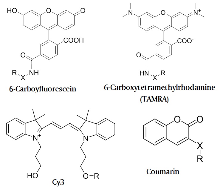
Several groups of chromophores consisting of conjugated unsaturated hydrocarbons and hetero-aromatic molecules have strong fluorescent properties. The most common fluorophores employed in fluorescent assays are derived from fluorescein, rhodamine, coumarin or cyanine type of chromophores which structures are illustrated in figure 1.

Figure 1. Structures of the most common fluorophores. Where X is a linker, R is an oligonucleotide.
Each of these molecules has a characteristic absorbance spectrum and a characteristic emission spectrum. The specific wavelength at which one of these molecules will most efficiently absorb energy is called the absorbance peak and the wavelength at which it will most efficiently emit energy is called the emission peak as illustrated in figure 2.

The difference between absorbance peak and emission peak is known as the Stokes Shift. Absorbance peak and emission peak wavelengths for most of the fluorophores used in molecular applications are shown in Table 1 (for a complete list of the fluorescent dyes please visit our website (6).
Table 1. Fluorescent properties of commonly used dyes.
Dye | Ab (nm) | Em (nm) | SS (nm) | e (M-1cm-1) |
Acridine | 362 | 462 | 100 | 11,000 |
Alexa 350 | 346 | 442 | 96 | 19,000 |
Alexa 488 | 495 | 519 | 24 | 71,000 |
Alexa 594 | 590 | 716 | 26 | 73,000 |
Alexa 610 | 612 | 628 | 16 | 144,000 |
Alexa 633 | 632 | 647 | 15 | 159,000 |
Alexa 700 | 696 | 719 | 23 | 196,000 |
AMCA | 353 | 442 | 89 | 19,000 |
ATTO 390 | 390 | 479 | 89 | 24,000 |
ATTO 425 | 436 | 486 | 50 | 45,000 |
ATTO 465 | 453 | 508 | 55 | 75,000 |
ATTO 488 | 501 | 523 | 22 | 90,000 |
ATTO 495 | 495 | 527 | 32 | 80,000 |
ATTO 590 | 594 | 624 | 30 | 120,000 |
ATTO 610 | 615 | 634 | 19 | 150,000 |
ATTO 633 | 629 | 657 | 28 | 130,000 |
ATTO 647 | 645 | 669 | 24 | 120,000 |
ATTO 700 | 700 | 719 | 19 | 120,000 |
BODIPY FL | 531 | 545 | 14 | 75,000 |
BODIPY TMR | 544 | 570 | 26 | 56,000 |
BODIPY TR | 588 | 616 | 28 | 68,000 |
Cascade Blue | 396 | 410 | 14 | 29,000 |
Cy2 | 489 | 506 | 17 | 150,000 |
Cy3 | 552 | 570 | 18 | 150,000 |
Cy3.5 | 581 | 596 | 15 | 150,000 |
Cy5 | 643 | 667 | 24 | 250,000 |
Cy5.5 | 675 | 694 | 19 | 250,000 |
Cy7 | 743 | 767 | 24 | 250,000 |
Edans | 335 | 493 | 158 | 5,900 |
Eosin | 521 | 544 | 23 | 95,000 |
Erythrosin | 529 | 553 | 24 | 90,000 |
6-FAM | 494 | 518 | 24 | 83,000 |
6-TET | 521 | 536 | 15 | - |
6-HEX | 535 | 556 | 21 | - |
JOE | 520 | 548 | 28 | 71,000 |
LightCycler 640 | 625 | 640 | 15 | 110,000 |
LightCycler 705 | 685 | 705 | 20 | - |
Lissamine | 558 | 583 | 25 | 88,000 |
NBD | 465 | 535 | 70 | 22,000 |
Rhodamine 6G | 524 | 550 | 26 | 102,000 |
Rhodamine Green | 504 | 532 | 28 | 78,000 |
Rhodamine Red | 560 | 580 | 20 | 129,000 |
TAMRA | 565 | 580 | 15 | 91,000 |
ROX | 585 | 605 | 20 | 82,000 |
Texas Red | 595 | 615 | 20 | 80,000 |
NED | 546 | 575 | 29 | - |
VIC | 538 | 554 | 26 | - |
The conjugation or addition of electron withdrawing groups (EWG) to a basic fluorophore moiety usually leads to a red shift resulting in a shift of the absorbance and emission peaks to longer wavelengths or lower energies.
Methods of incorporation
The most common and convenient method for the attachment of a fluorescent dye to an oligonucleotide is the phosphoramidite method. This method makes it possible to use commercially available fluorescent phosphoramidites for the conjugation or incorporation of one or more fluorophores into or to both, the 5' and/or 3' end, of the oligonucleotide. However, if the fluorophore is not stable in basic conditions needed for the oligonucleotide base deprotection step, the attachment to an oligonucleotide has to be done using a post-synthetic method after the base deprotection step is completed. In this situation, it is best that the oligonucleotide contains a functional group that will react with a reactive moiety on the selected fluorophore resulting in a stable covalent bond between the fluorophore and the oligonucleotide.
Several chemo-selective methods are available that can be used for the post-synthetic oligonucleotide labeling. One of the commonly chemo-selective labeling method used employs amino modified oligonucleotides together with the corresponding NHS esters or similar amine reactive synthons as illustrated in figure 3.
 Figure 3. Oligonucleotide labeling using TAMRA NHS ester.
More recently “click chemistry” was successfully employed to label oligonucleotides with various fluorescent reporter molecules (7-9). This chemical process is outlined in figure 4.

Figure 4. Huisgen’s 1,3-dipolar cycloaddition between an alkyne modified oligonucleotide (1) and an azide modified reporter molecule (2). Where R1 and R2 are a hydrogen atom (H) or an extension of the oligonucleotide chain, X is a linker, and Y is a reporter molecule.
The advantage of this chemistry is that it is completely orthogonal to any other attachment method. This chemistry can also be used in addition to any type of Michael-Addition reaction or chemistry as well as any other active esters that are reactive towards alkylamino modified oligonucleotides.
Polymerase dependent polynucleotide labeling using fluorescently labeled deoxynucleoside-5’-triphosphates (NTP) can be considered to be the major method for cDNA labeling (10). This method uses DNA polymerase or terminal deoxynucleotidyl transferase in order to incorporate fluorescently labeled nucleobases with the help of the corresponding NTPs into polynucleotides. These types of oligonucleotides may be used further in any type of micro-array applications.
Dark quenchers suitable for ultrasensitive probes
In recent years Dabcyl, TAMRA and other fluorescent acceptor molecules used in qPCR probes, have been replaced with one or more of the growing family of dark quencher molecules. For this reason, fluorophore-quencher dual-labeled probes have become a standard in kinetic qPCR assay. The properties of dark quencher dyes are provided in table 2.
Table 2. Characteristic properties of quencher dyes.
Quencher | lmax, nm | e, M-1cm-1 |
Dabcyl | 470 | 32,000 |
BHQ2 | 578 | 38,000 |
IB FQ | 531 | 38,000 |
IQ4 | 585 | 59,000 |
However, even the most efficient quencher dyes show a narrow and limited range of quenching that is predetermined by their narrow absorbance spectra. Therefore, each of the quencher dyes requires a fluorophore within a certain fluorescence emission spectrum range in order to have an efficient energy transfer between the two dyes or chromophors. The broad absorbance spectrum of our new generation of quencher dyes, for Instant for the Quencher dyes (IQ4), makes these probes suitable for multiplexing (11). Their highly efficient quenching characteristics lead to a higher sensitivity expressed by the probe. These significantly improved novel quencher dyes, also showing improved Ct values, now allow for the design of new linear highly sensitive probes. A comparison of the UV spectral properties for standard mono-labeled oligonucleotides are illustrated in the figure 4.

Figure 4. UV Spectra of standard mono-labeled decamer oligonucleotides labeled with the leading quencher dyes.
Biosynthesis, Inc. now offers all types of fluorescently labeled oligonucleotides including their conjugates with peptides proteins and various nanoparticles.
References:
- Zimmerman, J.; Voss, H.; Schwanger, C.; Stegemann, J.; Erfle, H.; Stucky, K.; Kristensen, T.; Ansorge, W., Nucleic Acids Res., 1990, 18, 1067.
- Agrawal, S.; Zamecnik, P. C.,. Nucleic Acids Res., 1990, 18, 5419.
- a) Landgraf, A.; Reckmann, B.; Pingoud, A., Anal. Biochem., 1991, 193, 231. b) Lee, L. G.; Connell, C. R. and Bloch, W. Nucleic Acids Res., 1993, 21, 3761. c) Tyagi, S.; Kramer, F. R., Nature Biotechnology, 1996, 14, 303.
- Fisher, T. L.; Terhorst, T.; Cao, X.; Wagner, R. W., Nucleic Acids Res., 1993, 21, 3857.
- Urdea, M. S.; Warner, B. D.; Running, J. A.; Stempien, M.; Clyne, J.; Horn, T., Nucleic Acids Res., 1988, 16, 4937.
- http://www.biosyn.com/oligonucleotide-modification-services.aspx
- A.V. Ustinov, et al, Tetrahedron, 2007, 64, 1467-1473.
- Agnew, B. et al., US Patent application 20080050731/A1.
- X. Ming, P. Leonard, D. Heindle and F. Seela, Nucleic Acid Symposium Series No. 52, 471-472, 2008.
- a) Hessner, M.J., X. Wang, K. Hulse, L. Meyer, Y. Wu, S. Nye, S.W. Guo, and S. Ghosh. 2003. Nucleic Acids Res., 2003, 31:e14. b) C. E. Guerra, BioTechniques, 2006, 41 (1), 53–56.
- Laikhter A. et al. US patent 7,956,169.
-.-