Quantcast
Channel: Latest Articles of Bio-Synthesis Inc.
Viewing all 591 articles
Browse latest View live

Synthesis of circular oligonucleotides

$
0
0

Synthesis of circular oligonucleotides

The polymerase chain reaction (PCR) is a technique that allows amplification of nucleic acids in-vitro. However, the reaction produces linear products. Many forms of DNA that are competent to duplicate, copy, or reproduce DNA contain closed circular deoxyoligonucleotides (DNA). Until the introduction of thermostable ligases, these molecules could only be amplified in-vivo in the appropriate host cells.

Since the introduction of the ligation-during-amplification (LDA) reaction, in-vitro synthesis of closed circular DNA is possible without the need for subcloning. Chen and Ruffer in 1998 elegantly showed how to do this. In their paper, the two scientists describe an in-vitro procedure for the selective amplification of closed circular DNA using  sequence-specific primers. 


Figure 1: Amplification of closed circular plasmids via the ligation-during-amplification (LDA) method. The graphic illustrates the primer configuration utilized during the amplification. The open arrows represent the primers. For site-directed mutagenesis, mutagenic primers can be used.


Chen and Ruffer demonstrated that LDA is a very useful method for site-directed mutagenesis, mutation detection, DNA modification, DNA library screening and the production of circular DNA. LDA uses a thermostable DNA ligase during the PCR reaction in which a circular DNA serves as the template. After a primer is fully extended along the circular template, the ligase closes the gap. This closing of the gap results in a double-stranded circular DNA. Thermal denaturation separates the two circular DNA strands that can now serve as the template for the next round of extension and amplification. The use of thermal cycling allows the exponential amplification of closed circular DNA. Chen and Ruffer used a circular plasmid of 1990 base pairs (bp), and two 5’ phosphorylated primers,16 and 17 nucleotides (nt) long, for the generation and amplification of closed circular DNA. The primers used were complementary to different strands of the plasmid. The primers, one containing a single G to A mismatch on an HphI site, were applied in the plasmid in an inward orientation. The reaction products were analyzed using agarose electrophoresis and tested for their function by their ability to transform bacterial cells. The scientists reported that the yields of transformation was very high. The researchers concluded that LDA can be used to amplify replicatively competent closed circular DNA directly from other circular DNA, such as circular plasmids, using sequence-specific primers without the need for subcloning.


Reference
 
Z Chen, D E Ruffner; Amplification of closed circular DNA in vitro. Nucleic Acids Res. 1998 February 15; 26(4): 1126–1127.

Patent: 6620597, "Method for in vitro amplification of circular DNA", Chen, Zhidong (Salt Lake City, UT), Ruffner, Duane E. (Salt Lake City, UT), 2003, September, http://www.freepatentsonline.com/6620597.html


Enzymatic synthesis of Circular Oligonucleotides

$
0
0

Enzymatic Synthesis of Circular Oligonucleotides


Chemical solid phase and solution phase synthesis methods allow the synthesis of circular oligonucleotides, both for the production of circular DNA and RNA molecules. However, the use of enzymatic approaches may of advantage in some cases. Small circular single-stranded oligonucleotides (<28 base pairs {bp}) can be chemically synthesized using solution and solid-phase methods from partially protected linear precursor molecules.  Longer circular oligos (>28 bp) are relatively easy to synthesize by both methods, chemical and enzymatic. The chemical circularization of linear oligonucleotides can be achieved using cyanogens bromide (BrCN) or the carbodiimide cross-linker 1-ethyl-3-(3-dimethylaminopropyl) carbodiimide (EDC) together with a template functioning as a bridge. However, small circular oligonucleotides, for example containing 28 to 50 bp, are readily synthesized using a single precursor segment. However, large-sized circles, containing more than 50 bp, can be prepared utilizing two or three smaller precursor oligonucleotide segments. Unfortunately, the preparation of large circular oligonucleotides from multiple precursor segments often results in low yields. In addition to chemical circularization methods, enzymatic ligation offers an alternative approach for the preparation of circular oligonucleotides. For this approach, T4 ligases are routinely utilized for the cyclization reactions.

For the enzymatic synthesis of circular DNA and RNA oligonucleotides, T4 ligases are commonly used. These enzymes can circularize the reactive 3’-hydroxyl (OH) and 5’-phosphate (PO4) groups of linear ssDNA using a complementary short template. The enzyme DNA ligase catalyzes the covalent bond formation between the 3’OH and the 5’PO4 on DNA phosphodiester bonds. The reaction requires two ends of double-stranded DNA and ATP. Bacteriophage T4 ligase is the enzyme of choice most often used for because it can ligate blunt-ended DNA as well as DNA with compatible cohesive ends. However, the optimal temperature may vary for different reaction conditions. T4 RNA ligase I can circularize linear ssRNA in the absence of a template. 


Figure 1: Models for the T4 RNA ligase 2 with nicked RNA. A synthetic construct of Enterobacteria phage T4 was used to generate crystal structures. The resulting structural models illustrate the stereochemistry of the nucleotidyl transfer. In addition, the remodeling of active-site contacts and conformational changes that propel the ligation reaction forward are revealed (Nandakumar et al., 2006).


T4 RNA ligase 2 (Rnl2) and kinetoplastid RNA editing ligases belong to a family of RNA repair enzymes. RNA ligases seal 3'-OH/5'-PO(4) nicks in duplex RNAs via ligase adenylylation, followed by a transfer of an adenosine monophosphate (AMP)  to the nick 5'PO(4) group. The attack by the nick 3'-OH on the 5'-adenylylated strand forms a phosphodiester bond.


Resulting products can be separated and visualized using denaturing polyacrylamide gel electrophoresis (PAGE). The slower migration mobility of circular oligonuleotides in comparison to their linear counterparts circular products allows the observation of these products as stained bands migrating with higher apparent molecular weights (figure 2). In addition, success of the circularization reaction can be determined with the help of endonuclease cleavage.

Figure 2: Quality control of enzymatic synthesis products of circular DNA and RNA using PAGE.


References


Beaudry D., Perreault J-P. An efficient strategy for the synthesis of circular RNA molecules. Nucleic Acids Research, 1995, Vol. 23, 3064-3066.

Diegelman A. M. and Kool E. T. Chemical and enzymatic methods for preparing circular single-stranded DNAs. Current Protocols in Nucleic Acid Chemistry, 2000, 5.2.1-5.2.27.

Dolinnaya N. G., Blumenfeld M., etc. Oligonucleotide circularization by template-directed chemical ligation. Nucleic Acids Research, 1993, Vol. 21, 5403-5407.

Nandakumar J, Shuman S, Lima CD;  Rna ligase structures reveal the basis for RNA specificity and conformational changes that drive ligation forward. Cell(Cambridge,Mass.) (2006) 127 p.71.

Pedroso E., Escaja N., Frieden M., and Gradas A. Solid-phase synthesis of circular oligonucleotides. Methods in Molecular Biology, Vol. 288, 2005: Oligonucleotide synthesis: Methods and Applications. Edited by: P. Herdewijn, Humana Press Inc., Totowa, NJ Page 101-125.

Applications for circular oligonucleotides

$
0
0
 Applications for circular oligonucleotides


Circularized or circular single-stranded deoxyribosnucleotides (ssDNAs), also often just called circular oligonucleotides, are more resistant to the attack by nucleases when compared to their linear oligonucleotides. These properties may make these circular oligonucleotides important tools for in vivo studies.  

In the past circular oligonucleotides have been investigated for their unique DNA binding properties and as useful models in studying DNA structures such as hairpin motifs by NMR. Currently, circular oligonucleotides are also being used for diagnostic applications, such as padlock probes, as well as the synthesis of concatemeric polypeptides. In addition, circular ssDNAs allow for both DNA and RNA amplification as these molecules are accepted as templates by both DNA and RNA polymerases.


Use and applications for circular oligonucleotides are

  • Anti sense using circular oligonucleotides
  • Binding of duplex DNA
  • Delivery vectors for miRNAs
  • Diagnostics
  • DNA fragment assembly using a nicking enzyme system
  • DNA polymerase inhibition
  • DNA structure studies
  • Efficient templates for DNA and RNA polymerases
  • Hairpin motif design
  • Hairpin studies
  • Ligation-independent cloning (LIC)
  • Manipulating gene expression with caged circular oligonucleotides
  • Mutation detection
  • Padlock probes
  • Probing DNA-protein interactions
  • Quantitation of sequence-dependent DNA bending and flexibility
  • RNA polymerase inhibition
  • Rolling Circle Amplification (RCA)
  • Single molecule counting
  • Specific gene expression
  • Study of noncanonical DNA structural motifs
  • Synthesis of concatemeric polypeptides
  • Topologic modifications
  • Triple helix formation
  • Unique DNA recognition properties.
  • Study of splicing events
  • Post-translational modifications


References

Diegelman, A. M. and Kool, E. T. (2000) Chemical and enzymatic methods for preparing circular single-stranded DNAs. In Current Protocols in Nucleic Acid Chemistry, vol. 2 (Beaucage, S. et al., eds.). John Wiley, New York, Chapter 5.2.


Oligonucleotide synthesis: methods and applications / edited by Piet Herdewijn. (Methods in molecular biology ; 288). ISBN 1-58829-233-9


Plasmids are autonomous circular oligonucleotides

$
0
0
 Plasmids are autonomous circular oligonucleotides


Modern recombinant technology produces large numbers of identical DNA molecules. The technology relays on the formation of 3’ -> 5’ phosphodiester bonds to link DNA fragments to a vector molecule. When introduced into a host cell the DNA fragment together with the vector DNA is produced in large numbers. Two types of vectors are used most commonly: E. coli plasmid vectors and bacteriophage λ vectors.


Plasmids are circular double stranded DNA oligonucleotides that are separate from but can replicate independently of the hosts cell’s chromosome. These extrachromosomal circular oligonucleotides or DNAs occur naturally in bacteria, yeast, and some higher eukaryotic cells, and exist in a parasitic or symbiotic relationship with their host cell. The size of plasmids can vary from a few thousand base pairs to more than 100 kilobases (kb). Similar to chromosomal DNA, plasmid DNA is duplicated before every cell division. At least one copy of plasmid DNA is segregated to each daughter cell during cell division. These self-replicating circular oligonucleotides are maintained in the host cell in a stable and characteristic number of copies. The copy number remains constant from one generation to the next. 


Plasmids contain genes that are beneficial to the host cell. Many of them contain ‘transfer genes” encoding proteins forming a pilus, or a macromolecular tube. Through this tube a copy of the plasmid can be transferred to other host cells of related bacterial species. However, most plasmid vectors contain just the essential nucleotide sequences required for their use in DNA cloning. Figure 1 shows diagrams of cloning vectors derived from plasmids.

Figure 1: Diagrams of cloning vectors derived from a plasmid. A diagram for a simple cloning vector is illustrated in A. A diagram for a pUC vector is depicted in B.

Plasmid vectors are approximately 1.2 to 3 kilobases (kb) in length and contain a replication origin (ori) sequence and a gene that permits selection. The gene used for the selection usually codes for a gene that is sensitive to antibiotics. The ampr gene that codes for the enzyme β–lactamase is an example. The enzyme β–lactamase inactivates ampicillin. Exogenic DNA can be inserted into regions that do not disturb the ability of the plasmid to replicate or express the ampr gene. pUC vectors such as the
pUC18 and pUC19 vectors are small, high copy number, E.coli plasmids, 2686 bp in length and are identical except that they contain multiple cloning sites (MCS) arranged in opposite orientations. The physical properties including the topologies of these vectors have been investigated (http://pubs.acs.org/doi/abs/10.1021/ma0711689). A collection of plasmids can be found at the plasmid repository (https://dnasu.org/DNASU/Resources/Plasmid.jsp).

Proteins are involved in the partitioning of plasmid DNA.


The replication origin (ori), a specific DNA sequence of 50 to 100 base pairs, must be present in a plasmid to allow for its replication. Proteins or enzymes in the host cell bind to this sequence motif and initiate replication of the circular plasmid.


Plasmids use two mechanisms for the replication of their DNA. These are the

  1. bidirectional replication of the plasmid and
  2.  the rolling circle replication mechanisms.

Most plasmids replicate like small bacterial chromosomes. They possess an origin of replication where the DNA opens and replication begins. Two replication forks move around the circular oligonucleotide or plasmid DNA in opposite directions. Some tiny plasmids have only one replication fork that moves around back to the origin.


The rolling circle replication mechanism is shared by some plasmids and a few viruses. In this mechanism, one strand of the double stranded DNA is nicked at the origin of replication. The other circular stand starts to roll away from the broken strand. The result is two single stranded regions of DNA, one belonging to the broken strand and one circular strand. DNA is than synthesized starting at the end of the broken strand. This strand is now elongated and the circular strand is used as a template. The gap left where the original strands rolled apart is filled in. The process of rolling and filling continues and eventually the original broken strand is completely unrolled. Finally, the circular oligonuclotides are all paired with a new strand of DNA. The result is a single strand of DNA hanging loose. However, the next steps of this process depend on circumstances and the nature of plasmids involved. Some plasmids can transfer themselves from one bacterium to the next, as is the case for the F-plasmid.     


Accurate and stable maintenance of DNA partition in bacteria requires proteins. This process can involve partition loci found on both chromosomes and plasmids. Bacterial low copy-number plasmids make simple DNA segregating machines that use an elongating protein filament between sister plasmids. The ParMR C system of Escherichia coli R1 plasmid, ParM, forms the spindle between ParR C complexes on sister plasmids. ParM filaments enable two ParR C–bound filaments to associate in an anti-parallel orientation to form a bipolar spindle. The spindle can elongate as a bundle of at least two anti-parallel filaments and pushes two plasmid clusters towards the poles.


During the partition of the Escherichia coli plasmid R1 a partition complex between the DNA-binding protein ParR and its cognate centromere site parC on the DNA is formed. This partition complex is recognized by a second partition protein, the ATPase ParM, that now forms filaments that allow the active bidirectional movement of DNA replicates.The plasmids P1and F are reported to employ a three-component system to partition replicated genomes. This system contains a partition site on the DNA target, typically called parS, a partition site binding protein, typically called ParB, and a Walker-type ATPase, typically called ParA. Par A also binds non-specific DNA. In vivo these protein family forms dynamic patterns over the nucleoid.


However, the process how the ATP-driven patterning works is not net very well understood and more research will be needed to decipherer all the details involved. The use of synthetic oligonucleotides, peptides and protein constructs may enable researchers to solve this puzzle in the future.

Figure 2: Crystal structures of the DNA-binding ParR protein which is a part of the plasmid partition complex (left) and the plasmid segregation protein Parm (right) are illustrated.


Moeller-Jensen et al. in 2007 reported the structures of a
family of dimeric ribbon–helix–helix (RHH) 2 site-specific DNA-binding proteins. The reported crystallographic and electron microscopic data indicated that ParR dimers assemble into a helix structure with DNA-binding sites facing outward. In addition, genetic and biochemical experiments reported by this research group supported a structural arrangement in which the centromere-like parC DNA is wrapped around a ParR protein scaffold.

Gayathri et sl. In 2012 investigated the ParMRC system of the Escherichia coli R1 plasmid, ParM, an actin like protein. This protein assembly forms the spindle between ParRC complexes on sister plasmids. The researchers showed that ParRC is bound and could accelerate growth at only one end of polar ParM filaments. The architecture of ParM filaments enabled two ParRC-bound filaments to associate in an antiparallel orientation forming a bipolar spindle. The spindle elongated as a bundle of at least two antiparallel filaments that pushes two plasmid clusters toward the poles.


Reference


Gayathri P, Fujii T, Moller-Jensen J, Van Den Ent F, Namba K, Lowe J.; A bipolar spindle of antiparallel parm filaments drives bacterial plasmid segregation.Science (2012) 338 p.1334.


Ling Chin Hwang, Anthony G Vecchiarelli, Yong-Woon Han, Michiyo Mizuuchi, Yoshie Harada, Barbara E Funnell and Kiyoshi Mizuuchi; ParA-mediated plasmid partition driven by protein pattern self-organization. The EMBO Journal (2013) 32, 1249. www.embojournal.org.


Molecular Cell Biology. 4th edition. Lodish H, Berk A, Zipursky SL, et al. New York: W. H. Freeman; 2000.


Jakob Moeller-Jensen, Simon Ringgaard, Christopher P Mercogliano, Kenn Gerdes and Jan Loewe; Structural analysis of the ParR/parC plasmid partition complex. The EMBO Journal (2007) 26, 4413–4422.

Jeanne Salje, Pananghat Gayathri and Jan Löwe;The ParMRC system: molecular mechanisms of plasmid segregation by actin-like filaments. http://www2.mrc-lmb.cam.ac.uk/groups/JYL/PDF/nrmicro2425.pdf, http://www.ncbi.nlm.nih.gov/pubmed/20844556


A circular ribo-oligonucleotide or RNA is a noncoding RNA

$
0
0
 A circular ribo-oligonucleotide or RNA is a noncoding RNA


Circular RNAs (circRNAs), or circular ribo-oligonucleotides, were recently added to the growing list of noncoding RNAs. However, since the last 20 years circular RNAs were known to biologists. These types of molecules were thought to be artefacts or molecular flukes resulting from aberrant RNA splicing or from the infection of host genes by specific pathogens. In the early years circular RNAs were found in plant viroids and the hepatitis δ virus. More recently circular RNAs have been detected in animal cells as well. However, their function was unclear. Recent findings from the laboratories of Nikolaus Rajewsky and Jørgen Kjems have now defined a function for one circular RNA that binds the microRNA miR-7. Their results indicated that this circular RNA is full of microRNA binding sites. Apparently this circular RNA can act as a miRNA 'sponge' capable of binding many miRNAs per circular RNA molecule. In addition, their results suggested a role for circular RNAs in post-transcriptional regulation.


More resently, in 2012, Salzman et al. reported that circular RNA isoforms are a prevalent feature of eukaryotic gene expression programs. Previously thought to be very rare, these observations suggested that the major RNA isoform from hundreds of human genes are circular RNAs. In the following year, in 2013, Salzman et al. used an improved computational approach for the identification of circular RNA. This new method allowed the scientists to detect a more extensive catalogue of circular RNA. Many, many more than previously reported. This also included small RNA circles formed by non-canonical splicing of short exons and noncoding RNAs. The research group found that the expression of circular RNAs in Drosophila melanogaster is widespread. In addition, the researchers estimated that in humans, circular RNAs may account for approximately 1%. That is as many molecules as poly(A) RNA. Furthermore, the researchers suggested that this newly found abundance of circular RNA could significantly alter our present perspective on post-transcriptional regulation as well as the roles that RNA can play in the cell.

The improved detection of circular RNA isoforms allowed the scientists to characterize how many of differential circular RNA splicing events happen within a single gene and also to study variations in alternative splicing of circular RNAs. These observations support the hypothesis that circular RNAs may have an evolutionarily conserved function. However, their exact nature and mechanisms are still to be discovered.

Already a database for circular RNAs is available for browsing and downloading information and the location on the chromosomes of identified circular RNAs: http://cirbase.org/.

Reference

http://cirbase.org/

Hansen, T.B. et al. Natural RNA circles function as efficient microRNA sponges.Nature 495, 384–388 (21 March 2013) doi:10.1038/nature11993.

William R Jeck Norman E Sharpless; Detecting and characterizing circular RNAs. Nature Biotechnology 32, 453–461 (2014). doi:10.1038/nbt.2890.


Kos, A., Dijkema, R., Arnberg, A.C., van der Meide, P.H. & Schellekens, H. The hepatitis delta (delta) virus possesses a circular RNA. Nature 323, 558–560 (1986).

Sebastian Memczak,  Marvin Jens, Antigoni Elefsinioti, Francesca Torti, Janna Krueger,Agnieszka Rybak, Luisa Maier, Sebastian D. Mackowiak, Lea H. Gregersen, Mathias Munschauer, Alexander Loewer, Ulrike Ziebold, Markus Landthaler, Christine Kocks,Ferdinand le Noble Nikolaus Rajewsky; Circular RNAs are a large class of animal RNAs with regulatory potency. Nature 495, 333–338 (21 March 2013) doi:10.1038/nature11928.


Mohan T. Bolisetty1 and Brenton R. Graveley1; Circuitous Route to Transcription Regulation. Molecular Cell 51, September 26, 2013, pp 705-706.   graveley@neuron.uchc.edu, http://dx.doi.org/10.1016/j.molcel.2013.09.012

Sanger, H.L., Klotz, G., Riesner, D., Gross, H.J. & Kleinschmidt, A.K. Viroids are single-stranded covalently closed circular RNA molecules existing as highly basepaired rod-like structures. Proc. Natl. Acad. Sci. USA 73, 3852–3856 (1976).

Salzman J, Gawad C, Wang PL, Lacayo N, Brown PO (2012) Circular RNAs Are the Predominant Transcript Isoform from Hundreds of Human Genes in Diverse Cell Types. PLoS ONE 7(2): e30733. doi:10.1371/journal.pone.0030733.

Salzman J, Chen RE, Olsen MN, Wang PL, Brown PO (2013) Correction: Cell-Type Specific Features of Circular RNA Expression. PLoS Genet 9(12): 10.1371/annotation/f782282b-eefa-4c8d-985c-b1484e845855. doi: 10.1371/annotation/f782282b-eefa-4c8d-985c-b1484e845855 View correction


Telomere repeat sequences

$
0
0

Telomere repeat sequences


Telomeres are DNA structures at the ends of eukaryotic chromosomes that protect them from degradation and DNA repair activities. Initially, telomeres were defined functionally as the natural ends of eukaryotic chromosomes. Without telomeres, a chromosome is unstable. Telomeric DNA consists of simple sequences repeated in tandem. These sequence stretchers have been found to be a conserved feature in throughout eukaryotes. The terminal DNA stretch can range in total length from under fifty base pairs in some protozoans, through a few hundred base pairs in yeasts and several other eukaryotes, to thousands of base pairs in mammalian cells. These sequence repeats appear to maintain a stable telomere. For this reason, telomeres can be functionally defined as regions of DNA at the end of linear chromosomes. Apparently these sequence repeat regions are needed for replication and stability of chromosomes. All known eukaryotic telomeres contain simple repeated sequences of G- and C-rich complementary strands. These telomere repeat sequences have the general structure (T or A)m(G)n. In Tetrahymena and Oxytricha the G-rich DNA strand, oriented in a 5' - 3' direction toward the end of the chromosome, is synthesized by an RNA-dependent "telomerase" activity.

Protein telomere with telomere repeat sequence complex in Sterkiella nova

In Sterkiella nova, alpha and beta telomere proteins bind cooperatively with single-stranded DNA to form a ternary alpha.beta.DNA complex. The association of telomere protein subunits is DNA-dependent. The alpha-beta association enhances DNA affinity. To further understand the molecular basis for binding cooperativity, Buczek and Horvath in 2006 characterized several possible stepwise assembly pathways using isothermal titration calorimetry.

The enzyme telomerase is a reverse transcriptase that elongates telomeres in the cells it is expressed in. Telomerase was found to be active in germ cells and stem cell populations but not in adult tissue cells. In normal adult tissue cells or somatic cells the telomere activity levels are not sufficient to prevent telomere shortening associated with cell division. During the lifetime of an organism including humans the number of times a normal human cell population will divide until cell division stops appear to be finite. However, cancer cell lines can almost live on forever and are considered to be immortal.

Chromosomes can be stained with fluorescently or otherwise labeled oligonucleotide probes directed against highly conserved telomere repeat sequences.

In general, to stain chromosomes during in-situ hybridization experiments labeled oligonucleotide probes directed against highly conserved mammalian telomere repeat sequences and against major satellite repeats are used. Popular probes of this type contain the fluorogenic dyes Cy3 or FITC at the 5’ or 3’ end.  Artificial nucleotides, such as bridged nucleic acids or BNAs, can be used to enhance the specificity of the probes.

For example molecular probes that target the (TTAGG)4 repeat are often used for the detection and staining of  highly conserved sequence repeats consisting of (TTAGGG)n and (CCCTTA)n sequences in human chromosomes. Human chromosomes contain stretches of up to 30,000 C and G bases repeating over and over. These sequence stretches often occur adjacent to gene-rich areas and form a barrier between the genes and the non-coding DNA. In general many FISH probes are targeted towards repetitive sequences found in the chromosomes. Scientists now believe that CpG islands are involved in regulating gene activities and more evidence has been accumulated recently to support the functional significance of satellite DNA sequences.

Structure of probes used for the staining of chromosomes


A typical non-radioactive probe consists of a fluorophore that is conjugated to the 5’- or 3’-end of the oligonucleotide used.


Telomer Probe:  Fluorophore-5’-CCC TAA CCC TAA CCC TAA-3’


Centromere Probe:  Fluorophore-5’-TCG CCA TAT TCC AGG TC-3’

Target:  Mouse major satellite repeats.

Structure of the human telomere in K+ solution

An intramolecular (3 + 1) G-quadruplex scaffold.


Ref.: Luu, K.N.,  Phan, A.T.,  Kuryavyi, V.V.,  Lacroix, L.,  Patel, D.J.; (2006) J.Am.Chem.Soc.128: 9963-9970.

Abstract

We present the intramolecular G-quadruplex structure of human telomeric DNA in physiologically relevant K(+) solution. This G-quadruplex, whose (3 + 1) topology differs from folds reported previously in Na(+) solution and in a K(+)-containing crystal, involves the following: one anti.syn.syn.syn and two syn.anti.anti.anti G-tetrads; one double-chain reversal and two edgewise loops; three G-tracts oriented in one direction and the fourth in the opposite direction. The topological characteristics of this (3 + 1) G-quadruplex scaffold should provide a unique platform for structure-based anticancer drug design targeted to human telomeric DNA.
 

Telomeric repeat sequences in eukaryotes

Organism

Telomeric repeat

(5' to 3' toward the end)

Reference

Vertebrates, human

TTAGGG n repeats

Meyne et al. 1989

Giardia lamblia

TAGGG

Adam et al. 1991

Human, mouse, Xenopus, Neurospora crassa, Trypanosoma brucci, Aspergillus nidulans

TTAGGG

Bhattacharyya and Blackburn 1997b

Neurospora crassa

Cryptococcus

TTAGGG

TTAGGGGG


Edman 1992

Physarum, Didymium,

TTAGGG


Dictyostelium

AG(1-8)


Trypanosoma, Crithidia

TTAGGG


Tetrahymena, Glaucoma

TTGGGG


Paramecium

TTGGG(T/G)


Oxytricha, Stylonychia, Euplotes

TTTTGGGG


Plasmodium

TTAGGG(T/C)

TT(T/C)AGGG


Arabidopsis thaliana

TTTAGGG


Chlamydomonas

TTTTAGGG


Bombyx mori

TTAGG


Ascaris lumbricoides

(C)TTAGG(C)

Muller et al. 1991

Schizosaccharomyces pombe

TTAC(A)(C)G(1-8)

TTAC(A)AG2-7


Saccharomyces cerevisiae

TGTGGGTGTGGTG

(from RNA template)
or G(2-3)(TG)(1-6)T

(consensus)

TG2-3(TG)1-3


Saccharomyces castellii

TCTGGGTG


Candida glabrata

GGGGTCTGGGTGCTG


Candida albicans

GGTGTACGGATGTCTAACTTCTT


Candida tropicalis

GGTGTA[C/A]GGATGTCACGATCATT


Candida maltose

GGTGTACGGATGCAGACTCGCTT


Candida guillermondii

GGTGTAC


Candida pseudotropicalis

GGTGTACGGATTTGATTAGTTATGT


Kluyveromyces lactis

GGTGTACGGATTTGATTAGGTATGT

McEachern and Blackburn 1996


Reference

Adam R.D., Nash T.E., and Wellems T.E., 1991. Telomeric location of Giardia rDNA genes. Mol. Cell. Biol. 11: 3326-3330.

Blackburn, E. H. & Szostak, J. W. (1984) Annu. Rev. Biochem. 53, 163-194.

Buczek P, Horvath MP; Structural reorganization and the cooperative binding of single-stranded telomere DNA in sterkiella nova.J.Biol.Chem. (2006) 281 p.40124.

The RNA World, Second edition. Gesteland, Chech, Atkins, CSHL Press.


Links to data bases

http://telomerase.asu.edu/

http://labs.fhcrc.org/bedalov/telomeredatabase.html

http://www.biophysica.com/telomere.html

http://www.rcsb.org/pdb/results/results.do?outformat=&qrid=31238D90&tabtoshow=Current


PEGylation of therapeutic proteins

$
0
0

PEGylation of Therapeutic Proteins


Therapeutic proteins such as 
monoclonal antibodies and cytokines, after delivery to the body, are eliminated by proteolysis and clearance in the kidney or liver, a process that limits their usefulness as therapeutic agents. In addition, many of these proteins can also trigger an antibody immune response that would block their beneficial effects. Hence, significant efforts have been dedicated developing methods that while extending these proteins’ circulating life and decreasing their immunogenicity do not interfere with their specific biological functions. Between the different methods explored,
pegylation or the covalent addition of polyethylene glycol (PEG) chains to a protein, has been the most successful. Pegylation is used in several types of  therapeutic proteins. Apparently it results in the increase of the hydrodynamic volume of a protein. However, addition of PEG can also have beneficial effects on nucleic acids by increasing their stability against nucleases.

PEGylation, or pegylation, refers to the covalent attachment of polyethylene glycol polymer chains to molecules such as proteins, peptides, oligonucleotides or other biomolecules.

The first FDA-approved pegylated therapeutic protein appeared on the market in 1990, which was Adagen®, a PEGylated form of adenosine deaminase. Following that, seven successful pegylated therapeutic proteins have been approved by the FDA, including Oncaspar®(Pegaspargase), PEGIntron ® (PEGylated IFN-α2b), Pegasys® (PEGylated IFN-α2a), Neulasta® (pegfilgrastm), Somavert® (Pegvisomant), Mircera® (PEGylatedepoetin-β), and Cimzia®(Certolizumabpegol). In addition to these already approved PEGylated biopharmaceuticals, many new products are currently under investigation and in different stages of clinical trials. Still, when considering pegylation as a strategy to extend the circulating life of a protein, several factors need to be considered, which will be discussed here.

  • Location of the PEG chains:

As a rule, PEG chains should be covalently linked at a site distant from the binding site of the protein, i.e. that region of the protein that is involved in binding to a cell receptor or an antigen; this way, the blocking of specific groups needed for binding and/or the steric hindrance of the binding process caused by the PEG are avoided. That means that at a minimum we should have amino acid sequence information about the protein as well as its binding site, post-translational modifications, such as glycosylation may be also helpful as they provide additional potential sites for the covalent binding of the PEG chains. Information about the tertiary structure of the protein would help in fine tuning the choice for a pegylation site. Indeed, a new approach is to modify the gene coding for a protein to add a new reactive group, like a cysteine or lysine, which is distal from the binding site, to allow the covalent binding of the PEG chain(s). 


One strategy commonly used is to add the PEG chain at the N-terminal group of a protein, as usually this amino acid residue is not part of the binding site and it is located far from that site. Specificity for the terminal amino group is based on the differences in the pKa values for the lysine α and ε amino groups, and that the N-terminal group is always α. Another strategy used with glycosylated proteins is to oxidize the sugar moiety with periodate and to link the PEG to the aldehyde groups; because glycosylation sites are usually distant from the binding site, this approach minimizes any potential steric hindrance due to the added PEG.

  • Size and number of PEG chains:

In general, the larger the PEG chain the lower number of chains needed per mole of protein; yet, the length of the chain would be limited by its effects on stability and interference with binding. An advantage of longer PEGs is that usually a single chain will do, while smaller PEGs would require more chains added, which may interfere with the binding of the proteins. The size of the PEG is usually found by experimentation, in the case of monoclonal antibodies and their Fab fragments, PEGs of 40 kDaltons have been used successfully. However, while increase of the PEG size may result in a longer life, the biological activity of the protein may decrease as a function of the PEG size. Thus, the reason for the need to asses experimentally each therapeutic protein performance after pegylation. A new development in pegylation of therapeutic proteins is the use of branched lower molecular weight PEGs; these PEGs having usually two or three chains linked to a single linker carrying also a reactive group for binding to a protein, apparently has lower side-effects while maintaining a good stability. Also a higher circulating life of a pegylated protein may result in the accumulation of breakdown products that can damage the kidney; hence, as indicated above, pegylation effects need to be evaluated case by case.

  • Chemistry of the linking process:

A number of PEGs are available that have a single terminal reactive group capable of forming a covalent bond with some of the functional groups from proteins. For instance, PEGs having an aldehyde group can be used to preferentially add a PEG chain at the protein N-terminal group. This goal can be achieved by adjusting the pH of the reaction to have most of α amino groups deprotonated and thus able to react with the aldehyde. Other reactive groups are carboxyl, amine, maleimide, succimidyl and triazinyl, which are able to react with many of the functional groups available in proteins. To prevent damage to the protein, the reactions are carried under aqueous conditions, avoiding extreme pH conditions and using a concentration of activated PEG closer to the one needed to deliver the desired degree of pegylation. Once the pegylation of therapeutic proteins has taken place the different conjugates showing different degrees of substitution can be separated by ion exchange chromatography using a salt gradient to remove the excess of activated PEG and resolve the proteins with different degrees of pegylation. The pegylated therapeutic proteins are then concentrated, filter sterilized, and stored in PBS at 4oC. It may be possible to lyophilize the pegylated proteins in the presence of an excipient like mannitol or sorbitol.

  • Analysis for the Pegylation of therapeutic proteins:

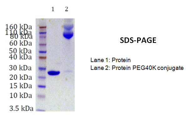
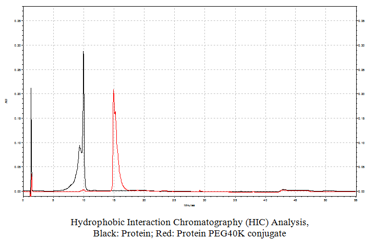
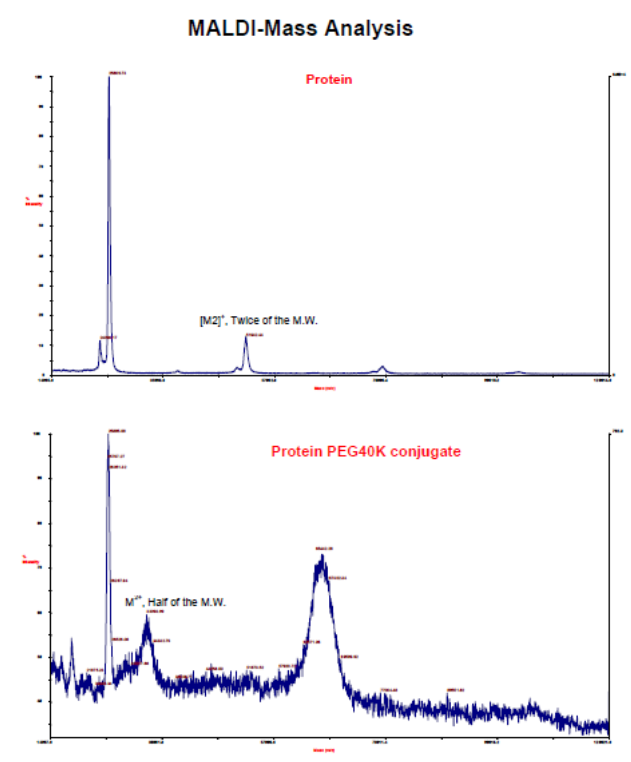
Several methods can be used to assess the quality of the final product, such as SDS-PAGE, hydrophopic interaction chromatography (HIC), size exclusion chromatography (SEC), and matrix assisted laser desorption time-of-flight mass spectrometry (MALDI-TOF-MS). 

SDS-PAGE:


This method delivers information about the different conjugates prepared during the process. However, the molecular weight information is usually not related to the actual molecular weight.


Hydrophobic Interaction Chromatography:


This method that allows resolving the different species according to their lipophilicity can be done using a butyl column, where the different conjugates are bound to the matrix using a high salt concentration and eluted in order of increasing lipophilicity by using a decreasing linear gradient of a salt, such as ammonium sulfate.


Size Exclusion Chromatography:


This method uses matrices like cross linked agarose gels, and the different conjugates are separated according to their hydrodynamic volume that is a result of the volume occupied by the protein plus the volume added by the PEG chain(s). This parameter is somewhat correlated with mol. wt. but it is not reliable to deliver the actual mol. wts.


MALDI-Mass spectrometry:


Under optimal conditions, this method delivers the actual molecular weight of the conjugate and it is quite useful to establish the actual molecular weight of the pegylated proteins thereby indicating the presence of the correct pegylated protein conjugate.

Storage:


Pegylated proteins are best stored under sterile conditions at neutral pH and at 4oC. The buffer can be the common phosphate buffered saline solution at pH 7.2.        


Reference

Ginn C, Khalili H, Lever R, Brocchini S.; PEGylation and its impact on the design of new protein-based medicines.Future Med Chem. 2014;6(16):1829-46. doi: 10.4155/fmc.14.125.

Jevsevar S, Kunstelj M, Porekar VG.; PEGylation of therapeutic proteins. Biotechnol J. 2010 Jan;5(1):113-28. doi: 10.1002/biot.200900218.

Veronese FM; Peptide and protein PEGylation: a review of problems and solutions. Biomaterials. 2001 Mar;22(5):405-17.

Yang T (2013) PEGylation – Successful Approach for Therapeutic Protein Conjugation. Mod Chem appl 1:e112. doi: 10.4172/2329-6798.1000e112


What are Circular Oligonucleotides?

$
0
0

 What are Circular Oligonucleotides?


Circular oligonucleotides are covalently closed singular DNA or RNA strands that are resistant to degradation by many cellular DNA and RNA decay machineries.


Models of circular DNA and RNA


[Figures were adapted from: Modelling DNA Phil. Trans. R. Soc. A-2006-Harris-3319-34; David Goodsell at http://www.rcsb.org/pdb/101/motm.do?momID=65: The self-splicing RNA in PDB entry 1u6b caught in the middle of its splicing reaction is illustrated].

Circular DNA


Modern biology focuses on the study of molecules within cells as well as the interaction between cells to understand and describe multi-cellular organisms most of which are visible to a human’s eye. Molecular cell biology concentrates on the study of macromolecules, such as carbohydrates, DNA, polypeptides, proteins, RNA, lipids as well as others, their biochemistry and processes that regulate metabolic pathways in these organisms.

Mitochondria and chloroplasts found in today’s eukaryotes contain circular DNAs encoding proteins. These proteins are essential for the function of organelles and the ribosomal and transfer RNAs required for their translation. 

Eukaryotic cells have multiple genetic systems: 

(1) a nuclear system, and 

(2) systems with their own DNA present in mitochondria and chloroplasts. 


Human mitochondrial DNA, mtDNA, is a circular DNA molecule which sequence is now completely known. This human mtDNA is among the smallest known mtDNAs. The circular mtDNA molecule is a compact, double-stranded circular genome contains 16,569 base pairs that encode two ribosomal RNAs (rRNAs). These are present in mitochondrial ribosomes and 22 transfer RNAs (tRNAs) needed to translate mitochondrial messenger RNAs (mRNAs). In addition, human mtDNA contains 13 sequences that begin with the ATG (methionine) codon and end with a stop codon. These DNA sequence stretches are long enough to encode polypeptides of more than 50 amino acids. All of the possible proteins encoded by these open reading frames have now been identified. Mammalian mtDNA has no introns and contains no long noncoding sequences. A single cell can have several hundred to thousands of copies of the circular mitochondrial genome. 

Since the 1970s scientists have known that, bacteriophages contain circular DNA. For example, Arnberg et al. in 1976 reported that a DNA-protein complex contains circular DNA in the bacillus bacteriophage GA-1. In addition, the researchers found that proteins are bound to DNA purified from bacillus bacteriophage GA-1. The removal of DNA-associated proteins by treatment with proteolytic enzymes resulted in 

(i)    a loss of transfecting activity of GA-1 DNA on competent Bacillus subtilis cells and 

(ii)    an increase (0.004 g/cm3) in the buoyant density of the DNA. 


GA-1 DNA, when analyzed by electron microscopy appeared as 6.4-µm-long circular and linear molecules. The analysis of 170 DNA molecules in the untreated GA-1 DNA preparation found 30 (18%) to be circular oligonucleotides. However, no circular molecules among 500 molecules analyzed were observed in trypsin-treated DNA. The researchers concluded that proteins are responsible for the circularization of GA-1 DNA.

Since the 1990’s researchers have set out to synthesize and study circular DNA constructs. For example, Prakash and Kool reported in 1992 the synthesis of circular DNA oligomers ranging in sizes from 24 to 46 nucleotides. In addition, the scientist reported that oligomers containing four, eight, twelve, and eighteen nucleotides can form strong complexes with these circular DNA. The melting temperatures (Tm) of these complexes were observed to be 17 to >33 °C higher than the ones for the corresponding Watson-Crick duplexes of the same length. In addition, melting temperature studies confirmed that the circular DNAs can bind to complementary sites within longer oligonucleotides. These data indicated that circular DNA molecules can act as hosts for molecular recognition and form a triple helix with single-stranded nucleic acids.   

Chen and Ruffner in 1998 reported that an in vitro procedure called ligation-during-amplification (LDA) can be used for the selective amplification of closed circular DNA using sequence-specific primers. Their report showed that LDA is a useful technique for site-directed mutagenesis, mutation detection, DNA modification, DNA library screening and the production of circular DNA. Scientists now know that triple helix formation can be used for the recognition of single-stranded DNA or RNA. Oligonucleotides that were designed to form intermolecular parallel triple helixes at a polypyrimidine RNA sequence stretch were shown to arrest protein synthesis from that RNA template in a cell-free system. Other types of circular oligonucleotides were synthesized in recent years either using  chemical or biological ligation. These circular oligonucleotides can also form a triple helix with single-stranded nucleic acid displaying higher mismatch discrimination for its complement as compared to normal DNA duplexes. Detailed kinetic studies showed that the rates of triple helix formation by circular oligonucleotides were approximately 100 times faster than that for normal triple helix formation.


Circular RNA


Circular ribonucleic acids (RNAs) are part of a newly revealed previously unexplored hidden world.


Over the last 20 years may forms of RNAs have been discovered, some of them were unexpected. What made these discoveries possible? Most RNA sequencing methods detect molecules that contain tails. The old sequencing methods did not allow detecting and analyzing circular RNA molecules. However, newer sequencing methods now are revealing the presence of these molecules in many organisms such as humans, mice and worms.Furthermore, recent research indicates that circRNA molecules may be more abundant than what we previously thought. For example, the RNA genome of the hepatitis D virus (HDV) is a single-stranded, negative sense, small circular RNA molecule containing approximately 1700 nucleotides. This circular, covalently closed RNA strand is rod-shaped because of extensive base pairing. The HDV genome is surrounded by the d antigen core encoded by HDV that is subsequently encased in an envelope embedded with Hepatitis B antigens (HBsAg) or virus envelope proteins. Circular transcripts were observed in rodents 20 years ago. For example, the mouse SRY gene consists of a single exon. This gene determines the sex in male rodents. During the development of the animal, the RNA exists as a linear transcript that is translated into protein. However, in the adult testes the RNA exists primarily as a circular product predominantly localized to the cytoplasm and apparently not translated. It has been found that inverted repeats in the genomic sequence flanking the SRY exon direct transcript circularization.   

Recently a new class of circRNAs has been reported to be abundant in mammalian cells. These circular RNAs are an enigmatic class of RNA with unknown function. However, emerging data indicate that these RNAs appear to regulate microRNAs (miRNAs). In recent years high-throughput sequencing has identified a large number of distinct RNAs generated from non-protein-coding regions of the genome. These noncoding RNAs can vary in length and like protein-coding RNAs appear to be linear molecules containing 5’ and 3’ termini. 

Wang and Kool in 1994 reported the synthesis and nucleic acid binding properties of two cyclic RNA oligonucleotides. These RNA molecules were designed to bind single-stranded nucleic acids by pyrimidine.purine.pyrimidine-type (pyr.pur.pyr-type) triple helix formation. The circular RNAs containing 34 nucleotides were cyclized employing a template-directed nonenzymatic ligation method. One nucleotide at the ligation site was a 2'-deoxyribose. These nucleotides was selected to ensure isomeric 3'-5' purity during the ligation reaction. One circular molecule was complementary to the sequence 5'-A12, and the second was complementary to the sequence 5'-AAGAAAGAAAAG. The researchers performed thermal denaturing experiments to show that both circular RNA molecules bind to complementary single-stranded DNA or RNA substrates by triple helix formation. According to the reported results two domains in a pyrimidine-rich circular RNA molecule sandwich a central purine-rich substrate. In addition, the measured affinities of these circular RNAs with their purine complements were much higher than the affinities of either the linear precursors or simple Watson-Crick DNA complements. The comparison of circular RNAs with previously synthesized circular DNA oligonucleotides of the same sequence showed that they were similar in binding to DNA, but very different in the binding behavior towards RNA. The relative order of thermodynamic stability for the four types of triplex studied were found to be DDD >> RRR > RDR >> DRD. The researchers argued that triplex-forming circular RNAs represent a novel and potentially useful strategy for high-affinity binding of RNA.

Wilusz and Sharp in 2013 (Science 26 April 2013) report that circular RNAs have covalently linked ends and are found in pathogens such as viroids, circular satellite viruses, and hepatitis B. 

Viroids are small, circular, single-stranded RNAs that cause several infectious plant diseases. The RNA genome of the viroids contains functional motifs that allowing them to spread in the plant by recruiting host proteins. Many of these circular RNAs are thought to replicate by a rolling-circle-based mechanism. 

In addition, a few circular RNAs generated from eukaryotic genomes have also been identified. However, until recently their exact role in the cells was unclear. The scientists report in this paper that a new method that combines high-throughput sequencing data with a new computational algorithms has recently revealed thousands of circular RNAs in a wide range of species ranging from humans to archaea. In addition, it was observed that human fibroblasts alone have more than 25,000 circular RNAs. These circular RNAs are derived from approximately 15% of actively transcribed genes, apparently mostly from exonic sequences. Large numbers of these RNAs accumulate in the cytoplasm of cells and sometimes exceed the abundance of associated linear mRNA by a factor of 10.

Fibroblasts are cells that synthesize the extracellular matrix and collagen as part of the structural framework of animal tissues. In addition, these cells play a critical role in wound healing and are the most common cells of connective tissue in animals.

Circular RNAs are resistant to degradation by many cellular RNA decay machineries since these recognize the ends of linear RNAs to function. The identification of a subset of these circular RNAs in humans and mice indicates that the circularization signals are evolutionarily conserved. It is hypothesized that the splicing machinery is involved in their biogenesis. However, the exact mechanism, how this works, will need to be elucidated in the future. 

Salzman et al. in 2012 report their deep-sequencing results of normal and malignant cells that showed that RNA transcripts from exons in many human genes were arranged in a non-canonical order. The analysis of these transcripts with statistical and biochemical assays provided strong evidence that different modes of RNA splicing resulted in circular RNAs. Their results suggest that many eukaryotic RNAs exist in a circular form that may be part of a general feature of the gene expression program. The researchers proposed models to explain how these circular RNAs are formed. One model attempts to explain the mechanism of exon scrambling and the others to explain circle splicing.

Memczak et al. recently sequenced and computationally analyzed human, mouse and nematode RNA. The research group detected thousands of well-expressed, stable circRNAs. Their sequence data and analysis indicated that the circRNAs showed tissue-developmental-stage-specific expression and that these RNAs may have important regulatory functions. Furthermore, the researchers report that a human circRNA that is antisense to the cerebellar degeneration-related protein 1 transcript (CDR1as) is densely bound by microRNA (miRNA) effector complexes and contains 63 conserved binding sites for the ancient miRNA miR-7. In summary, the researchers data provided evidence that circRNas form a large class of post-transcriptional regulators and that numerous circRNA form by head-to-tail splicing of exons. The researcher argue that their data support the notion that animal genomes express thousands of circRNAs from diverse genomic locations from coding and non-coding exons, intergenic regions or transcripts antisense to 5’ and 3’ untranslated regions (UTRs). In addition to binding miRNAs circRNAs could function to store, sort, or localize ribonucleotide biding proteins (RBPs).

Eric T. Kool in 1996, 17 year ago, reported in the journal “Annual Reviews in Biophysics and Biomolecular Structure” the synthesis of both circular DNA and RNA oligonucleotides as useful biological tools and potential therapeutics. The paper discusses synthetic methods to prepare circular oligonucleotides as well as the effect RNA versus DNA on the backbone of the formed helices and the stability and half-live of these molecules in human serum. Furthermore, the paper reported that small circular oligonucleotides can encode information by serving as a template for polymerase enzymes and that rolling circle synthesis can be used to form long DNA and RNA multimer strands using these circular molecules as catalytic templates.

Seidl and Ryan in 2011 published a paper in which they showed how to use circular single-stranded DNA templates as synthetic DNA delivery Vectors for the production of miRNAs in human cells that express the proper RNApolymerases.


Proposed functions of circRNAs


 Circular RNA appears to have the following functions:

  • Bind miRNAs
  • Store miRNAs as well as ribonucleotide biding proteins (RBPs)
  • Sort miRNAs as well as RBPs 
  • Localize miRNA as well as RBPs


Whatever their function, more research in the future will hopefully more clearly define how these RNAs function. However, in recent years it has become increasingly clear that the cell can use a myriad of ways to process and stabilize RNA molecules.


Caged circular antisense oligonucleotides can be used to modulate RNA digestion and gene expression in cells


Wu et al. in 2012 report the chemical synthesis of 20mer caged circular antisense oligonucleotides. The researchers used the circular oligonucleotides to test their usefulness to perform photomodulated gene expression using ribonuclease H and non-enzyme antisense strategies. A photomodulated expression of green fluorescent protein (GFP) in HeLa cells to test the efficacy of the approach was used for this. Their results showed that the three synthetic caged circular antisense oligonucleotides containing 2’OMe modified RNA and phosphorothioate modifications were capable of photoregulating GFP expression in cells. 


The oligonucleotides were synthesized using two 10mer oligonucleotides. A photocleavable linker and an amide bond linker were inserted between the two nucleotides to form the circular construct. The 3’-end of the linear oligonucleotide was modified with an amine group and the 5’-end contained a carboxylic acid group. After cyclization by forming an amide bond the caged circular oligonucleotide can be photo-cleaved in the middle of the previous linear oligonucleotide. The key feature is the resulting linear oligonucleotide strand that can hybridize with the complementary target RNA with the flexible linker in the middle.

The scientists argue that this strategy can overcome the problems observed for cleaved caged moieties or inhibitor strands used in earlier studies. One interesting finding reported was that the size of the ring of the caged antisense oligonucleotide is the most important factor to enable photomodulation of target RNA digestion by ribonuclease H. Furthermore, the research group proposed that this design may help to minimize the binding of caged circular oligonucleotides with target sequences due to the non-base linkers present in the circular oligonucleotides. However, upon light activation, the binding ability can be restored. 

The same research group used caged circular morpholino oligomers to photo modulate β–catenin-2 and no tail expression in zebrafish embryos.

Morpholino antisense oligonucleotides have been used in the past, however, it has been noted that their use can generate misleading results. Eisen and Smith in 2008 discussed in their review published in the same year year how the use of morpholinos can lead to misleading results, including off-target effects.

The main observed difficulties encountered in interpreting experiments using morpholino oligonucleotides are as follows: 

  • The effectively of the knock-down is hard to determine;
  • The possibility of “off-target” effects has to be addressed as well;
  • it can be difficult to inject precise and reproducible volumes of morpholino oligonucleotides.   

It is expected that the use of caged circular antisense oligonucleotides will help to block protein translation through a non-enzymatic antisense strategy. The researchers further suggested that the circular oligonucleotides could be made more functional by using other artificial nucleotides, such as bridged nucleic acids (BNAs).

Reference


BSI Blog: http://blog-biosyn.com/2013/09/11/what-are-circular-oligonucleotides/ 

Judith S. Eisen and James C. Smith; Controlling morpholino experiments: don't stop making antisense. May 15, 2008 Development 135, 1735-1743. Review.


Thomas B. Hansen, Trine I. Jensen, Bettina H. Clausen, Jesper B. Bramsen, Bente Finsen, Christian K. Damgaard & Jørgen Kjems; Natural RNA circles function as efficient microRNA sponges. Nature 495, 384–388 (21 March 2013) doi:10.1038/nature11993.

Kool ET.; Circular oligonucleotides: new concepts in oligonucleotide design. Annu Rev Biophys Biomol Struct. 1996;25:1-28.


Sebastian Memczak, Marvin Jens, Antigoni Elefsinioti, Francesca Torti, Janna Krueger, Agnieszka Rybak, Luisa Maier, Sebastian D. Mackowiak, Lea H. Gregersen, Mathias Munschauer, Alexander Loewer, Ulrike Ziebold, Markus Landthaler, Christine Kocks, Ferdinand le Noble & Nikolaus Rajewsky; Circular RNAs are a large class of animal RNAs with regulatory potency.
Nature 495, 333–338 (21 March 2013).


Enrique Pedroso, Nuria Escaja, Miriam Frieden, Anna Grandas; Solid-Phase Synthesis of Circular Oligonucleotides.
Methods in Molecular Biology Volume 288, 2005, pp 101-125.


Martin Tabler and Mina Tsagris. Viroids: petite RNA pathogens with distinguished talents. TRENDS in Plant Science Vol.9 No.7 July 2004.


Seidl CI, Ryan K; Department of Chemistry, City College of New York, New York, New York, United States of America.
PloS onedoi:10.1371/journal.pone.0016925.g001.


Yuan Wang, Li Wu, Peng Wang, Cong Lv, Zhenjun Yang and Xinjing Tang; Manipulation of gene expression in zebrafish using caged circular morpholino oligomers. Nucleic Acids Research, 2012, Vol. 40, No. 21 11155–11162 
doi:10.1093/nar/gks840.  http://dev.biologists.org/content/135/10/1735.full


Wu L, Wang Y, Wu J, Lv C, Wang J, Tang X.; Caged circular antisense oligonucleotides for photomodulation of RNA digestion and gene expression in cells. Nucleic Acids Res. 2013 Jan 7;41(1):677-86. doi: 10.1093/nar/gks996. Epub 2012 Oct 26.

-.-


Synthesis of circular oligonucleotides

$
0
0

Synthesis of circular oligonucleotides

The control of biomolecular functions has become an important research subject in biology and biomedicine in recent years.  Chemical approaches useful for this type ofr research often utilize different types of synthetic oligonucleotides such as circular oligonucleotides, either modified or unmodified. The ligation-during-amplification (LDA) reaction can be used for the synthesis of circular oligonucleotides.

The polymerase chain reaction (PCR) is a technique that allows amplification of nucleic acids in-vitro. However, the reaction produces linear products. Many forms of DNA that are competent to duplicate, copy, or reproduce DNA contain closed circular deoxyoligonucleotides (DNA). Until the introduction of thermostable ligases, these molecules could only be amplified in-vivo in the appropriate host cells.

Since the introduction of the ligation-during-amplification (LDA) reaction, in-vitro synthesis of closed circular DNA is possible without the need for subcloning. Chen and Ruffer in 1998 elegantly showed how to do this. In their paper, the two scientists describe an in-vitro procedure for the selective amplification of closed circular DNA using  sequence-specific primers. 


Figure 1: Amplification of closed circular plasmids via the ligation-during-amplification (LDA) method. The graphic illustrates the primer configuration utilized during the amplification. The open arrows represent the primers. For site-directed mutagenesis, mutagenic primers can be used.


Chen and Ruffer demonstrated that LDA is a very useful method for site-directed mutagenesis, mutation detection, DNA modification, DNA library screening and the production of circular DNA. LDA uses a thermostable DNA ligase during the PCR reaction in which a circular DNA serves as the template. After a primer is fully extended along the circular template, the ligase closes the gap. This closing of the gap results in a double-stranded circular DNA. Thermal denaturation separates the two circular DNA strands that can now serve as the template for the next round of extension and amplification. The use of thermal cycling allows the exponential amplification of closed circular DNA. Chen and Ruffer used a circular plasmid of 1990 base pairs (bp), and two 5’ phosphorylated primers,16 and 17 nucleotides (nt) long, for the generation and amplification of closed circular DNA. The primers used were complementary to different strands of the plasmid. The primers, one containing a single G to A mismatch on an HphI site, were applied in the plasmid in an inward orientation. The reaction products were analyzed using agarose electrophoresis and tested for their function by their ability to transform bacterial cells. The scientists reported that the yields of transformation was very high. The researchers concluded that LDA can be used to amplify replicatively competent closed circular DNA directly from other circular DNA, such as circular plasmids, using sequence-specific primers without the need for subcloning.


Reference
 
Z Chen, D E Ruffner; Amplification of closed circular DNA in vitro. Nucleic Acids Res. 1998 February 15; 26(4): 1126–1127.

Patent: 6620597, "Method for in vitro amplification of circular DNA", Chen, Zhidong (Salt Lake City, UT), Ruffner, Duane E. (Salt Lake City, UT), 2003, September, http://www.freepatentsonline.com/6620597.html


Enzymatic synthesis of Circular Oligonucleotides

$
0
0

Enzymatic Synthesis of Circular Oligonucleotides


Chemical solid phase and solution phase synthesis methods allow the synthesis of circular oligonucleotides, both for the production of circular DNA and RNA molecules. However, the use of enzymatic approaches may of advantage in some cases. Small circular single-stranded oligonucleotides (<28 base pairs {bp}) can be chemically synthesized using solution and solid-phase methods from partially protected linear precursor molecules.  Longer circular oligos (>28 bp) are relatively easy to synthesize by both methods, chemical and enzymatic. The chemical circularization of linear oligonucleotides can be achieved using cyanogens bromide (BrCN) or the carbodiimide cross-linker 1-ethyl-3-(3-dimethylaminopropyl) carbodiimide (EDC) together with a template functioning as a bridge. However, small circular oligonucleotides, for example containing 28 to 50 bp, are readily synthesized using a single precursor segment. However, large-sized circles, containing more than 50 bp, can be prepared utilizing two or three smaller precursor oligonucleotide segments. Unfortunately, the preparation of large circular oligonucleotides from multiple precursor segments often results in low yields. In addition to chemical circularization methods, enzymatic ligation offers an alternative approach for the preparation of circular oligonucleotides. For this approach, T4 ligases are routinely utilized for the cyclization reactions.

For the enzymatic synthesis of circular DNA and RNA oligonucleotides, T4 ligases are commonly used. These enzymes can circularize the reactive 3’-hydroxyl (OH) and 5’-phosphate (PO4) groups of linear ssDNA using a complementary short template. The enzyme DNA ligase catalyzes the covalent bond formation between the 3’OH and the 5’PO4 on DNA phosphodiester bonds. The reaction requires two ends of double-stranded DNA and ATP. Bacteriophage T4 ligase is the enzyme of choice most often used for because it can ligate blunt-ended DNA as well as DNA with compatible cohesive ends. However, the optimal temperature may vary for different reaction conditions. T4 RNA ligase I can circularize linear ssRNA in the absence of a template. 


Figure 1: Models for the T4 RNA ligase 2 with nicked RNA. A synthetic construct of Enterobacteria phage T4 was used to generate crystal structures. The resulting structural models illustrate the stereochemistry of the nucleotidyl transfer. In addition, the remodeling of active-site contacts and conformational changes that propel the ligation reaction forward are revealed (Nandakumar et al., 2006).


T4 RNA ligase 2 (Rnl2) and kinetoplastid RNA editing ligases belong to a family of RNA repair enzymes. RNA ligases seal 3'-OH/5'-PO(4) nicks in duplex RNAs via ligase adenylylation, followed by a transfer of an adenosine monophosphate (AMP)  to the nick 5'PO(4) group. The attack by the nick 3'-OH on the 5'-adenylylated strand forms a phosphodiester bond.


Resulting products can be separated and visualized using denaturing polyacrylamide gel electrophoresis (PAGE). The slower migration mobility of circular oligonuleotides in comparison to their linear counterparts circular products allows the observation of these products as stained bands migrating with higher apparent molecular weights (figure 2). In addition, success of the circularization reaction can be determined with the help of endonuclease cleavage.

Figure 2: Quality control of enzymatic synthesis products of circular DNA and RNA using PAGE.


References


Beaudry D., Perreault J-P. An efficient strategy for the synthesis of circular RNA molecules. Nucleic Acids Research, 1995, Vol. 23, 3064-3066.

Diegelman A. M. and Kool E. T. Chemical and enzymatic methods for preparing circular single-stranded DNAs. Current Protocols in Nucleic Acid Chemistry, 2000, 5.2.1-5.2.27.

Dolinnaya N. G., Blumenfeld M., etc. Oligonucleotide circularization by template-directed chemical ligation. Nucleic Acids Research, 1993, Vol. 21, 5403-5407.

Nandakumar J, Shuman S, Lima CD;  Rna ligase structures reveal the basis for RNA specificity and conformational changes that drive ligation forward. Cell(Cambridge,Mass.) (2006) 127 p.71.

Pedroso E., Escaja N., Frieden M., and Gradas A. Solid-phase synthesis of circular oligonucleotides. Methods in Molecular Biology, Vol. 288, 2005: Oligonucleotide synthesis: Methods and Applications. Edited by: P. Herdewijn, Humana Press Inc., Totowa, NJ Page 101-125.

Applications for circular oligonucleotides

$
0
0
 Applications for circular oligonucleotides


Circularized or circular single-stranded deoxyribosnucleotides (ssDNAs), also often just called circular oligonucleotides, are more resistant to the attack by nucleases when compared to their linear oligonucleotides. These properties may make these circular oligonucleotides important tools for in vivo studies.  

In the past circular oligonucleotides have been investigated for their unique DNA binding properties and as useful models in studying DNA structures such as hairpin motifs by NMR. Currently, circular oligonucleotides are also being used for diagnostic applications, such as padlock probes, as well as the synthesis of concatemeric polypeptides. In addition, circular ssDNAs allow for both DNA and RNA amplification as these molecules are accepted as templates by both DNA and RNA polymerases.


Use and applications for circular oligonucleotides are

  • Anti sense using circular oligonucleotides
  • Binding of duplex DNA
  • Delivery vectors for miRNAs
  • Diagnostics
  • DNA fragment assembly using a nicking enzyme system
  • DNA polymerase inhibition
  • DNA structure studies
  • Efficient templates for DNA and RNA polymerases
  • Hairpin motif design
  • Hairpin studies
  • Ligation-independent cloning (LIC)
  • Manipulating gene expression with caged circular oligonucleotides
  • Mutation detection
  • Padlock probes
  • Probing DNA-protein interactions
  • Quantitation of sequence-dependent DNA bending and flexibility
  • RNA polymerase inhibition
  • Rolling Circle Amplification (RCA)
  • Single molecule counting
  • Specific gene expression
  • Study of noncanonical DNA structural motifs
  • Synthesis of concatemeric polypeptides
  • Topologic modifications
  • Triple helix formation
  • Unique DNA recognition properties.
  • Study of splicing events
  • Post-translational modifications


References

Diegelman, A. M. and Kool, E. T. (2000) Chemical and enzymatic methods for preparing circular single-stranded DNAs. In Current Protocols in Nucleic Acid Chemistry, vol. 2 (Beaucage, S. et al., eds.). John Wiley, New York, Chapter 5.2.


Oligonucleotide synthesis: methods and applications / edited by Piet Herdewijn. (Methods in molecular biology ; 288). ISBN 1-58829-233-9


Plasmids are autonomous circular oligonucleotides

$
0
0
 Plasmids are autonomous circular oligonucleotides


Modern recombinant technology produces large numbers of identical DNA molecules. The technology relays on the formation of 3’ -> 5’ phosphodiester bonds to link DNA fragments to a vector molecule. When introduced into a host cell the DNA fragment together with the vector DNA is produced in large numbers. Two types of vectors are used most commonly: E. coli plasmid vectors and bacteriophage λ vectors.


Plasmids are circular double stranded DNA oligonucleotides that are separate from but can replicate independently of the hosts cell’s chromosome. These extrachromosomal circular oligonucleotides or DNAs occur naturally in bacteria, yeast, and some higher eukaryotic cells, and exist in a parasitic or symbiotic relationship with their host cell. The size of plasmids can vary from a few thousand base pairs to more than 100 kilobases (kb). Similar to chromosomal DNA, plasmid DNA is duplicated before every cell division. At least one copy of plasmid DNA is segregated to each daughter cell during cell division. These self-replicating circular oligonucleotides are maintained in the host cell in a stable and characteristic number of copies. The copy number remains constant from one generation to the next. 


Plasmids contain genes that are beneficial to the host cell. Many of them contain ‘transfer genes” encoding proteins forming a pilus, or a macromolecular tube. Through this tube a copy of the plasmid can be transferred to other host cells of related bacterial species. However, most plasmid vectors contain just the essential nucleotide sequences required for their use in DNA cloning. Figure 1 shows diagrams of cloning vectors derived from plasmids.

Figure 1: Diagrams of cloning vectors derived from a plasmid. A diagram for a simple cloning vector is illustrated in A. A diagram for a pUC vector is depicted in B.

Plasmid vectors are approximately 1.2 to 3 kilobases (kb) in length and contain a replication origin (ori) sequence and a gene that permits selection. The gene used for the selection usually codes for a gene that is sensitive to antibiotics. The ampr gene that codes for the enzyme β–lactamase is an example. The enzyme β–lactamase inactivates ampicillin. Exogenic DNA can be inserted into regions that do not disturb the ability of the plasmid to replicate or express the ampr gene. pUC vectors such as the
pUC18 and pUC19 vectors are small, high copy number, E.coli plasmids, 2686 bp in length and are identical except that they contain multiple cloning sites (MCS) arranged in opposite orientations. The physical properties including the topologies of these vectors have been investigated (http://pubs.acs.org/doi/abs/10.1021/ma0711689). A collection of plasmids can be found at the plasmid repository (https://dnasu.org/DNASU/Resources/Plasmid.jsp).

Proteins are involved in the partitioning of plasmid DNA.


The replication origin (ori), a specific DNA sequence of 50 to 100 base pairs, must be present in a plasmid to allow for its replication. Proteins or enzymes in the host cell bind to this sequence motif and initiate replication of the circular plasmid.


Plasmids use two mechanisms for the replication of their DNA. These are the

  1. bidirectional replication of the plasmid and
  2.  the rolling circle replication mechanisms.

Most plasmids replicate like small bacterial chromosomes. They possess an origin of replication where the DNA opens and replication begins. Two replication forks move around the circular oligonucleotide or plasmid DNA in opposite directions. Some tiny plasmids have only one replication fork that moves around back to the origin.


The rolling circle replication mechanism is shared by some plasmids and a few viruses. In this mechanism, one strand of the double stranded DNA is nicked at the origin of replication. The other circular stand starts to roll away from the broken strand. The result is two single stranded regions of DNA, one belonging to the broken strand and one circular strand. DNA is than synthesized starting at the end of the broken strand. This strand is now elongated and the circular strand is used as a template. The gap left where the original strands rolled apart is filled in. The process of rolling and filling continues and eventually the original broken strand is completely unrolled. Finally, the circular oligonuclotides are all paired with a new strand of DNA. The result is a single strand of DNA hanging loose. However, the next steps of this process depend on circumstances and the nature of plasmids involved. Some plasmids can transfer themselves from one bacterium to the next, as is the case for the F-plasmid.     


Accurate and stable maintenance of DNA partition in bacteria requires proteins. This process can involve partition loci found on both chromosomes and plasmids. Bacterial low copy-number plasmids make simple DNA segregating machines that use an elongating protein filament between sister plasmids. The ParMR C system of Escherichia coli R1 plasmid, ParM, forms the spindle between ParR C complexes on sister plasmids. ParM filaments enable two ParR C–bound filaments to associate in an anti-parallel orientation to form a bipolar spindle. The spindle can elongate as a bundle of at least two anti-parallel filaments and pushes two plasmid clusters towards the poles.


During the partition of the Escherichia coli plasmid R1 a partition complex between the DNA-binding protein ParR and its cognate centromere site parC on the DNA is formed. This partition complex is recognized by a second partition protein, the ATPase ParM, that now forms filaments that allow the active bidirectional movement of DNA replicates.The plasmids P1and F are reported to employ a three-component system to partition replicated genomes. This system contains a partition site on the DNA target, typically called parS, a partition site binding protein, typically called ParB, and a Walker-type ATPase, typically called ParA. Par A also binds non-specific DNA. In vivo these protein family forms dynamic patterns over the nucleoid.


However, the process how the ATP-driven patterning works is not net very well understood and more research will be needed to decipherer all the details involved. The use of synthetic oligonucleotides, peptides and protein constructs may enable researchers to solve this puzzle in the future.

Figure 2: Crystal structures of the DNA-binding ParR protein which is a part of the plasmid partition complex (left) and the plasmid segregation protein Parm (right) are illustrated.


Moeller-Jensen et al. in 2007 reported the structures of a
family of dimeric ribbon–helix–helix (RHH) 2 site-specific DNA-binding proteins. The reported crystallographic and electron microscopic data indicated that ParR dimers assemble into a helix structure with DNA-binding sites facing outward. In addition, genetic and biochemical experiments reported by this research group supported a structural arrangement in which the centromere-like parC DNA is wrapped around a ParR protein scaffold.

Gayathri et sl. In 2012 investigated the ParMRC system of the Escherichia coli R1 plasmid, ParM, an actin like protein. This protein assembly forms the spindle between ParRC complexes on sister plasmids. The researchers showed that ParRC is bound and could accelerate growth at only one end of polar ParM filaments. The architecture of ParM filaments enabled two ParRC-bound filaments to associate in an antiparallel orientation forming a bipolar spindle. The spindle elongated as a bundle of at least two antiparallel filaments that pushes two plasmid clusters toward the poles.


Reference


Gayathri P, Fujii T, Moller-Jensen J, Van Den Ent F, Namba K, Lowe J.; A bipolar spindle of antiparallel parm filaments drives bacterial plasmid segregation.Science (2012) 338 p.1334.


Ling Chin Hwang, Anthony G Vecchiarelli, Yong-Woon Han, Michiyo Mizuuchi, Yoshie Harada, Barbara E Funnell and Kiyoshi Mizuuchi; ParA-mediated plasmid partition driven by protein pattern self-organization. The EMBO Journal (2013) 32, 1249. www.embojournal.org.


Molecular Cell Biology. 4th edition. Lodish H, Berk A, Zipursky SL, et al. New York: W. H. Freeman; 2000.


Jakob Moeller-Jensen, Simon Ringgaard, Christopher P Mercogliano, Kenn Gerdes and Jan Loewe; Structural analysis of the ParR/parC plasmid partition complex. The EMBO Journal (2007) 26, 4413–4422.

Jeanne Salje, Pananghat Gayathri and Jan Löwe;The ParMRC system: molecular mechanisms of plasmid segregation by actin-like filaments. http://www2.mrc-lmb.cam.ac.uk/groups/JYL/PDF/nrmicro2425.pdf, http://www.ncbi.nlm.nih.gov/pubmed/20844556


A circular ribo-oligonucleotide or RNA is a noncoding RNA

$
0
0
 A circular ribo-oligonucleotide or RNA is a noncoding RNA


Circular RNAs (circRNAs), or circular ribo-oligonucleotides, were recently added to the growing list of noncoding RNAs. However, since the last 20 years circular RNAs were known to biologists. These types of molecules were thought to be artefacts or molecular flukes resulting from aberrant RNA splicing or from the infection of host genes by specific pathogens. In the early years circular RNAs were found in plant viroids and the hepatitis δ virus. More recently circular RNAs have been detected in animal cells as well. However, their function was unclear. Recent findings from the laboratories of Nikolaus Rajewsky and Jørgen Kjems have now defined a function for one circular RNA that binds the microRNA miR-7. Their results indicated that this circular RNA is full of microRNA binding sites. Apparently this circular RNA can act as a miRNA 'sponge' capable of binding many miRNAs per circular RNA molecule. In addition, their results suggested a role for circular RNAs in post-transcriptional regulation.


More resently, in 2012, Salzman et al. reported that circular RNA isoforms are a prevalent feature of eukaryotic gene expression programs. Previously thought to be very rare, these observations suggested that the major RNA isoform from hundreds of human genes are circular RNAs. In the following year, in 2013, Salzman et al. used an improved computational approach for the identification of circular RNA. This new method allowed the scientists to detect a more extensive catalogue of circular RNA. Many, many more than previously reported. This also included small RNA circles formed by non-canonical splicing of short exons and noncoding RNAs. The research group found that the expression of circular RNAs in Drosophila melanogaster is widespread. In addition, the researchers estimated that in humans, circular RNAs may account for approximately 1%. That is as many molecules as poly(A) RNA. Furthermore, the researchers suggested that this newly found abundance of circular RNA could significantly alter our present perspective on post-transcriptional regulation as well as the roles that RNA can play in the cell.

The improved detection of circular RNA isoforms allowed the scientists to characterize how many of differential circular RNA splicing events happen within a single gene and also to study variations in alternative splicing of circular RNAs. These observations support the hypothesis that circular RNAs may have an evolutionarily conserved function. However, their exact nature and mechanisms are still to be discovered.

Already a database for circular RNAs is available for browsing and downloading information and the location on the chromosomes of identified circular RNAs: http://cirbase.org/.

Reference

http://cirbase.org/

Hansen, T.B. et al. Natural RNA circles function as efficient microRNA sponges.Nature 495, 384–388 (21 March 2013) doi:10.1038/nature11993.

William R Jeck Norman E Sharpless; Detecting and characterizing circular RNAs. Nature Biotechnology 32, 453–461 (2014). doi:10.1038/nbt.2890.


Kos, A., Dijkema, R., Arnberg, A.C., van der Meide, P.H. & Schellekens, H. The hepatitis delta (delta) virus possesses a circular RNA. Nature 323, 558–560 (1986).

Sebastian Memczak,  Marvin Jens, Antigoni Elefsinioti, Francesca Torti, Janna Krueger,Agnieszka Rybak, Luisa Maier, Sebastian D. Mackowiak, Lea H. Gregersen, Mathias Munschauer, Alexander Loewer, Ulrike Ziebold, Markus Landthaler, Christine Kocks,Ferdinand le Noble Nikolaus Rajewsky; Circular RNAs are a large class of animal RNAs with regulatory potency. Nature 495, 333–338 (21 March 2013) doi:10.1038/nature11928.


Mohan T. Bolisetty1 and Brenton R. Graveley1; Circuitous Route to Transcription Regulation. Molecular Cell 51, September 26, 2013, pp 705-706.   graveley@neuron.uchc.edu, http://dx.doi.org/10.1016/j.molcel.2013.09.012

Sanger, H.L., Klotz, G., Riesner, D., Gross, H.J. & Kleinschmidt, A.K. Viroids are single-stranded covalently closed circular RNA molecules existing as highly basepaired rod-like structures. Proc. Natl. Acad. Sci. USA 73, 3852–3856 (1976).

Salzman J, Gawad C, Wang PL, Lacayo N, Brown PO (2012) Circular RNAs Are the Predominant Transcript Isoform from Hundreds of Human Genes in Diverse Cell Types. PLoS ONE 7(2): e30733. doi:10.1371/journal.pone.0030733.

Salzman J, Chen RE, Olsen MN, Wang PL, Brown PO (2013) Correction: Cell-Type Specific Features of Circular RNA Expression. PLoS Genet 9(12): 10.1371/annotation/f782282b-eefa-4c8d-985c-b1484e845855. doi: 10.1371/annotation/f782282b-eefa-4c8d-985c-b1484e845855 View correction


Extrachromosomal circular DNA is found in Eukaryotic Cells

$
0
0

Extrachromosomal circular DNA is found in Eukaryotic Cells 


Various eukaryotic cells, including human cells, contain extrachromosomal circular DNA (eccDNA). Genomic plasticity, the ability of eukaryotic organisms of the same genotype to vary in developmental pattern or phenotype, is depending on different environmental conditions and is associated with changes in extrachromosomal circular DNA. Recent findings indicate that this extrachromosomal circular DNA  can vary in size, sequence complexity, and copy number. However, the best characterized eccDNAs contain sequences homologous to chromosomal DNA. These findings may indicate that eccDNA may arise from genetic rearrangements, for example, from homologous recombination events. Elevated levels of eccDNA are now thought to correlate with genomic instability and exposure to carcinogens. Cohen et al. in 2003 showed that the use of two-dimensional gel electrophoresis allows for the detection and characterization of eccDNA in Drosophila. The reported findings showed that eccDNA is present in cells throughout the fly's life cycle. In addition, the data revealed that extrachromosomal circular DNA comprise up to 10% of the total repetitive DNA content. Reported ranges in sizes where from <1 kb to >20 kb. Further analysis showed that eccDNA populations contain circular multimers of tandemly repeated genes such as histones, rDNA, Stellate, a star-shaped molecular pattern, and the Suppressor of Stellate. The study detected multimers of centromeric heterochromatin sequences as well.

Note: In Drosophila Melanogaster a 30-kb cluster comprising close to 20 copies of tandemly repeated Stellate genes is found that was localized by Tulin et al. in 1997 in the distal heterochromatin of the X chromosome.  [A. V. Tulin, G. L. Kogan, D. Filipp, M. D. Balakireva, and V. A. Gvozdev; Heterochromatic Stellate Gene Cluster in Drosophila Melanogaster: Structure and Molecular Evolution. Genetics. 1997 May; 146(1): 253–262. PMCID: PMC1207940.]

Grinsted et al., in 1972, find that the acquisition of multiple drug resistance in Pseudomonas aeruginosa, Escherichia coli, and Proteus mirabilis, as specified by RP1 genes in these strains, was accompanied by the acquisition of an extrachromosomal satellite of covalently closed circular deoxyribonucleic acid. The observed molecular weight was approximately 40 million daltons and had a buoyant density of 1.719 g/cm(3) (60% guanine plus cytosine). This finding is the earliest mention of the existence of extrachromosomal circular DNA I have found so far in the reported literature resulting from a Pubmed search.


Selected References


George P. Rédei
; Encyclopedia of Genetics, Genomics, Proteomics and Informatics. 2008. ISBN: 978-1-4020-6753-2 (Print) 978-1-4020-6754-9.

http://ghr.nlm.nih.gov/gene/RP1


1972


Grinsted J, Saunders JR, Ingram LC, Sykes RB, Richmond MH. Properties of an R Factor Which Originated in Pseudomonas aeruginosa 1822. Journal of Bacteriology. 1972;110(2):529-537.

Abstract

RP1, a group of genes specifying resistance to carbenicillin, neomycin, kanamycin, and tetracycline and originating in a strain of Pseudomonas aeruginosa, was freely transmissible between strains of P. aeruginosa, Escherichia coli, and Proteus mirabilis. Acquisition of the multiple drug resistance specified by RP1 by these strains was accompanied by acquisition of an extrachromosomal satellite of covalently closed circular deoxyribonucleic acid of molecular weight about 40 million daltons and of buoyant density 1.719 g/cm(3) (60% guanine plus cytosine). PMID: 4336689 [PubMed - indexed for MEDLINE]  PMCID: PMC247445


1973


Timmis K, Winkler U. Isolation of Covalently Closed Circular Deoxyribonucleic Acid from Bacteria Which Produce Exocellular Nuclease. Journal of Bacteriology. 1973;113(1):508-509.

Abstract

Reproducible yields of covalently closed circular (plasmid) deoxyribonucleic acid were obtained from mutants defective for extracellular nuclease but not from the corresponding wild-type strain of Serratia marcescens

PMID: 4569698 [PubMed - indexed for MEDLINE] PMCID: PMC251656


1983


Kunisada T
Yamagishi HSekiguchi T.; Intracellular location of small circular DNA complexes in mammalian cell lines. Plasmid. 1983 Nov;10(3):242-50.

Abstract

For determination of the cellular location of small polydisperse circular DNA complexes, rat myoblastic L6 cells, HeLa cells, and mouse L cells were enucleated and processed by the micapress-adsorption method for electron microscopy (H. Yamagishi, T. Kunisada, and T. Tsuda, 1982, Plasmid 8, 299-306). Small circular DNA complexes from intact cells showed a heterogeneous size distribution of from 0.1 to more than 2 micron with a mean contour length of 0.6 to 0.8 micron, like that of covalently closed circularDNAs. Cells contained 400 to 1200 copies. The size distribution in the cytoplasts was narrow and the number-average length was 0.3 to 0.4 micron, whereas that in L6 karyoplasts was wide and the average length was 0.9 micron. The longer circular complexes appeared to be absent from the cytoplasts. The origin and biological functions of these complexes are discussed in relation to the cellular locations of the complexes.

PMID: 6657776 [PubMed - indexed for MEDLINE]


1985


Karl T. Riabowol, Robert J. Shmookler Reis, Samuel Goldstein; Properties of extrachromosomal covalently closed circular DNA isolated and cloned from aged human fibroblasts. October 1985, Volume 8, 
Issue 4, pp 114-121.

Abstract

Extrachromosomal molecules of covalently closed cirular DNA (cccDNAs) were isolated from human fibroblasts near the end of their in vitro replicative lifespan and cloned into plasmid pBR322. Uncloned cccDNAs varied from several hundred to several thousand base pairs in size and contained a higher proportion of sequences homologous to the interspersed repetitive sequences AluI (SINES) and Kpnl (LINES), than to human alphoid and satellite III sequences that are tandemly repeated in the genome. After molecular cloning into pBR322, cccDNA inserts also showed a 3 to 4 fold over-representation of sequences homologous to Kpnl. There was also a strong age-dependent decline in the number of fibroblast RNA transcripts homologous to one of the cccDNAs containing a Kpnl sequence. The average size of cloned fibroblast cccDNAs was 2.52 kilobase pairs (Kbp) which is several fold larger than that reported for permanent mammalian cell lines. This may reflect fundamental differences in the mechanisms of generation of cccDNAs between mortal and immortal cells.


Lumpkin CK Jr
McGill JRRiabowol KTMoerman EJShmookler Reis RJGoldstein S.; Extrachromosomal circular DNA and aging cells. Adv Exp Med Biol. 1985;190:479-93.

Abstract

A DNA sequence situated in the human genome between Alu-repeat clusters ("Inter-Alu" DNA) is progressively amplified inextrachromosomal DNA, including covalently closed DNA circles, during serial passage of diploid fibroblasts. A single size-class of Inter-Alu circles is also amplified in lymphocytes from 16 of 24 old donors and yet is not detected in cells from 18 young donors. PMID:  3002151 [PubMed - indexed for MEDLINE]


Riabowol K
Shmookler Reis RJGoldstein S.; Interspersed repetitive and tandemly repetitive sequences are differentially represented inextra-chromosomal covalently closed circular DNA of human diploid fibroblasts. Nucleic Acids Res. 1985 Aug 12;13(15):5563-84.

Abstract

Extrachromosomal covalently closed circular DNA (cccDNA) was isolated from human diploid fibroblasts by alkaline denaturation/renaturation and CsCl-ethidium bromide isopycnic centrifugation. Probing across these gradient fractions showed a higher proportion of cccDNA sequences homologous to the interspersed highly repetitive Alu I and Kpn I sequences than to the human tandemly-repetitive Eco RI (alphoid) DNA. Cloning of these cccDNAs was then carried out following digestion with restriction endonucleases Hind III, Bam HI or Pst I, and ligation into plasmid pBR322. Many isolated recombinant clones were unstable as seen by a high rate of loss over four cycles of antibiotic selection, and frequent plasmid modifications including deletions adjoining the site of insertion. Of 107 cloned sequences which appeared relatively stable, i.e., survived four cycles of antibiotic selection without incurring detectable deletions, 28% and 11% showed homology to Alu I and Kpn I families, respectively, while 4% contained sequences homologous to both. In contrast, less than one percent hybridized to probes for tandemly-repetitive sequences, Eco RI and Satellite III. The average insert size of cloned cccDNA derived from human fibroblasts, 2.52 Kbp, was larger than previously reported for similar clones derived from genetically less stable permanent lines, which may reflect differences in the process of cccDNA generation.

PMID: 2994003 [PubMed - indexed for MEDLINE]  PMCID:  PMC321890


1990


Gaubatz JW; Extrachromosomal circular DNAs and genomic sequence plasticity in eukaryotic cells.
Mutat Res. 1990 Sep-Nov;237(5-6):271-92. 

Abstract

The ability of eukaryotic organisms of the same genotype to vary in developmental pattern or in phenotype according to varying environmental conditions is frequently associated with changes in extrachromosomal circular DNA (eccDNA) sequences. Although variable in size, sequence complexity, and copy number, the best characterized of these eccDNAs contain sequences homologous to chromosomal DNA which indicates that they might arise from genetic rearrangements, such as homologous recombination. The abundance of repetitive sequence families in eccDNAs is consistent with the notion that tandem repeats and dispersed repetitive elements participate in intrachromosomal recombination events. There is also evidence that a fraction of this DNA has characteristics similar to retrotransposons. It has been suggested that eccDNAs could reflect altered patterns of gene expression or an instability of chromosomal sequences during development and aging. This article reviews some of the findings and concepts regarding eccDNAs and sequence plasticity in eukaryotic genomes.

PMID:  2079966  [PubMed - indexed for MEDLINE]


2003 


Cohen S, Yacobi K, Segal D. Extrachromosomal Circular DNA of Tandemly Repeated Genomic Sequences in  Drosophila. Genome Research 2003;13(6a):1133-1145. doi:10.1101/gr.907603.

Abstract

One characteristic of genomic plasticity is the presence of extrachromosomal circular DNA (eccDNA). This DNA is found in various eukaryotes from yeast to humans, and its levels are elevated by exposure to carcinogens. eccDNA is heterogeneous in size and composed of chromosomal sequences. In this study we used two-dimensional gel electrophoresis to detect and characterize eccDNA in Drosophila. We found eccDNA throughout the fly's life cycle. These molecules comprise up to 10% of the total repetitive DNA content, and their size ranges from <1 kb to >20 kb. The eccDNA population contains circular multimers of tandemly repeated genes such as histones, rDNA, Stellate, and the Suppressor of Stellate. Multimers of centromeric heterochromatin sequences are included in eccDNA as well. Our findings are consistent with the hypothesis that intramolecular homologous recombination between direct tandem repeats is a favorite mechanism for eccDNA formation. The level of eccDNA increased following MMS treatment of wild-type larvae, consistent with phenomena observed in cultured mammalian cells. This shows mutagen-induced eccDNA formation in the context of the whole organism for the first time. Mutations in the genesokra, mus309, and mei41 did not affect eccDNA under normal conditions or following mutagen treatment, implying that eccDNA formation is different from known pathways of DNA repair.


2008


Alice Navrátilová, Andrea Koblížková and Jiří Macas; Survey of extrachromosomal circular DNA derived from plant satellite repeats. BMC Plant Biology 2008, 8:90  doi:10.1186/1471-2229-8-90.

Abstract

Background: Satellite repeats represent one of the most dynamic components of higher plant genomes, undergoing rapid evolutionary changes of their nucleotide sequences and abundance in a genome. However, the exact molecular mechanisms driving these changes and their eventual regulation are mostly unknown. It has been proposed that amplification and homogenization of satellite DNA could be facilitated by extrachromosomal circular DNA (eccDNA) molecules originated by recombination-based excision from satellite repeat arrays. While the models including eccDNA are attractive for their potential to explain rapid turnover of satellite DNA, the existence of satellite repeat-derived eccDNA has not yet been systematically studied in a wider range of plant genomes.

Results: We performed a survey of eccDNA corresponding to nine different families and three subfamilies of satellite repeats in ten species from various genera of higher plants (Arabidopsis, Oryza, Pisum, Secale, Triticum and Vicia). The repeats selected for this study differed in their monomer length, abundance, and chromosomal localization in individual species. Using two-dimensional agarose gel electrophoresis followed by Southern blotting, eccDNA molecules corresponding to all examined satellites were detected. EccDNA occurred in the form of nicked circles ranging from hundreds to over eight thousand nucleotides in size. Within this range the circular molecules occurred preferentially in discrete size intervals corresponding to multiples of monomer or higher-order repeat lengths.

Conclusion: This work demonstrated that satellite repeat-derived eccDNA is common in plant genomes and thus it can be seriously considered as a potential intermediate in processes driving satellite repeat evolution. The observed size distribution of circular molecules suggests that they are most likely generated by molecular mechanisms based on homologous recombination requiring long stretches of sequence similarity.


2009


Cohen S, Segal D.; Extrachromosomal circular DNA in eukaryotes: possible involvement in the plasticity of tandem repeats.
Cytogenet Genome Res. 2009;124(3-4):327-38. doi: 10.1159/000218136. Epub 2009 Jun 25.

Abstract

Extrachromosomal circular DNA (eccDNA) is ubiquitous in eukaryotic organisms, and has been noted for more than 3 decades. eccDNA occurs in normal tissues and in cultured cells, is heterogeneous in size, consists of chromosomal sequences and reflects plasticity of the genome. Two-dimensional (2D) gel electrophoresis has been adapted for the detection and characterization of eccDNA. It shows that most eccDNA consists of chromosomal tandem repeats, both coding genes and satellite DNA and is organized as circular multimers of the repeating sequence. 2D gels were unable to detect dispersed repeats within the population of eccDNA. eccDNA, organized as circular multimers, can be formed de novo in Xenopus egg extracts, in the absence of DNA replication. These findings support a mechanism for the formation of eccDNA that involves intra-chromosomal homologous recombination between tandem repeats and looping-out. Furthermore, eccDNA appears to undergo extrachromosomal replication via a rolling circle mechanism. Hence, the formation of eccDNA from arrays of tandem repeats may cause deletions, and the possible re-integration of rolling-circle replication products could expand these arrays. This review summarizes recent experimental data which characterizes eccDNA in several organisms using 2D gel electrophoresis, and discusses its possible implications on the dynamics of chromosomal tandem repeats.

(c) 2009 S. Karger AG, Basel.  PMID: 19556784  [PubMed - indexed for MEDLINE]

Cohen, Zoya; Sara Lavi (2009). Sullivan, Beth A, ed. "Replication of Independent Formation of Extrachromosomal Circular DNA in Mammalian Cell-Free System". Plos ONE 4 (7): 1–8. doi:10.1371/journal.pone.0006126.

Abstract

Extrachromosomal circular DNA (eccDNA) is a pool of circular double stranded DNA molecules found in all eukaryotic cells and composed of repeated chromosomal sequences. It was proposed to be involved in genomic instability, aging and alternative telomere lengthening. Our study presents novel mammalian cell-free system for eccDNA generation. Using purified protein extract we show that eccDNA formation does not involve de-novo DNA synthesis suggesting that eccDNA is generated through excision of chromosomal sequences. This process is carried out by sequence- independent enzymes as human protein extract can produce mouse- specific eccDNA from high molecular weight mouse DNA, and vice versa. EccDNA production does not depend on ATP, requires residual amounts of Mg2+ and is enhanced by double strand DNA breaks.


2010


Cohen, Sarit; Neta Agmon; Olga Sobol; Daniel Segal (2010). "Extrachromosomal circles of satellite repeats and 5S ribosomal DNA in human cells". Mobile DNA 1 (1): 1–11.
doi:10.1186/1759-8753-1-11

Abstract

BACKGROUND: Extrachomosomal circular DNA (eccDNA) is ubiquitous in eukaryotic organisms and was detected in every organism tested, including in humans. A two-dimensional gel electrophoresis facilitates the detection of eccDNA in preparations of genomic DNA. Using this technique we have previously demonstrated that most of eccDNA consists of exact multiples of chromosomal tandemly repeated DNA, including both coding genes and satellite DNA.

RESULTS: Here we report the occurrence of eccDNA in every tested human cell line. It has heterogeneous mass ranging from less than 2 kb to over 20 kb. We describe eccDNA homologous to human alpha satellite and the SstI mega satellite. Moreover, we show, for the first time, circular multimers of the human 5S ribosomal DNA (rDNA), similar to previous findings in Drosophila and plants. We further demonstrate structures that correspond to intermediates of rolling circle replication, which emerge from the circular multimers of 5S rDNA and SstI satellite.

CONCLUSIONS: These findings, and previous reports, support the general notion that every chromosomal tandem repeat is prone to generate eccDNA in eukryoric organisms including humans. They suggest the possible involvement of eccDNA in the length variability observed in arrays of tandem repeats. The implications of eccDNA on genome biology may include mechanisms of centromere evolution, concerted evolution and homogenization of tandem repeats and genomic plasticity.

PMID:  20226008 [PubMed] PMCID: PMC3225859 

2012


Yoshiyuki Shibata, Pankaj Kumar, Ryan Layer, Smaranda Willcox, Jeffrey R. Gagan, Jack D. Griffith, and Anindya Dutta; Extrachromosomal microDNAs and chromosomal microdeletions in normal tissues.
Science. 2012 Apr 6; 336(6077): 82–86.  Published online 2012 Mar 8. doi:  10.1126/science.1213307

Abstract
We have identified tens of thousands of short extrachromosomal circular DNAs (microDNA) in mouse tissues as well as mouse and human cell lines. These microDNAs are 200–400 bp long, derived from unique non-repetitive sequence and are enriched in the 5' untranslated regions of genes, exons and CpG islands. Chromosomal loci that are enriched sources of microDNA in adult brain are somatically mosaic for micro-deletions that appear to arise from the excision of microDNAs. Germline microdeletions identified by the "Thousand Genomes" project may also arise from the excision of microDNAs in the germline lineage. We have thus identified a new DNA entity in mammalian cells and provide evidence that their generation leaves behind deletions in different genomic loci.

Single nucleotide polymorphisms and copy number variations are known sources of genetic variation between individuals (), but there is also great interest in variations that arise during generation of somatic tissues like the mammalian brain, leading to genetic mosaicism between somatic cells. To identify sites of intramolecular homologous recombination during brain development, we searched for extrachromosomal circular DNA (eccDNA) derived from excised chromosomal regions in normal mouse embryonic brains.

-.-

xxx

$
0
0

 Extrachromosomal circular DNA is found in Eukaryotic Cells 


Various eukaryotic cells, including human cells, contain extrachromosomal circular DNA (eccDNA). Genomic plasticity, the ability of eukaryotic organisms of the same genotype to vary in developmental pattern or phenotype, is depending on different environmental conditions and is associated with changes in extrachromosomal circular DNA. Recent findings indicate that this extrachromosomal circular DNA  can vary in size, sequence complexity, and copy number. However, the best characterized eccDNAs contain sequences homologous to chromosomal DNA. These findings may indicate that eccDNA may arise from genetic rearrangements, for example, from homologous recombination events. Elevated levels of eccDNA are now thought to correlate with genomic instability and exposure to carcinogens. Cohen et al. in 2003 showed that the use of two-dimensional gel electrophoresis allows for the detection and characterization of eccDNA in Drosophila. The reported findings showed that eccDNA is present in cells throughout the fly's life cycle. In addition, the data revealed that extrachromosomal circular DNA comprise up to 10% of the total repetitive DNA content. Reported ranges in sizes where from <1 kb to >20 kb. Further analysis showed that eccDNA populations contain circular multimers of tandemly repeated genes such as histones, rDNA, Stellate, a star-shaped molecular pattern, and the Suppressor of Stellate. The study detected multimers of centromeric heterochromatin sequences as well.

Note: In Drosophila Melanogaster a 30-kb cluster comprising close to 20 copies of tandemly repeated Stellate genes is found that was localized by Tulin et al. in 1997 in the distal heterochromatin of the X chromosome.  [A. V. Tulin, G. L. Kogan, D. Filipp, M. D. Balakireva, and V. A. Gvozdev; Heterochromatic Stellate Gene Cluster in Drosophila Melanogaster: Structure and Molecular Evolution. Genetics. 1997 May; 146(1): 253–262. PMCID: PMC1207940.]

Grinsted et al., in 1972, find that the acquisition of multiple drug resistance in Pseudomonas aeruginosa, Escherichia coli, and Proteus mirabilis, as specified by RP1 genes in these strains, was accompanied by the acquisition of an extrachromosomal satellite of covalently closed circular deoxyribonucleic acid. The observed molecular weight was approximately 40 million daltons and had a buoyant density of 1.719 g/cm(3) (60% guanine plus cytosine). This finding is the earliest mention of the existence of extrachromosomal circular DNA I have found so far in the reported literature as a result of a Pubmed search.

Selected References


George P. Rédei
 ; Encyclopedia of Genetics, Genomics, Proteomics and Informatics. 2008. ISBN: 978-1-4020-6753-2 (Print) 978-1-4020-6754-9 (Online)

http://ghr.nlm.nih.gov/gene/RP1


1972


Grinsted J, Saunders JR, Ingram LC, Sykes RB, Richmond MH. Properties of an R Factor Which Originated in Pseudomonas aeruginosa 1822. Journal of Bacteriology. 1972;110(2):529-537.

Abstract

RP1, a group of genes specifying resistance to carbenicillin, neomycin, kanamycin, and tetracycline and originating in a strain of Pseudomonas aeruginosa, was freely transmissible between strains of P. aeruginosa, Escherichia coli, and Proteus mirabilis. Acquisition of the multiple drug resistance specified by RP1 by these strains was accompanied by acquisition of an extrachromosomal satellite of covalently closed circular deoxyribonucleic acid of molecular weight about 40 million daltons and of buoyant density 1.719 g/cm(3) (60% guanine plus cytosine). PMID: 4336689 [PubMed - indexed for MEDLINE]  PMCID: PMC247445


1973


Timmis K, Winkler U. Isolation of Covalently Closed Circular Deoxyribonucleic Acid from Bacteria Which Produce Exocellular Nuclease. Journal of Bacteriology. 1973;113(1):508-509.

Abstract

Reproducible yields of covalently closed circular (plasmid) deoxyribonucleic acid were obtained from mutants defective for extracellular nuclease but not from the corresponding wild-type strain of Serratia marcescens

PMID: 4569698 [PubMed - indexed for MEDLINE] PMCID: PMC251656


1983


Kunisada T
Yamagishi HSekiguchi T.; Intracellular location of small circular DNA complexes in mammalian cell lines. Plasmid. 1983 Nov;10(3):242-50.

Abstract

For determination of the cellular location of small polydisperse circular DNA complexes, rat myoblastic L6 cells, HeLa cells, and mouse L cells were enucleated and processed by the micapress-adsorption method for electron microscopy (H. Yamagishi, T. Kunisada, and T. Tsuda, 1982, Plasmid 8, 299-306). Small circular DNA complexes from intact cells showed a heterogeneous size distribution of from 0.1 to more than 2 micron with a mean contour length of 0.6 to 0.8 micron, like that of covalently closed circularDNAs. Cells contained 400 to 1200 copies. The size distribution in the cytoplasts was narrow and the number-average length was 0.3 to 0.4 micron, whereas that in L6 karyoplasts was wide and the average length was 0.9 micron. The longer circular complexes appeared to be absent from the cytoplasts. The origin and biological functions of these complexes are discussed in relation to the cellular locations of the complexes.

PMID: 6657776 [PubMed - indexed for MEDLINE]


1985


Karl T. Riabowol, Robert J. Shmookler Reis, Samuel Goldstein; Properties of extrachromosomal covalently closed circular DNA isolated and cloned from aged human fibroblasts.October 1985, Volume 8, 
Issue 4, pp 114-121.

Abstract


Extrachromosomal molecules of covalently closed cirular DNA (cccDNAs) were isolated from human fibroblasts near the end of their in vitro replicative lifespan and cloned into plasmid pBR322. Uncloned cccDNAs varied from several hundred to several thousand base pairs in size and contained a higher proportion of sequences homologous to the interspersed repetitive sequences AluI (SINES) and Kpnl (LINES), than to human alphoid and satellite III sequences that are tandemly repeated in the genome. After molecular cloning into pBR322, cccDNA inserts also showed a 3 to 4 fold over-representation of sequences homologous to Kpnl. There was also a strong age-dependent decline in the number of fibroblast RNA transcripts homologous to one of the cccDNAs containing a Kpnl sequence. The average size of cloned fibroblast cccDNAs was 2.52 kilobase pairs (Kbp) which is several fold larger than that reported for permanent mammalian cell lines. This may reflect fundamental differences in the mechanisms of generation of cccDNAs between mortal and immortal cells.


Lumpkin CK Jr
McGill JRRiabowol KTMoerman EJShmookler Reis RJGoldstein S.; Extrachromosomal circular DNA and aging cells. Adv Exp Med Biol. 1985;190:479-93.

Abstract

A DNA sequence situated in the human genome between Alu-repeat clusters ("Inter-Alu" DNA) is progressively amplified inextrachromosomal DNA, including covalently closed DNA circles, during serial passage of diploid fibroblasts. A single size-class of Inter-Alu circles is also amplified in lymphocytes from 16 of 24 old donors and yet is not detected in cells from 18 young donors. PMID:  3002151 [PubMed - indexed for MEDLINE]


Riabowol K
Shmookler Reis RJGoldstein S.; Interspersed repetitive and tandemly repetitive sequences are differentially represented inextra-chromosomal covalently closed circular DNA of human diploid fibroblasts. Nucleic Acids Res. 1985 Aug 12;13(15):5563-84.

Abstract

Extrachromosomal covalently closed circular DNA (cccDNA) was isolated from human diploid fibroblasts by alkaline denaturation/renaturation and CsCl-ethidium bromide isopycnic centrifugation. Probing across these gradient fractions showed a higher proportion of cccDNA sequences homologous to the interspersed highly repetitive Alu I and Kpn I sequences than to the human tandemly-repetitive Eco RI (alphoid) DNA. Cloning of these cccDNAs was then carried out following digestion with restriction endonucleases Hind III, Bam HI or Pst I, and ligation into plasmid pBR322. Many isolated recombinant clones were unstable as seen by a high rate of loss over four cycles of antibiotic selection, and frequent plasmid modifications including deletions adjoining the site of insertion. Of 107 cloned sequences which appeared relatively stable, i.e., survived four cycles of antibiotic selection without incurring detectable deletions, 28% and 11% showed homology to Alu I and Kpn I families, respectively, while 4% contained sequences homologous to both. In contrast, less than one percent hybridized to probes for tandemly-repetitive sequences, Eco RI and Satellite III. The average insert size of cloned cccDNA derived from human fibroblasts, 2.52 Kbp, was larger than previously reported for similar clones derived from genetically less stable permanent lines, which may reflect differences in the process of cccDNA generation.

PMID: 2994003 [PubMed - indexed for MEDLINE]  PMCID:  PMC321890


1990


Gaubatz JW; Extrachromosomal circular DNAs and genomic sequence plasticity in eukaryotic cells. 
Mutat Res. 1990 Sep-Nov;237(5-6):271-92. 

Abstract

The ability of eukaryotic organisms of the same genotype to vary in developmental pattern or in phenotype according to varying environmental conditions is frequently associated with changes in extrachromosomal circular DNA (eccDNA) sequences. Although variable in size, sequence complexity, and copy number, the best characterized of these eccDNAs contain sequences homologous to chromosomal DNA which indicates that they might arise from genetic rearrangements, such as homologous recombination. The abundance of repetitive sequence families in eccDNAs is consistent with the notion that tandem repeats and dispersed repetitive elements participate in intrachromosomal recombination events. There is also evidence that a fraction of this DNA has characteristics similar to retrotransposons. It has been suggested that eccDNAs could reflect altered patterns of gene expression or an instability of chromosomal sequences during development and aging. This article reviews some of the findings and concepts regarding eccDNAs and sequence plasticity in eukaryotic genomes.

PMID:  2079966  [PubMed - indexed for MEDLINE]


2003


Cohen S, Yacobi K, Segal D. Extrachromosomal Circular DNA of Tandemly Repeated Genomic Sequences in  Drosophila. Genome Research 2003;13(6a):1133-1145. doi:10.1101/gr.907603.

Abstract

One characteristic of genomic plasticity is the presence of extrachromosomal circular DNA (eccDNA). This DNA is found in various eukaryotes from yeast to humans, and its levels are elevated by exposure to carcinogens. eccDNA is heterogeneous in size and composed of chromosomal sequences. In this study we used two-dimensional gel electrophoresis to detect and characterize eccDNA in Drosophila. We found eccDNA throughout the fly's life cycle. These molecules comprise up to 10% of the total repetitive DNA content, and their size ranges from <1 kb to >20 kb. The eccDNA population contains circular multimers of tandemly repeated genes such as histones, rDNA, Stellate, and the Suppressor of Stellate. Multimers of centromeric heterochromatin sequences are included in eccDNA as well. Our findings are consistent with the hypothesis that intramolecular homologous recombination between direct tandem repeats is a favorite mechanism for eccDNA formation. The level of eccDNA increased following MMS treatment of wild-type larvae, consistent with phenomena observed in cultured mammalian cells. This shows mutagen-induced eccDNA formation in the context of the whole organism for the first time. Mutations in the genesokra, mus309, and mei41 did not affect eccDNA under normal conditions or following mutagen treatment, implying that eccDNA formation is different from known pathways of DNA repair.


2008


Alice Navrátilová, Andrea Koblížková and Jiří Macas; Survey of extrachromosomal circular DNA derived from plant satellite repeats. BMC Plant Biology 2008,8:90  doi:10.1186/1471-2229-8-90.

Abstract

Background

Satellite repeats represent one of the most dynamic components of higher plant genomes, undergoing rapid evolutionary changes of their nucleotide sequences and abundance in a genome. However, the exact molecular mechanisms driving these changes and their eventual regulation are mostly unknown. It has been proposed that amplification and homogenization of satellite DNA could be facilitated by extrachromosomal circular DNA (eccDNA) molecules originated by recombination-based excision from satellite repeat arrays. While the models including eccDNA are attractive for their potential to explain rapid turnover of satellite DNA, the existence of satellite repeat-derived eccDNA has not yet been systematically studied in a wider range of plant genomes.

Results

We performed a survey of eccDNA corresponding to nine different families and three subfamilies of satellite repeats in ten species from various genera of higher plants (Arabidopsis, Oryza, Pisum, Secale, Triticum and Vicia). The repeats selected for this study differed in their monomer length, abundance, and chromosomal localization in individual species. Using two-dimensional agarose gel electrophoresis followed by Southern blotting, eccDNA molecules corresponding to all examined satellites were detected. EccDNA occurred in the form of nicked circles ranging from hundreds to over eight thousand nucleotides in size. Within this range the circular molecules occurred preferentially in discrete size intervals corresponding to multiples of monomer or higher-order repeat lengths.

Conclusion

This work demonstrated that satellite repeat-derived eccDNA is common in plant genomes and thus it can be seriously considered as a potential intermediate in processes driving satellite repeat evolution. The observed size distribution of circular molecules suggests that they are most likely generated by molecular mechanisms based on homologous recombination requiring long stretches of sequence similarity.


2009


Cohen S, Segal D.; Extrachromosomal circular DNA in eukaryotes: possible involvement in the plasticity of tandem repeats. 
Cytogenet Genome Res. 2009;124(3-4):327-38. doi: 10.1159/000218136. Epub 2009 Jun 25.

Abstract

Extrachromosomal circular DNA (eccDNA) is ubiquitous in eukaryotic organisms, and has been noted for more than 3 decades. eccDNA occurs in normal tissues and in cultured cells, is heterogeneous in size, consists of chromosomal sequences and reflects plasticity of the genome. Two-dimensional (2D) gel electrophoresis has been adapted for the detection and characterization of eccDNA. It shows that most eccDNA consists of chromosomal tandem repeats, both coding genes and satellite DNA and is organized as circular multimers of the repeating sequence. 2D gels were unable to detect dispersed repeats within the population of eccDNA. eccDNA, organized as circular multimers, can be formed de novo in Xenopus egg extracts, in the absence of DNA replication. These findings support a mechanism for the formation of eccDNA that involves intra-chromosomal homologous recombination between tandem repeats and looping-out. Furthermore, eccDNA appears to undergo extrachromosomal replication via a rolling circle mechanism. Hence, the formation of eccDNA from arrays of tandem repeats may cause deletions, and the possible re-integration of rolling-circle replication products could expand these arrays. This review summarizes recent experimental data which characterizes eccDNA in several organisms using 2D gel electrophoresis, and discusses its possible implications on the dynamics of chromosomal tandem repeats.

(c) 2009 S. Karger AG, Basel.  PMID: 19556784  [PubMed - indexed for MEDLINE]

Cohen, Zoya; Sara Lavi (2009). Sullivan, Beth A, ed. "Replication of Independent Formation of Extrachromosomal Circular DNA in Mammalian Cell-Free System". Plos ONE 4 (7): 1–8.doi:10.1371/journal.pone.0006126.

Abstract

Extrachromosomal circular DNA (eccDNA) is a pool of circular double stranded DNA molecules found in all eukaryotic cells and composed of repeated chromosomal sequences. It was proposed to be involved in genomic instability, aging and alternative telomere lengthening. Our study presents novel mammalian cell-free system for eccDNA generation. Using purified protein extract we show that eccDNA formation does not involve de-novo DNA synthesis suggesting that eccDNA is generated through excision of chromosomal sequences. This process is carried out by sequence- independent enzymes as human protein extract can produce mouse- specific eccDNA from high molecular weight mouse DNA, and vice versa. EccDNA production does not depend on ATP, requires residual amounts of Mg2+ and is enhanced by double strand DNA breaks.


2010


Cohen, Sarit; Neta Agmon; Olga Sobol; Daniel Segal (2010). "Extrachromosomal circles of satellite repeats and 5S ribosomal DNA in human cells". Mobile DNA 1 (1): 1–11.
doi:10.1186/1759-8753-1-11

Abstract

BACKGROUND: 

Extrachomosomal circular DNA (eccDNA) is ubiquitous in eukaryotic organisms and was detected in every organism tested, including in humans. A two-dimensional gel electrophoresis facilitates the detection of eccDNA in preparations of genomic DNA. Using this technique we have previously demonstrated that most of eccDNA consists of exact multiples of chromosomal tandemly repeated DNA, including both coding genes and satellite DNA.

RESULTS:

Here we report the occurrence of eccDNA in every tested human cell line. It has heterogeneous mass ranging from less than 2 kb to over 20 kb. We describe eccDNA homologous to human alpha satellite and the SstI mega satellite. Moreover, we show, for the first time, circular multimers of the human 5S ribosomal DNA (rDNA), similar to previous findings in Drosophila and plants. We further demonstrate structures that correspond to intermediates of rolling circle replication, which emerge from the circular multimers of 5S rDNA and SstI satellite.

CONCLUSIONS:


These findings, and previous reports, support the general notion that every chromosomal tandem repeat is prone to generate eccDNA in eukryoric organisms including humans. They suggest the possible involvement of eccDNA in the length variability observed in arrays of tandem repeats. The implications of eccDNA on genome biology may include mechanisms of centromere evolution, concerted evolution and homogenization of tandem repeats and genomic plasticity.

PMID:  20226008 [PubMed] PMCID: PMC3225859 

2012


Yoshiyuki Shibata, Pankaj Kumar, Ryan Layer, Smaranda Willcox, Jeffrey R. Gagan, Jack D. Griffith, and Anindya Dutta; Extrachromosomal microDNAs and chromosomal microdeletions in normal tissues. 
Science. 2012 Apr 6; 336(6077): 82–86.  Published online 2012 Mar 8. doi:  10.1126/science.1213307


Abstract


We have identified tens of thousands of short extrachromosomal circular DNAs (microDNA) in mouse tissues as well as mouse and human cell lines. These microDNAs are 200–400 bp long, derived from unique non-repetitive sequence and are enriched in the 5' untranslated regions of genes, exons and CpG islands. Chromosomal loci that are enriched sources of microDNA in adult brain are somatically mosaic for micro-deletions that appear to arise from the excision of microDNAs. Germline microdeletions identified by the "Thousand Genomes" project may also arise from the excision of microDNAs in the germline lineage. We have thus identified a new DNA entity in mammalian cells and provide evidence that their generation leaves behind deletions in different genomic loci.

Single nucleotide polymorphisms and copy number variations are known sources of genetic variation between individuals (), but there is also great interest in variations that arise during generation of somatic tissues like the mammalian brain, leading to genetic mosaicism between somatic cells. To identify sites of intramolecular homologous recombination during brain development, we searched for extrachromosomal circular DNA (eccDNA) derived from excised chromosomal regions in normal mouse embryonic brains.

-.-


ZINC FINGER PEPTIDES

$
0
0
 Zinc finger peptides


Zinc fingers are polypeptides that are part of small protein domains in which the zinc complex structure contributes to the overall stability of the domain. However, zinc fingers are structurally diverse polypeptides and a variety of zinc finger peptide motifs or zinc finger structural motifs have now been identified and classified. Since the discovery of the first zinc finger polypeptides or proteins intensive research has revealed over the years that zinc fingers are involved in a broad range of cellular processes. For example, zinc fingers are a part of nuclear receptor proteins including receptors for steroid hormones, thyroid hormone, and retinoic acid, and many others. In summary zinc binding polypeptide motifs are ubiquitous structures found in a multitude of proteins.


In a typical zinc finger, as present in natural transcription factors, a zinc ion is held by a pair of histidines and a pair of cysteines. This zinc ion amino acid complex stabilizes the packing of an anti-parallel β-sheet against an α-helix. The α-helix is also called the “recognition helix”. This helix interacts with a triplet of bases in the DNA indicating that the DNA recognition is a one to one interaction between the three amino acids in the protein helix and the nucleotide bases. Apparently zinc fingers can function as independent modules. In recent years the engineering of unique non-natural zinc fingers has led to the development of the zinc finger technology.


A comprehensive classification of zinc finger spatial structures was published by Krishna et al. in 2003. According to this classification scheme each available zinc finger structure can be placed into one of eight fold groups that are based on the structural properties in the vicinity of the zinc-binding site. The majority of zinc fingers are found in the three fold groups, C2H2-like finger, treble clef finger and the zinc ribbon.


A whole family of synthetic peptides that interact with zinc ions have been modeled and designed using zinc figure domains. This has led to the production of fluorescent peptide-based sensor molecules for divalent zinc. The design for the chemosensor relies on a synthetic peptide sequence template covalently attached to a fluorescence reporter which is sensitive to metal induced changes in the conformation of the polypeptide construct.


Zinc is a metallic element with the atomic number 30 and a molecular weight of 65.37. Zinc is an important and essential element in many organisms including human. A number of zinc salts are used in medicine and many proteins use zinc as a cofactor.


Zinc-finger Proteins


The transcription factor IIIA (TFIIIA) was the first well-characterized eukaryotic positive-acting regulator protein. The cloning and sequencing of the gene encoding for this transcription factor, TFIIIA, led to the discovery of a recurring cysteine and/or histidine amino acid residue motif spaced at regular intervals along the amino acid sequence of this protein. The protein was found to have nine repeated domains that contain cysteines and histidines spaced at regular intervals. These amino acids can bind zinc ions. In addition, it was found that the purified native protein contained an amount of zinc equal to the cystine-histidine clusters present in the transcription factor. These findings led to the proposal that TFIIIA contains ‘zinc finger” folds that protrude from the surface of the protein and bind to DNA. In the proposed zinc-finger folded structure of TFIIIA the repeated domains form loops in such as way that zinc ions are bound between a pair of cysteines and a pair of histidines. Furthermore, a phenylalanine or tyrosine residue and a leucine residue occur at nearly constant positions in the peptide loops. Mutational studies showed that these amino acid residues are required for DNA binding. Many regulatory proteins have now been found to contain zinc fingers, and because of the rapid growth of structural information on proteins and their folding a multitude of structures are now available for study.  

BNA Based Digital PCR application in modern research

$
0
0

Digital PCR

Digital PCR (dPCR) enables accurate, highly sensitive quantification of nucleic acids (DNA/RNA). Traditional PCR is an end-point analysis that is semi-quantitative because the amplified product is detected by agarose gel electrophoresis. Real-time PCR (or qPCR) uses fluorescence-based detection to allow the measurement of accumulated amplified product as the reaction progresses. qPCR requires normalization to controls (either to a reference or to a standard curve), allowing only relative quantification.

Digital PCR

Furthermore, variations in amplification efficiency may affect qPCR results. Digital PCR builds on traditional PCR amplification and fluorescent-probe–based detection methods to provide highly sensitive absolute quantification of nucleic acids without the need for standard curves. In the Droplet Digital™ PCR System, a PCR sample is partitioned into 20,000 droplets. After amplification, droplets containing target sequence are detected by fluorescence and scored as positive, and droplets without fluorescence are scored as negative. Poisson statistical analysis of the numbers of positive and negative droplets yields absolute quantitation of the target sequence.

There are following describes some of the most relevant areas of application such as.

  • Copy Number Variation (CNV)
  • Rare Sequence Detection
  • Gene Expression
  • Single-Cell Analysis
  • Pathogen Detection and Microbiome Analysis
  • Next-Generation Sequencing (NGS)

Bridged Nucleic Acids (BNA) Technology

Natural nucleic acids have a higher degree of liberty in their chemical structure. This characteristic is thermodynamically unfavorable for DNA-DNA and RNA-RNA double strand formation (hybridization) and is often subject to degradation by both endonucleases and exonucleases. Improving binding affinity (hybridizing capability) is yet unresolved for highly sensitive gene-targeting applications.

Bridged nucleic acid 2',4'-BNANC (2'-O,4'-aminoethylene bridged nucleic acid) is a compound containing a six-member bridged structure with an N-O linkage. This novel nucleic acid analogue can be synthesized and incorporated into oligonucleotides. When compared to the earlier generation of LNA, BNA was found to possess:

  • Higher binding affinity against an RNA complement
  • Excellent single-mismatch discriminating power
  • Enhanced RNA selective binding
  • Stronger and more sequence selective triplex-forming characters
  • Stronger nuclease resistance to endo and exo-nucleases, even higher than the S(p)-phosphorothioate analogue.

BNA for applications in antisense and antigene technologies.

BNA can be easily incorporated into oligonucleotide strands. This feature allows for designing BNA hybrid oligos to satisfy the need for very high and sequence-specific hybridization with nucleic acids. Additionally, BNA possesses a strong nuclease-resistant property. Due to these outstanding properties, Bio-Synthesis Inc. now offers the third generation BNA (BNANC).

BNA Oligonucleotides Synthesis:

BNA oligos allow greater flexibility in the design of primers and probes ideal for the detection of short RNA and DNA targets. . They can be mixed with DNA, RNA and other nucleic acid analogs using standard phosphoramidite synthesis chemistry. BNA oligonucleotides are also easily labeled or modified with standard oligonucleotide tags such as DIG, fluorescent dyes, biotin, amino-linkers etc.

Multi Functional Bridged Nucleic Acid (BNA), An Alternative aspect to LNA and PNA

A novel bead-based suspension assay using Bridged Nucleic Acid, BNA-NC, probes allows quantitative detection of DNMT3A p.R882C/H/R/S mutations. The comparison with LNA based probes revealed the superior hybridization characteristics of the BNA based probes. The researchers demonstrated for the first time the benefit of BNA-NC probes coupled to fluorescently labeled beads for quantitative detection of DNMT3A R882 mutations. This type of assay adds to the list of molecular diagnostics techniques used for the analysis of biological markers in genomic and proteomic research. The specific diagnosis and monitoring of diseases enables detection and evaluation of disease risks for individual patients. By analyzing the specifics of a patient’s disease, molecular diagnostics offers the promise to enable and optimize personalized medicine.

Probe Design for BNA/DNA Probes.

Probe Design for BNA/DNA Probes.

  • Primers are designed to allow for the amplification of a DNA sequence fragment that contains the mutated sequence codon. One of the primers is labeled with biotin.
  • First, human genomic DNA is extracted from blood.
  • The exon 23 of the human DNMT3A gene is amplified using the selected forward and reverse primer.
  • To determine the exact sequence the purified and amplified DNA can be sequenced using Big Dye terminator cycle-sequencing.
  • Next, the exon 23 DNMT3A fragments are amplified from either genomic or plasmic DNA samples using a 5’-biotinylated forward primer.
  • Genotyping is performed with the BNA-NC modified oligonucleotide probes connected to microsphere beads, specific for the wild type or the mutant alleles, by direct hybridization.
  • The captured DNA fragment containing biotin on its 5’-end is detected with the help of streptavidin-phycoerythrine (SAPE) in the hybridization buffer using the LabScan200 flow platform from Luminex (USA).

Advantages of the BNA Technology

  • Improved hybridization selectivity and specificity over PNA & LNA
  • Ideal for the detection of short RNA and DNA targets
  • Facilitate Tm normalization
  • Increased thermal duplex stability of duplexes
  • Increased thermal stability of triplexes
  • Superior antisense inhibition and potency
  • Strand invasion enable detection of "hard to access" sample
  • Flexible probe designs regardless of GC content
  • Easily adaptable to many DNA or RNA detection system
  • Capable of single nucleotide discrimination
  • Resistance to exonuclease and endonucleases resulting in high biological stability for in vivo and in vitro application

Applications of BNA in modern research

  • Antigene inhibition
  • Gapmer antisense research
  • In vivo, in vitro delivery
  • Biosensor and more..
  • Bio-Synthesis, home of BNATM oligonucleotides
  • Inhibition of RNA function
  • Real-time/ qPCR
  • SNP detection /allele specific PCR
  • RNAi research
  • In situ hybridization
  • DNAzymes and Ribozymes
References
  1. Boettger LM et al. (2012). Structural haplotypes and recent evolution of the human 17q21.31 region. Nat Genet 44, 881–885. PMID: 22751096
  2. Karlin-Neumann G et al. (2011). Probing copy number variations using Bio-Rad's QX100™ Droplet Digital™ PCR System. Bio-Rad Bulletin 6277.
  3. Soo Jin Kim, Hongbo Zhao, Swanand Hardikar, Anup Kumar Singh, Margaret A. Goodell, and Taiping Chen; A DNMT3A mutation common in AML exhibits dominant-negative effects in murine ES cells. December 12, 2013; Blood: 122 (25).
  4. Shivarov V, Ivanova M, Naumova E; Rapid Detection of DNMT3A R882 Mutations in Hematologic Malignancies Using a Novel Bead-Based Suspension Assay with BNA-NC(NC) Probes. PLoS One. 2014 Jun 10;9(6):e99769. doi: 10.1371/journal.pone.0099769. eCollection 2014
  5. 1.Zon, G.; Geiser, T. G., “Phosphorothioate oligonucleotides: chemistry, purification, analysis, scale-up and future directions”, Anticancer Drug Des. 1991, 6, 539-568.
  6. 2.Prakash, T. P.; Bhat, B., “2'-Modified oligonucleotides for antisense therapeutics”, Curr. Top. Med. Chem. 2007, 7, 641-649.
  7. 3.Manoharan, M., “2'-carbohydrate modifications in antisense oligonucleotide therapy: importance of conformation, configuration and conjugation”, Biochim. Biophys. Acta 1999, 1489, 117-130.
  8. 4.Dellinger, D. J.; Sheehan, D. M.; Christensen, N. K.; Lindberg, J. G.; and. Caruthers, M. H., “Solid-phase chemical synthesis of phosphonoacetate and thiophosphonoacetate oligodeoxynucleotides”J. Am. Chem. Soc., 2003, 125, 940950.
  9. 5.Summerton, J., Weller, D., “Morpholino antisense oligomers: design, preparation, and properties” Antisense Nucleic Acid Drug Dev., 1997, 7:187-195.
  10. 6.Hyrup, B.; Nielsen, P. E., “Peptide nucleic acids (PNA): synthesis, properties and potential applications”, Bioorg. Med. Chem. 1996, 4, 5-23.
  11. 7.Stein, C.A; Cohen, J. S., “Oligodeoxynucleotides as inhibitors of gene expression: a review”, Cancer Res., 1988, 48, 2659-2668.
  12. 8.Sanghvi, Y. S., “A status update of modified oligonucleotides for chemotherapeutics applications”, Curr. Protoc. Nucleic Acid Chem., 2011, Chapter 4, Unit 4.1.1-22.
  13. 9.Prakash, T. P.,“An overview of sugar-modified oligonucleotides for antisense therapeutics”, Chem. Bioliers., 2011, 8, 1616-1641.
  14. 10.Yamamoto, T.; Nakatani, M.; Narukawa, K.; Obika, S., “Antisense drug discovery and development”, Future Med. Chem. 2011, 3, 339-365.
  15. 11.Veedu, R. N.; Wengel, J., “Locked nucleic acids: promising nucleic acid analogs for therapeutic applications”, Chem. Bioliers., 2010, 7, 536-542.
  16. 12.Rahman, S.M.; Seki, S.; Utsuki, K.; Obika, S.; Miyashita, K.; Imanishi, T., “2',4'-BNA(NC): a novel bridged nucleic acid analogue with excellent hybridizing and nuclease resistance profiles”, Nucleosides Nucleotides Nucleic Acids., 2007, 26, 1625-1628.
  17. 13.Rahman, S. M.; Seki, S.; Obika, S.; Yoshikawa, H.; Miyashita, K.; Imanishi, T., “Design, synthesis, and properties of 2',4'-BNA(NC): a bridged nucleic acid analogue”, J. Am. Chem. Soc., 2008, 130, 4886-4896.
  18. Yamamoto, T.; Harada-Shiba, M.; Nakatani, M.; Wada, S.; Yasuhara, H.; Narukawa, K.; Sasaki, K.; Shibata, M.-A.; Torigoe, H.; Yamaoka, T.; Imanishi, T.; Obika, S., “Cholesterol-lowering Action of BNA-based Antisense Oligonucleotides Targeting PCSK9 in Atherogenic Diet-induced Hypercholesterolemic Mice”, Mol. Ther. Nucleic Acids, 2012, 1, e22.
  19. 1.Miyashita, K.; Abdur Rahman, S. M.; Seki, S.; Obika, S.; Imanishi, T., "N-Methyl substituted 2',4'-BNANC: a highly nuclease-resistant nucleic acid analogue with high-affnity RNA selective hybridization
  20. 2.Yamamoto, T.; Harada-Shiba, M.; Nakatani, M.; Wada, S.; Yasuhara, H.; Narukawa, K.; Sasaki, K.; Shibata, M.-A.; Torigoe, H.; Yamaoka, T.; Imanishi, T.; Obika, S., “Cholesterol-lowering Action of BNA-based Antisense Oligonucleotides Targeting PCSK9 in Atherogenic Diet-induced Hypercholesterolemic Mice”, Mol. Ther. Nucleic Acids, 2012, 1, e22.
  21. 3.Abdur Rahman, S. M.; Sato, H.; Tsuda, N.; Haitani, S.; Narukawa, K.; Imanishi, T.; Obika, S., " RNA interference with 2',4'-bridged nucleic acid analogues", Bioorganic & Mdicinal Chemistry 18 (010) 3474-3480
  22. Tsuyoshi Yamamoto, et al.: Superior Silencing by 2',4'-BNA NC-based Short Antisense Oligonucleotides Compared to 2',4'-BNA/LNA-based Apolipoprotein B Antisense Inhibitors; Journal of Nucleic Acid, Vol. 2012
  23. T. Yamamoto et al.: Cholesterol-lowering Action of BNA-based Antisense Oligonucleotides Targeting PCSK9 in Atherogenic Diet-induced Hypercholersterolemic Mic; Molecular TherapyNucleic Acid Researech. 2012.
  24. M. Nishida. et al.: Synthesis, RNA selective hybridization and high nuclease resistance of an oligonucleotide containing novel bridged nucleic acid with cyclic urea structure; ChemComm 2012.
  25. S.M. Abdur Rahman et al.: Highly Stable Pyrimidine-Motif Triplex Formation at Physiological pH Values by a Bridged Nculeci acid Analogues; Angeu. Chem. Int. Ed. 2007.
  26. S.M. Abdur Rahman et al.: Design, Synthesis, and Properties of 2',4'-BNANC: A Bridged Nculeic acid Analogue; JACS, 2008.

Protein Characterization and Purification Methods

$
0
0

Protein Characterization and Purification Methods


Protein characterization involves the use of experimental methods that allow for the detection and isolation of a protein and its purification, as well as the characterization of its structure and function. The success of newer advanced, sensitive methods and techniques was the result of recent advancements made in biochemistry, biotechnology, molecular biology, molecular medicine and other related sciences.  These were specially developed to allow the purification of amino acids, peptides, proteins, and metabolites.  The scope of methods used for protein purification can vary widely ranging from simple one-step precipitation procedures to large-scale validated production processes. To reach the desired purity more than one step is often necessary. The key to a successful purification is the selection of the most appropriate techniques, to optimize and combine them in a logical way to minimize required purification steps and to maximize yields. Most purification approaches involve some form of chromatography. Therefore, chromatography has become an essential tool for protein purification. The combination of different chromatography techniques with different selectivity allows for the design of powerful purification schemes to purify any biomolecule.


The development of recombinant DNA techniques revolutionized the production of proteins in large quantities. Often recombinant proteins are produced such this facilitates their subsequent chromatographic purification. This approach has made purification simpler but has not removed all challenges. Often contaminants are still present, and problems related to solubility, structural integrity, and biological activity can still exist. However, in many purification schemes, a protein maybe is first purified to a homogeneous product.

The analysis of the resulting protein products can be complex and challenging. However, accurate characterization of the final protein that was either purified from natural sources or expressed in different cell cultures or as a part of a drug development process is an essential step. The complex structure and larger size of proteins, as well as the intrinsic nature of each protein, makes the characterization of proteins inherently more complicated than the characterization of smaller molecules. Furthermore, micro heterogeneities are commonly observed even in highly purified protein fractions. Process- and product-related impurities or contaminants can make the purification process difficult. Monitoring the final protein product for all potential contaminations is mandatory. Each new purification protocol requires the implementation of the appropriate specifications. Sometimes, to ensure reproducibility, even a simple analytical assay, like quantification, can become extremely complex. During protein expression, purification and functional in-vitro and in-vivo assays many analytical questions and quality control issues will need to be addressed.

In general, a protein purification protocol involves the isolation of proteins from their source, either from plants, animals, bacteria, viruses, and other sources. For example, serum albumins, antibodies, and other proteins can be purified from serum, ascites fluid, culture supernatant of a cell line, and others. To use the purification of antibodies as an example, purification protocols can cover a wide range. To achieve a final product or formulation that fits the desired purity criteria, very crude to highly specific preparations will be needed. For example, a crude preparation protocol may include the precipitation of a subset of total serum proteins that may include the presence of immunoglobulins as well. However, a more general protocol may include the use of an affinity purification step to purify the desired antibody classes selectively, for example, IgGs.  For a more specific approach, the knowledge of the  antigen specificity will allow to tailor the affinity purification step needed to only purification of only those antibodies in a sample that bind to a particular antigen molecule.  However, the desired purity level depends upon the intended
application(s).

The availability of powerful computers allows now researchers to use computational methods for the analysis of proteins in-silico starting with the known protein sequence. Recent progress made in the field of genomics made the sequences of whole genomes available that now allows in-silico analysis of gene products. Selected sequences derived from nucleic acid information with the help of bioinformatics software can be sequences of known or theoretical proteins. Furthermore, the increase in computation power enable computational analysis of many modern experimental methods to speed up the interpretation of the raw experimental data.

Biochemistry studies the structure, composition, and chemical reactions of substances in living systems the majority of which contain proteins. The sciences of molecular biology, immunochemistry, neurochemistry, bioinorganic, bioorganic, and biophysical chemistry are all included in biochemistry. The term “biochemistry” appears to have been first used in 1882. The notion "biochemistry" or the word "biochemistry" was first proposed in 1903 by the German chemist Carl Neuberg. However, starting in the mid-20th century many new techniques have been developed. These include techniques such as chromatography, electron microscopy, NMR spectroscopy, radioisotopic labelling, protein identification and sequencing, UV spectroscopy, X-ray diffraction, and molecular dynamics simulations and many more. Protein sequencing is now possible with the help of the classical Edman chemistry or the more recently developed technique called "liquid chromatography tandem mass spectrometry (LC-MS/MS)". These methods were used to discover and analyze in more detail many molecules and metabolic pathways of the cell including glycolysis and the Krebs cycle (citric acid cycle). 

The metabolic health of the proteome directly influences the health of the cell and the lifespan of an organism. Multiple and diverse challenges during the life of an organism can perturb the homeostasis of the proteome. The term "proteome" was first coined by Marc Wilkins in 1994 during a symposium on "2D Electrophoresis: from protein maps to genomes" held in Siena in Italy. He and others published a paper in 1995 that included parts of Wilkins's Ph.D. thesis. In this publication, a protein map of the smallest known self-replicating organism, Mycoplama genitalium (Class: Mollicutes) was described, revealing a high content of acidic proteins. The term, proteome, was used to describe the entire complement of proteins expressed by a genome, cell, tissue or organism. Therefore, the science of proteomics aims to determine and characterize the set of proteins expressed by the genetic material present in an organism under a set of environmental conditions. In other words, proteomics aims to study the complete protein complement expressed by the genome of an organism at a given time, now also called the complete proteome. Therefore, classical protein analysis has evolved now to modern proteomic analysis or “proteomics”. 

A complete characterization of a given protein may involve the use of multiple techniques. A thorough characterization of the studied protein can be achieved by the quantitative determination of the protein concentration, for example by using amino acid analysis. Or, the measuring of the absorbance of the protein in solution at 280 nm using an ultraviolet (UV) spectrophotometer. The intact molecular weight can be measured using mass spectrometry. Next, the protein sequence can be elucidated using N-terminal  Edman chemistry based protein sequencing, and N- and C-terminal sequencing by mass spectrometry. Additional techniques for further characterizations are peptide mapping using chemical or enzymatic digest in combination with reversed phase liquid chromatography and tandem mass spectrometry. For this, often the protein has first to be isolated or separated from other proteins, metabolites or impurities present in the source. Routinely techniques such as polyacrylamide based gel electrophoresis (1D or 2D PAGE) in combination with blotting techniques (Electro-blotting or Western blotting) are used. Alternatively, liquid chromatographic methods (1D or 2D, ion exchange, reversed phase, etc.) may offer a more detailed analysis.

References

Steven A. Berkowitz, John R. Engen, Jeffrey R. Mazzeo & Graham B. Jones; Analytical tools for characterizing biopharmaceuticals and the implications for biosimilars. Nature Reviews Drug Discovery 11, 527-540 (July 2012).

Vidya Dhar Pandey and Santosh Kumar Singh;  Microbial Toxins and Toxigenic Microbes.  2012 Studium Press LLC.


Patterson SD, Aebersold RH., Proteomics: the first decade and beyond.
Nat Genet. 2003 Mar;33 Suppl:311-23. PMID:  12610541  [PubMed - indexed for MEDLINE]

"Protein Methods", 2nd Edition by Daniel M. Bollag, Michael D. Rozycki and Stuart J. Edelstein (1996) Published by Wiley Publishers ISBN 0-471-11837-0.


Protein Purification Handbook; Amersham Biosciences.


Wasinger VC, Cordwell SJ, Cerpa-Poljak A, Yan JX, Gooley AA, Wilkins MR, Duncan MW, Harris R, Williams KL, Humphery-Smith I. (1995). "Progress with gene-product mapping of the Mollicutes: Mycoplasma genitalium". Electrophoresis 7 (7): 1090–94. doi:10.1002/elps.11501601185. PMID 
7498152.

Wilkins, Marc (Dec. 2009). "Proteomics data mining".
Expert review of proteomics (England) 6 (6): 599–603. doi:10.1586/epr.09.81. PMID 19929606.


Harmonisation Guidelines for Pharmaceuticals for Human Use

$
0
0

 Harmonisation Guidelines for Pharmaceuticals for Human Use


or

Guidelines for Harmonisation of Technical Requirements for Registration of Pharmaceuticals for Human Use


The mission of the "International Conference on Harmonisation of Technical Requirements for Registration of Pharmaceuticals for Human Use" is to achieve greater harmonization for the pharmaceutical industry of Europe, Japan and the US and the business world as a whole. The goal is to ensure that safe, effective, and high-quality medicines are developed and registered in the most resource-efficient manner. To achieve this many guidelines were developed that  can be downloaded from the ICH website
.


Physicochemical properties 


The ICH Q6B guidelines on physicochemical properties cover biochemical techniques to assess the degree of molecular homo- and heterogeneity of biomolecules such as therapeutic protein drugs. The use of structural analyses is recommended that can include the assessment of any changes in secondary, tertiary, quaternary and higher-order structures.  These dedicated guidelines are described in the ICH Q6B document. The guidelines suggest that relevant biological assays can be used to confirm or support conformational equivalency between two products if higher-order structural information cannot be obtained.


Biological activity 


All manufacturers of peptide, protein or other biosimilar products are encouraged to provide meaningful and insightful bioassay data. The data should highlight or confirm the absence of any effects indicating any changes in process. Selecting the proper assays should allow the  confirmation of higher-order structures for all products. Non-clinical and clinical studies may be necessary in cases where physicochemical or biological assays are not sufficient.


Immunochemical properties 


For antibodies and antibody-based products, a manufacturer should confirm that specific attributes of the products are maintained and are comparable by using the appropriate assays. For example, since it is known that small differences in glycosylation and deglycosylation are known to have immunogenic consequences, a special interest of the conference is the development of guidelines for biosimilars and interchangeable biologics.


Purity, impurities and contaminants 


The guidelines on purities, impurities and contaminants are intended to ensure that isoforms and degradation products are detected. The guidelines recommend the use of a combination of analytical procedures to allow confirmation of the purity profile for the product or products and to guaranty that it has not changed. It is the duty of the manufacturer to take measures to prevent the formation of contaminants or to identify and characterize them by using appropriate methods.


Reference


Link to ICH website; 
http://www.ich.org/)

.

Guidelines for Protein Purification

$
0
0

 Guidelines for Protein Purification


Protein characterization involves the use of experimental methods that allow for the detecting and isolation of a protein and its purification. The characterization of the structure and function of a protein or gene product is usually the next step. The success of the methods and techniques used often depends on the proficiency of the person performing the purification as well as the proper selection of the purification approach. 

These general guidelines listed here are applicable to any purification process.

Define goal or objectives   

Determine purity, activity and quantity required for the final product to avoid using the wrong method. 


Define properties of target protein and critical impurities 

Needed to simplify technique selection and optimization.

Develop analytical assays

Needed for fast detection of protein activity, recovery and critical contaminants.


Minimize sample handling at every stage


Important to avoid lengthy procedures which may risk losing activity or reduce recovery.

Minimize use of additives


Additives may need to be removed in an extra purification step or may interfere with activity assays.
 

Remove damaging contaminants early


For example, proteases that can break down the target protein may need to be removed or inactivated.
 

Use a different technique at each step


This takes advantage of sample characteristics that can be used for separation by combining methods that separate by size, charge, hydrophobicity,  or ligand specificity.
 

Minimize number of steps


Extra steps will reduce yield and increase time therefore combine steps logically.


=== Keep it simple!  ===


Apply the rule of three:


Minimize sample handling

Minimize use of additives

Remove damaging contaminants early


In addition, the requirement for how pure a protein needs to be is determined by the application it is indented to be used for.

Examples are shown below.


Purity

%

Application

Extremely high

> 99

Therapeutic use, in vivo studies

High

95 to 99

X-ray crystallography and most physico-chemical characterization methods

Moderate

< 95

Antigen for antibody production or N-terminal sequencing

-.-

Viewing all 591 articles
Browse latest View live