Quantcast
Channel: Latest Articles of Bio-Synthesis Inc.
Viewing all 591 articles
Browse latest View live

Potential use of mRNA encoding scFv recognizing amyloid-beta peptide to treat Alzheimer's disease

$
0
0
The last century has witnessed a considerable increase in life expectancy.  Compared to the 1900s, the average lifespan increased by nearly 30 years for those born in the 2000s in the U. S.  Multiple factors have contributed to the increased longevity, which includes the advances in medicine, food industry, food preservation, access to health care, etc.  For instance, the use of refrigerators has led to a significant decline in gastric cancer globally (Yan et al., 2018).  While certain changes in physiology constitute a normal aging process, the increase in aging population has led to a rise in aging-associated disorders such as cardiovascular disease, diabetes and cancer. 

Likewise, the incidence rate of the neurodegenerative disorder Alzheimer's disease has also been rising and is expected to affect 131 million individuals by 2050 worldwide (47 million affected currently) (Jaul et al., 2017).  In the U.S., Alzheimer's disease (6th leading cause of death) affects ~5.8 million individuals and is expected to reach 14 million by 2050.  The highest incidence rates were reported in Western Europe or North America, followed by Latin America, China, and Western Pacific.  It affects principally those over 75-85 years of age and the risk factors include diet (dyslipedimia, elevation of lipid level in the blood), obesity, diabetes, and reduced physical activity.  The principal symptom associated with Alzheimer's disease is dementia (loss of memory) and cognitive impairment; as such, engaging in intellectual activities may lessen the risk.  Currently, there is no cure for Alzheimer's disease.

Though the underlying cause remains unknown, several mechanisms have been proposed.  As with cancer, Alzheimer's disease can be classified as early-onset or late-onset type.  Whereas the late-onset cases are sporadic, some of the early-onset cases (occurring before 65 y) could be hereditary.  Genetic studies of the familial cases (symptoms occurring in 40's-50's) have identified several predisposing genes, which include APP (chromosome 21), PSEN1 (chromosome 14) and PSEN2 (chromosome 1).  They encode proteins that affect the processing 'amyloid precursor protein' (APP).   

One hypothesis suggests that the build-up of amyloid-beta plaques outside the neurons may cause chronic inflammation (when immune cells could no longer clear the toxins), causing the death of neuronal cells.   The plaques are generated when the cell membrane-bound APP protein is cleaved at several points along the polypeptide, liberating amyloid-beta peptide (ABP), which then aggregates to form oligomers (which, in turn, form fibrils) within the brain.  One of the enzymes involved in the proteolytic cleavage of APP is gamma-secretase, which is comprised of 4 subunits including presenilin-1 (encoded by PSEN-1 gene) (Chen et al., 2019). 

Another gene implicated in Alzheimer's disease is ApoE gene, which is involved in trafficking lipids within the brain, and may facilitate the degradation of amyloid-beta peptide (Jiang et al., 2008).  The other contributing factor to Alzheimer's disease is the accumulation of Tau protein (microtubule associated protein) within the neuronal cells (Tiwari et al., 2019).
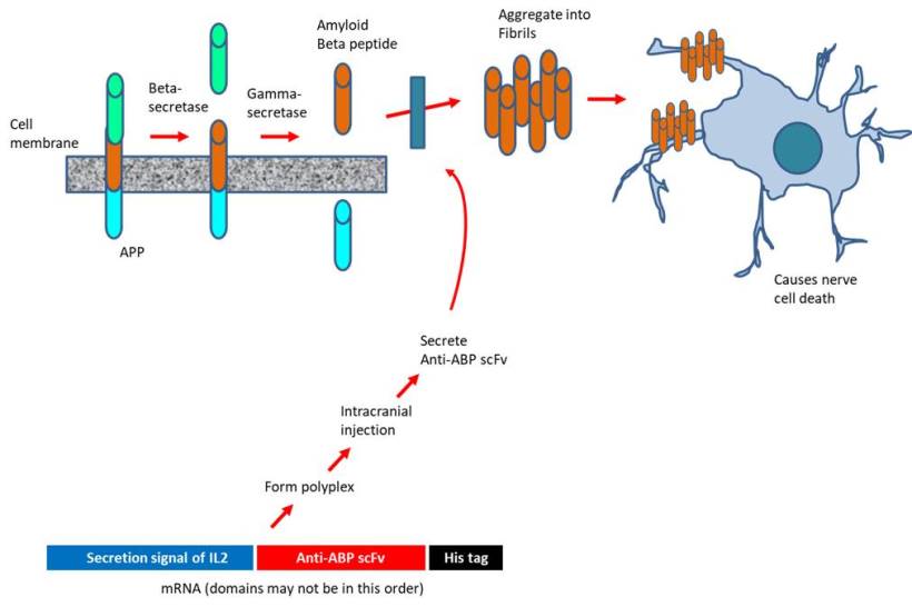


An innovative way exploiting the latest advances in mRNA technology to treat Alzheimer's disease is proposed.  In the past, pharmaceutical industries have focused on injecting high doses of antibodies targeting amyloid-beta peptide or administering recombinant adenoviruses to express such antibodies.  The approach is marred by the extremely low distribution to the brain (less than 1% of the systemically injected) or the side effects associated with drugs inhibiting the amyloid beta pathway. To address, the investigators at the University of Tokyo (Japan) designed mRNA encoding 3 distinct scFv (single-chain variable fragment that bind to amyloid-beta peptide) to be secreted by the expressing cells.  The scFV is comprised of the variable regions of the light and heavy chains of immunoglobulins connected through a linker.  The mRNA construct was delivered as polyplexes formed with copolymer PEG-PAsp (DET), i.e polyethylene glycol-poly[N'-[N-(2-aminoethyl)-2-aminoethyl] aspartamide] (Perche et al., 2017).  Intriguingly, scFv expression occurred following the transfection with mRNA (but not DNA) in neurons.  Upon intracranial injection, the scFv was able to reduce the level of amyloid beta peptide by 40% in an acute amyloidosis model although no such decline was observed in a transgenic model of Alzheimer's disease.  Nevertheless, the authors suggest that the drug design may be applicable for the immunological treatment of other neurological disorders.

The key to preventing epidemic is the ability to diagnose the infected early to preempt further propagation.  For this, Bio-Synthesis, Inc. provides primers and probes (as well as synthetic RNA control) for COVID-19 diagnosis via RT-PCR assay.  It specializes in oligonucleotide modification and provides an extensive array of chemically modified nucleoside analogues (over ~200) including bridged nucleic acid (BNA).  A number of options are available to label oligonucleotides (DNA or RNA) with fluorophoreseither terminally or internally as well as conjugate to peptidesIt recently acquired a license from BNA Inc. of Osaka, Japan, for the manufacturing and distribution of BNANC, a third generation of BNA oligonucleotides.  To meet the demands of therapeutic application, its oligonucleotide products are approaching GMP grade.  Bio-Synthesis, Inc. has recently entered into collaborative agreement with Bind Therapeutics, Inc. to synthesize miR-21 blocker using BNA for triple negative breast cancer.  The BNA technology provides superior, unequalled advantages in base stacking, binding affinity, aqueous solubility and nuclease resistance.  It also improves the formation of duplexes and triplexes by reducing the repulsion between the negatively charged phosphates of the oligonucleotide backbone.  Its single-mismatch discriminating power is especially useful for diagnosis (ex. FISH using DNA probe).  For clinical application, BNA oligonucleotide exhibits lesser toxicity than other modified nucleotides.


https://www.biosyn.com/oligo-flourescent-labeling.aspx

https://www.biosyn.com/tew/Speed-up-Identification-of-COVID19.aspx

https://www.biosyn.com/covid-19.aspx

https://www.biosyn.com/tew/Messenger-RNA-(mRNA)-for-Vaccine-Development-Against-Coronavirus.aspx

https://www.biosyn.com/mrna.aspx


References

Chen XQ, Mobley WC. Alzheimer Disease Pathogenesis: Insights From Molecular and Cellular Biology Studies of Oligomeric Aβ and Tau Species.  Front Neurosci. 13:659 (2019).  PMID: 31293377

Jaul E, Barron J. Age-Related Diseases and Clinical and Public Health Implications for the 85 Years Old and Over Population.  Front Public Health. 5:335 (2017).  PMID: 29312916

Jiang Q, Lee CY, Mandrekar S, et al. ApoE promotes the proteolytic degradation of Abeta.  Neuron. 58:681-93 (2008). PMID: 18549781

Perche F, Uchida S, et al. Improved Brain Expression of Anti-Amyloid β scFv by Complexation of mRNA Including a Secretion Sequence with PEG-based Block Catiomer.  Curr Alzheimer Res. 14:295-302 (2017).  PMID: 27829339

Tiwari S, Atluri V, et al. Alzheimer's disease: pathogenesis, diagnostics, and therapeutics.  Int J Nanomedicine. 14:5541-5554. (2019).  PMID: 31410002

Yan S, Gan Y, et al. Association between refrigerator use and the risk of gastric cancer: A systematic review and meta-analysis of observational studies.  PLoS One  13:e0203120. (2018)  PMID: 30161245


Antisense targeting of SARS-CoV-2 virus with BNAs

$
0
0

Can antisense oligonucleotides containing bridged nucleic acids (BNAs) be used for COVID-19 therapies?

Recent research suggests that bridged nucleic acid (BNA) antisense oligonucleotides (ASOs) enable the design of SARS-CoV-2 (COVID-19) targeting therapeutics. Many vaccines and drugs are now worldwide in development to stop COVID-19 infections and to treat infected people. In general, drug design is mostly based on protein targets since 3D structures for virus proteins are more readily available. 

The SARS-CoV-2 (COVID-19) virus requires the frameshift stimulation element (FSE) for a balanced expression of essential viral proteins. 

The coronavirus, SARS-CoV-2, has a positive sense (+), single-stranded RNA (ssRNA) genome. The first two open reading frames (ORFs) 1a and 1b encode for non-structural proteins. The ORFs include coding for the RNA-dependent polymerase and partial overlap. The precise stoichiometric expression of ORF 1a and ORF 1b throughout the viral replication cycles is vital for viral fitness. An essential regulatory mechanism regulates the two reading frames expression ratio called a -1 programmed ribosomal frameshifting (- PRF). A structured RNA motif at the 3’-end of ORF 1a called the frameshift stimulation element (FSE) initiates the PRF mechanism. FSE directs elongating ribosomes to randomly shift their reading frames by one base in the 5’-direction. The frameshift enables the readthrough past the ORF 1a stop codon needed to maintain appropriate levels of ORF 1a to ORF 1ab expression. FSE has a 5’-heptanucleotide “slippery site” UUUAAAC followed by an RNA element thought to form a three-stem pseudoknot.

Zhang et al. recently reported the 3D structure of the synthetic 28 kDA FSE from the SARS-CoV-2 RNA genome. The research team used cryo-electron microscopy and model-building to determine the 3D structure of the SARS-CoV-2 FSE with a 6.9 Å (0.69 nm) resolution.


Structural model of the 28-kDa Frameshift Stimulation Element from the SARS-CoV-2 RNA Genome

Figure 1: Models of the Frameshift Stimulation Element from the SARS-CoV-2 RNA Genome (6XRZ). A space fill model (left), a cartoon model (middle), and a surface model (right) is shown. The sequence TTAAAC is highlighted in yellow and as space fill residues.

GTTTTTAAACGGGTTTGCGGTGTAAGTGCAGCCCGTCTTACACCGTGCGGCACAGGCACTAGTACTGATGTCGTATACAGGGCTTTTG

SARS-CoV-2 FSE targeting BNA ASOs

The team utilized bridged nucleic acid (BNA/LNA) modified antisense oligonucleotides for the targeted disruption of the RNA frames shifting element. A few selected BNA ASOs were also synthesized with phosphorothioate (PS) internucleotide linkages. Binding pockets identified in the FSE 3D structure as targets to interrupt the element allowed the guided design of ASOs, including BNA ASOs.   

Frameshift Stimulation Element Sequences and targeting ASOs

>pdb|6XRZ|A Chain A, Frameshift Stimulation Element from the SARS-CoV-2 RNA Genome

GTTTTTAAACGGGTTTGCGGTGTAAGTGCAGCCCGTCTTACACCGTGCGGCACAGGCACTAGTACTGATGTCGTATACAGGGCTTTTG

MW390853.1

GTTTTTAAACGGGTTCGCGGTGTAAGTGCAGCCCGTCTTACACCGTGCGGCACAGGCACTAGTACTGATGTCGTATACAGGGCTTTTG

LC528233.2 

GTTTTTAAACGGGTTTGCGGTGTAAGTGCAGCCCGTCTTACACCGTGCGGCACAGGCACTAGTACTGATGTCGTATACAGGGCTTTTG

----AATTTGCCCAAGCGCCA----------------------------------------------TACAGCATATGTCCC(S2D)-

----AATTTGCCCAAACGCCA(SS2)-------------------------------TGATCATGACTACA(S3D1)-----------
----------------------------------------------------GTCCGTGATCATGACT(S3D2)--------------


NNN = BNAs

The UUUAAAC sequence is highly conserved in coronavirus genomes.

ASO and BNA ASOs used in the study.

ASOs

 Sequence

 Linkage

 Stem 2 Disruptor (S2D)

5′-+CCCTG+TA+TA+CGACA+T-3′

 -

 Stem 3 Disruptor-1 (S3D1)

5′-+A+C+ATCAGTACT+A+G+T-3′

PS linkages

 Stem 3 Disruptor-2 (S3D2)

5′-+T+C+AGTACTAGTG+C+C+T+G-3′

PS linkages

 Slippery site 2 (SS2)

5′-+A+C+C+GCGAACCCGTT+T+A+A-3′

PS linkages


Reference

Ref: Zhang K, Zheludev IN, Hagey RJ, Wu MT, Haslecker R, Hou YJ, Kretsch R, Pintilie GD, Rangan R, Kladwang W, Li S, Pham EA, Bernardin-Souibgui C, Baric RS, Sheahan TP, D Souza V, Glenn JS, Chiu W, Das R. Cryo-electron Microscopy and Exploratory Antisense Targeting of the 28-kDa Frameshift Stimulation Element from the SARS-CoV-2 RNA Genome. bioRxiv [Preprint]. 2020 Jul 20:2020.07.18.209270. doi: 10.1101/2020.07.18.209270. PMID: 32743589; PMCID: PMC7386510. [PMC]


---...---


RNA nucleotide repeats, Poly(A) RNA, and phase separation

$
0
0

The expansion of short nucleotide repeats is the cause of several neurological and neuromuscular disorders. Disease-causing short nucleotide repeats occur in the coding or non-coding regions of RNA transcripts. However, in all of the nucleotide repeat expansion disorders identified to date, only a critical number of repeats appear to cause the disease. Expanded repeats in transcripts can cause cellular toxicity and neurodegeneration by altering the splicing machinery.

Presently it is thought that toxic RNAs interact with different RNA binding proteins to produce disease, known as the "trans-dominant" model of RNA toxicity. The model proposes that the interaction of mutant RNA with RNA binding proteins interferes with the functions of the interacting proteins, leading to abnormalities in the pathways regulated by the RNA binding proteins.


Since 1992, scientists know that a CTG repeat expansion in the 3’-untranslated region (3’-UTR) of a protein kinase gene causes myotonic dystrophy type 1 (DM1), the most common form of adult muscular dystrophy. Southern blot, polymerase chain reaction (PCR)-based methods, and direct sequencing of PCR amplified CTG repeats allow the detection of these repeats in patients diagnosed with congenital myotonic dystrophy. 

The expansion of a hexanucleotide microsatellite DNA repeat (GGGGCC) in an intron of the C90RF72 gene is the cause of two neurodegenerative diseases. These are frontotemporal dementia (FTD) and amyotrophic lateral sclerosis (ALS). Unfortunately, presently there is no cure for these two diseases.

Jain & Vale in 2017 showed that repeat expansions create templates for multivalent base-pairing. In purified RNA of this type, a sol-gel transition occurs at a similar critical repeat number as that found in diseases. According to the researchers, in cells, RNA foci form by phase separation of the repeat-containing RNA. However, agents that disrupt RNA gelation in vitro can dissolve repeat-containing RNAs. Jain & Vale's suggested that sequence-specific gelation of RNAs may contribute to a specific neurological disease like in protein aggregation disorders.

Earlier studies by Eisenberg & Felsenfeld found that polyriboadenylic acid (poly (A) RNA) undergoes reversible phase separation at neutral pH. Poly (A) RNA is soluble at low and high temperatures but only partially miscible in a range of 35 to 40 °C. However, the range extent varies with molecular weight and polymer and salt concentrations. Poly(A) RNA precipitate at 35 to 40 °C but will remain soluble outside of that range. At a high temperature, the poly(A) polymer appears as a compact molecule. At a low temperature as an extended molecule.


This phenomenon's driving force appears to be base stacking, in which nucleic acid bases stack with their planes parallel to one another. However, Eisenberg & Felsenfeld's observation suggested structure formed by a non-co-operative process. 
As a result, poly(A) molecules of different lengths phase-separated at different conditions allowing the fractionation of poly(A) mixtures into homogenous solutions.

More recently, phase separation is emerging as a ubiquitous process for the compartmentalization and concentration of biomolecules at specific cell locations. Many many proteins crucial for cell growth and development undergo phase separation. However, the observation that RNA too can experience phase separation is a recent concept in RNA biochemistry. Apparently, RNA can also instruct its distribution in cells independently of other cellular components.

Poly (A) can also be used as a carrier molecule for the purification of DNA and RNA from a variety of samples.

In 2018 Treeck et al. suggested that RNA self-assembly also contributes to the formation of stress granules. 

What are stress granules?

Stress granules form during the inhibition of translation initiation. Stress granules are higher-order assemblies of non-translating mRNAs. These are ubiquitous, non-membrane-bound assemblies of protein and RNA forming during the inhibition of translation initiation. Stress granules appear to play a role in stress response and gene regulation. Related ribonucleoprotein (RNP) granules exist in neurons and can affect synaptic plasticity. Mutations in RNA binding proteins or stress granule-remodeling complexes leading to an increased formation of stress granules appear to cause amyotrophic lateral sclerosis (ALS) and other degenerative disorders. The formation of stress granules can influence both tumor progression and viral infection.

Reference

DeJesus-Hernandez M, et al.; Expanded GGGGCC hexanucleotide repeat in non-coding region of C9ORF72 causes chromosome 9p-linked frontotemporal dementia and amyotrophic lateral sclerosis. Neuron. 2011;72(2):245–256. [PMC]

Jain A, Vale RD. RNA phase transitions in repeat expansion disorders. Nature. 2017 Jun 8;546(7657):243-247. doi: 10.1038/nature22386. Epub 2017 May 31. PMID: 28562589. [PMC]

Ohya K, Tachi N, Sato T, Kon S, Kikuchi K, Chiba S. Detection of the CTG repeat expansion in congenital myotonic dystrophy. Jpn J Hum Genet. 1997 Mar;42(1):169-80. [PubMed]

Orr HT. Toxic RNA as a driver of disease in a common form of ALS and dementia. Proc Natl Acad Sci U S A. 2013 May 7;110(19):7533-4. [PMC]

Renton AE, Majounie E, Waite A, Simón-Sánchez J, Rollinson S, Gibbs JR, Schymick JC, Laaksovirta H, van Swieten JC, Myllykangas L, Kalimo H, Paetau A, Abramzon Y, Remes AM, Kaganovich A, Scholz SW, Duckworth J, Ding J, Harmer DW, Hernandez DG, Johnson JO, Mok K, Ryten M, Trabzuni D, Guerreiro RJ, Orrell RW, Neal J, Murray A, Pearson J, Jansen IE, Sondervan D, Seelaar H, Blake D, Young K, Halliwell N, Callister JB, Toulson G, Richardson A, Gerhard A, Snowden J, Mann D, Neary D, Nalls MA, Peuralinna T, Jansson L, Isoviita VM, Kaivorinne AL, Hölttä-Vuori M, Ikonen E, Sulkava R, Benatar M, Wuu J, Chiò A, Restagno G, Borghero G, Sabatelli M; ITALSGEN Consortium, Heckerman D, Rogaeva E, Zinman L, Rothstein JD, Sendtner M, Drepper C, Eichler EE, Alkan C, Abdullaev Z, Pack SD, Dutra A, Pak E, Hardy J, Singleton A, Williams NM, Heutink P, Pickering-Brown S, Morris HR, Tienari PJ, Traynor BJ. A hexanucleotide repeat expansion in C9ORF72 is the cause of chromosome 9p21-linked ALS-FTD. Neuron. 2011 Oct 20;72(2):257-68. [PMC]

Trcek, T. The curious behaviour of poly(A) RNA. Nat Rev Mol Cell Biol (2021). [Nature reviews]


Van Damme P, Robberecht W, Van Den Bosch L. Modelling amyotrophic lateral sclerosis: progress and possibilities. Dis Model Mech. 2017 May 1;10(5):537-549. [PMC]

Van Treeck, B. et al.; RNA self-assembly contributes to stress granule formation and defining the stress granule transcriptome. Proc. Natl Acad. Sci. USA 115, 2734–2739 (2018). [PNAS]


---...---


FDA approves second RNA interference-inducing siRNA oligonucleotide drug for treating acute hepatic porphyria, raising the prospect for cancer therapy

$
0
0

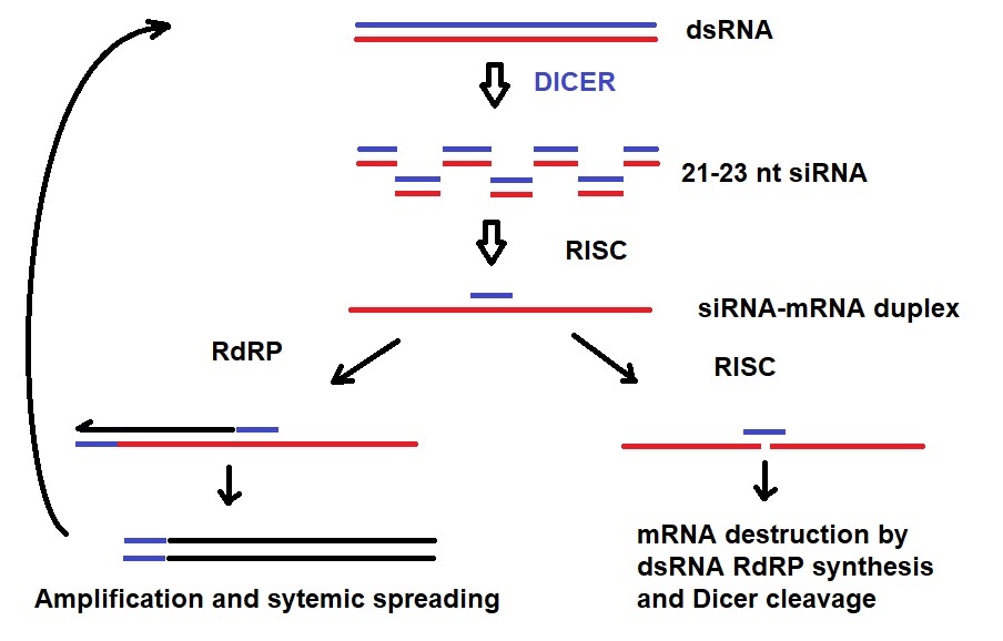
The post-transcriptional mechanism RNA interference silences gene expression by cleaving long double stranded RNA (dsRNA) into short dsRNA fragments, which are then unwound into single stranded RNAs to hybridize to target mRNA for degradation via the silencing complex (Agrawal et al., 2003; Li, 2008; Wilson et al, 2013).  RNA interference provides a unique opportunity to suppress gene expression without permanently inactivating the gene or introducing mutation (by integrating into the genomic DNA).  The ability of siRNAs to suppress gene expression by repeatedly targeting mRNAs for degradation constitutes a distinct pharmacological advantage.

Several notable advances have been made in the last decade.  In 2010, a phase I clinical trial showed that systemically delivered self-assembled nanoparticle containing siRNA targeting ribonucleotide reductase induced RNA interference in the tumor tissue of melanoma patients (Davis et al, 2010).   In 2013, the results from a Phase I clinical trial of ALN-VSP, a lipid nanoparticle carrying double siRNAs targeting vascular endothelial growth factor A (VEGF-A) and kinesin spindle protein KSP, in cancer patients was reported (Tabernero et al., 2013). 

In 2018, Onpattro was the first siRNA based drug developed by Alnylam, Inc. to be approved by Food & Drug Administration (FDA) for treating polyneuropathy.  It targets the mRNA encoding transthyretin (TTR), which transports thyroid hormone and retinol binding protein.

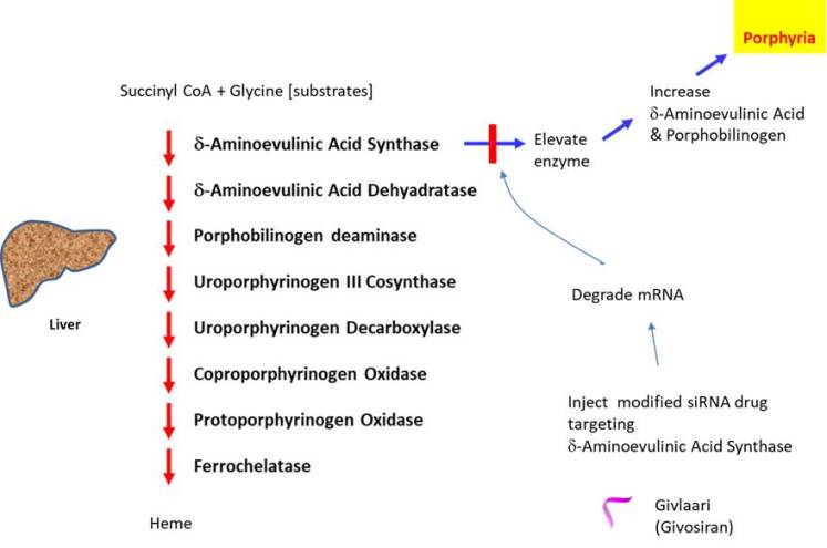
Acute hepatic porphyria is a rare inherited disorder that causes short lasting but rapid onset of life-threatening symptoms such as severe abdominal pain, high blood pressure, fever, neuropathic pain, etc.  It is caused by abnormal buildup of porphyrin, which forms the heme group of hemoglobin found in red blood cells to transport oxygen, in nervous system or skin.  Heme is also essential for the function of catalase, peroxidase, p450 liver cytochromes, etc.  The disorder could result from deficiency (i.e. genetic mutation is inherited) in any of the enzymes in the heme biosynthesis pathway, ex. acute intermittent porphyria, variegate porphyria, hereditary coproporphyria.  However, the symptoms are not caused by the reduction in heme (as it can be compensated by other enzymes) but rather due to the toxicity associated with the high level of porphyrins.   For instance, the acute attack may occur when aminolaevulinic acid synthase is induced by alcohol, reduced carbohydrate intake, etc.


In 2019, Givlaari was the second siRNA based drug to be approved by FDA (also called Givosiran: approved by European Medicines Agency in 2020), which was developed by Alnylam Pharmaceuticals (the Netherlands).  It was developed for treating acute hepatic porphyria.  Givlaari is comprised of synthetic double stranded siRNA oligonucleotide targeting the mRNA encoding delta-aminolevulinic acid synthase 1 enzyme.  The siRNA is conjugated to a ligand containing 3 N-acetylgalactosamine residues targeting galactose receptor (Wang et al., 2018).  Unlike Onpattro, to ensure stability, Givlaari has been modified to contain phosphorothioate backbone linkages at the termini and contains 2'-O-fluorourine and 2'-O-methyl groups in the pentose moiety.  Givlaari has demonstrated the ability to significantly decrease the rate of porphyria attacks requiring hospitalizations, urgent healthcare or IV hemin administration.  

The key to preventing epidemic is the ability to diagnose the infected early to preempt further propagation.  For this, Bio-Synthesis, Inc. provides primers and probes (as well as synthetic RNA control) for COVID-19 diagnosis via RT-PCR assay.  It specializes in oligonucleotide modification and provides an extensive array of chemically modified nucleoside analogues (over ~200) including bridged nucleic acid (BNA).  A number of options are available to label oligonucleotides (DNA or RNA) with fluorophoreseither terminally or internally as well as conjugate to peptidesIt recently acquired a license from BNA Inc. of Osaka, Japan, for the manufacturing and distribution of BNANC, a third generation of BNA oligonucleotides.  To meet the demands of therapeutic application, its oligonucleotide products are approaching GMP grade.  Bio-Synthesis, Inc. has recently entered into collaborative agreement with Bind Therapeutics, Inc. to synthesize miR-21 blocker using BNA for triple negative breast cancer.  The BNA technology provides superior, unequalled advantages in base stacking, binding affinity, aqueous solubility and nuclease resistance.  It also improves the formation of duplexes and triplexes by reducing the repulsion between the negatively charged phosphates of the oligonucleotide backbone.  Its single-mismatch discriminating power is especially useful for diagnosis (ex. FISH using DNA probe).  For clinical application, BNA oligonucleotide exhibits lesser toxicity than other modified nucleotides.  Site-specific labeling of RNA molecules is a valuable tool with multiple applications, and Bio-Synthesis, Inc. provides site-specific biotin labeling of long RNA at 5’-end.  

https://www.biosyn.com/oligo-flourescent-labeling.aspx

https://www.biosyn.com/tew/Speed-up-Identification-of-COVID19.aspx

https://www.biosyn.com/covid-19.aspx

https://www.biosyn.com/sirna-synthesis.aspx

https://www.biosyn.com/custom-rna-transcription-services.aspx

https://www.biosyn.com/tew/RNA-network-analysis-using-biotinylated-RNA-affinity-probes..aspx#


References

Agrawal N, Dasaradhi PV, et al. RNA interference: biology, mechanism, and applications.  Microbiol Mol Biol Rev. 67: 657-85 (2003).    PMID:14665679  

Davis ME, Yen Y, et al. Evidence of RNAi in humans from systemically administered siRNA via targeted nanoparticles. Nature. 464: 1067-70 (2010).  PMID: 20305636   

Li LC.  Small RNA-Mediated Gene Activation. RNA and the Regulation of Gene Expression: A Hidden Layer of Complexity. Caister Academic Press. ISBN 978-1-904455-25-7 (2008).

Tabernero J, Shapiro GI, et al. First-in-humans trial of an RNA interference therapeutic targeting VEGF and KSP in cancer patients with liver involvement.  Cancer Discov. 3: 406-17 (2013).  PMID: 23358650

Wang B, Rudnick S, et al.  Acute Hepatic Porphyrias: Review and Recent Progress.  Hepatol Commun  3:193-206 (2018) PMID: 30766957

Wilson RC, Doudna JA.  Molecular mechanisms of RNA interference.  Annu Rev Biophys. 42:217-39 (2013). PMID: 23654304

Site specific labeling of long RNA with biotin for diagnostic or other purposes

$
0
0
For experimental biomedical science, the ability of biotin to bind to streptavidin with high affinity has been exploited for both purification and detection purposes.  This has also been the case with RNA molecules as labeling with biotin allows it to be immobilized on avidin-based supports for purification.  As the biotinylated RNA could be detected by streptavidin coupled to chemiluminescent or fluorescent marker, it could also be used to detect the labeled RNA itself or its interacting target in various milieu.

The biotin labeling of RNA could be done site-specifically or randomly.  To label RNA randomly, modified nucleotides could be incorporated during the in vitro transcription reaction.  To label randomly in a single-step reaction, biotinylated UTP or biotinylated CTP can be incorporated during the co-transcriptional biotinylation step.  The disadvantage with this method is that the labeling position(s) as well as the extent of labeling cannot be adequately controlled.  Further, the biological function or property of the RNA may be altered or compromised through the biotin labeling.

This has led to the development of methods that enable site-specific labeling of RNA with biotin.  To biotin label at a specific position located internally along RNA, a transcription-based method utilizing unnatural bases was developed (Moriyama et al., 2005).  It relies on the ability of 2-amino-6-(2-thienyl)purine or 2-amino-6-(2-thiazolyl)purine to base pair with 2-oxo(1H)pyridine during transcription.  The latter (conjugated to biotin) can be incorporated by T7 RNA polymerase during the transcription.  A caveat to the method is that it requires one to prepare a template DNA containing unnatural bases beforehand.

To biotin label site-specifically at the 3' end of RNA, several methods are available.  For chemical biotinylation, 3' terminal ribose of RNA can be oxidized to dialdehyde using periodate, which is susceptible to nucleophilic attack by amino group of a suitable biotin derivative (Willkomm and Hartmann 2005).  Here, the disadvantage lies with the loss of 3' nucleotide through a competing reaction (beta-elimination) (Proudnikov and Mirzabekov 1996).  Another method employs T4 RNA ligase to add a biotinylated pCp derivative to the 3' end of RNA (Kore et al., 2009).  But the method is hampered by inefficient ligation reaction as well as the requirement for a high concentration of substrates.  Other methods prescribed include the use of poly(A) polymerase to extend RNA at 3' end with N6-biotin-ATP; alternatively, phi29 DNA polymerase could be used to extend RNA after priming with an oligodeoxynucleotide (Moritz et al, 2013).


Various techniques have been devised to biotin label site-specifically at the 5' end of RNA.  To selectively label at the 5' end (without simultaneously labeling the 3' end), RNA was incubated with aminopurine riboside triphosphate (to form a conjugate with RNA using T4 RNA ligase).  The adenylated intermediate could then be labeled with biotin at the 5' end of RNA (Kinoshita et al., 1997).  An alternate technique utilizes biotin-labeled nucleotide as the 'initiator nucleotide' for RNA transcription.  To biotin label at the 5' end of RNA, a biotin-AMP conjugate (biotin-HDAAMP) can be used as transcription initiator under the T7 phi2.5 promoter (Huang et al., 2008) or biotinyl-guanosine 5'-monophosphate under the conventional T7 promoter (Collett et al 2005).  Nevertheless, their main limitation is that neither method allows capping of the RNA.

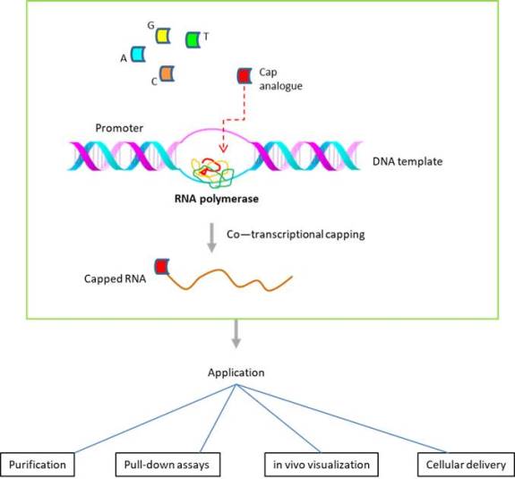
To address the above issue, additional approaches have been adopted.  One method is based on 'co-translational capping', wherein a cap analogue is incorporated at the first nucleotide position (of the RNA transcript generated). The cap analogue could be further modified to incorporate in the correct orientation.  To generate RNAs labeled with biotin via the 5' cap structure, the investigators at the University of Warsaw (Poland) developed biotin-labeled cap analogues modified within the triphosphate bridge, which could be incorporated for co-transcriptional capping (Bednarek et al., 2018).

Site-specific labeling of RNA molecules is a valuable tool with multiple applications, and Bio-Synthesis, Inc. provides site-specific biotin labeling of long RNA at 5’-end with nearly 100% efficiency.

References

Bednarek S, Madan V, et al. mRNAs biotinylated within the 5' cap and protected against decapping: new tools to capture RNA-protein complexes.  Philos Trans R Soc Lond B Biol Sci.  373:20180167 (2018).  PMID: 30397103

Collett,J.R., Cho,E.J., et al.  Functional RNA microarrays for high-throughput screening of antiprotein aptamers. Anal. Biochem., 338, 113–123 (2005).  PMID:  15707941  

Huang F, He J, Zhang Y, Guo Y.  Synthesis of biotin–AMP conjugate for 5′ biotin labeling of RNA through one-step in vitro transcription. Nat Protoc 3: 1848–1861 (2008).  PMID: 18989262

Kinoshita, Y., Nishigaki, K., et al. Y.   Fluorescence-, isotope- or biotin-labelinbeling of the 5-end of single-stranded DNA/RNA using T4 RNA ligase. 25:3747-8 (1997).  PMID: 9278501

Kore AR, Charles I, et al. Synthesis and activity of modified cytidine 5′-monophosphate probes for T4 RNA ligase 1.  Nucleosides Nucleotides Nucleic Acids 28: 292–302 (2009). PMID:  20183582  

Moritz B, Wahle E. Simple methods for the 3' biotinylation of RNA.  RNA. 20:421-7 (2014).  PMID: 24448448

Moriyama K, Kimoto M, et al. Site-specific biotinylation of RNA molecules by transcription using unnatural base pairs.  Nucleic Acids Res. 33:e129 (2005).  PMID: 16113238

Proudnikov D, Mirzabekov A. Chemical methods of DNA and RNA fluorescent labeling. Nucleic Acids Res 24: 4535–4542 (1996). PMID:  8948646  

Willkomm DK, Hartmann RK. 2005. 3′-Terminal attachment of fluorescent dyes and biotin. In Handbook of RNA biochemistry (ed. Hartmann RK, et al.), pp. 86–94. Wiley-VCH, Weinheim, Germany.


Applications for circular oligonucleotides

$
0
0

A closed-loop configuration formed from a linear nucleic oligonucleotide by an intramolecular covalent linkage of their two ends characterizes circular nucleic acids.

Circular nucleic acids (CNAs) exist in nature as circular DNAs and RNAs that vary in size and function. Naturally occurring circular DNA exists as bacterial genomes, plasmids, mitochondrial DNA, chloroplast DNA, bacteriophage DNA, and eukaryotic viral DNA. Viroids, virusoids, spliced introns or exons of eukaryotes, bacteria, and archaea contain circular RNA.

In general, theta replication, rolling circle replication, or back-splicing produces CNAs. The fields of biotechnology, disease therapy and nanotechnology already utilize CNAs.


Circularized or circular single-stranded deoxyribosnucleotides (cssDNAs) are more resistant to the attack by nucleases than their linear oligonucleotides. These properties make these circular oligonucleotides important tools for in vivo studies.

Circular DNAs have been investigated for their unique DNA binding properties, for example, as useful models in studying DNA structures such as hairpin motifs by NMR. Also, circular DNAs are used for diagnostic applications, such as padlock probes and the synthesis of concatemeric polypeptides. Also, circular ssDNA allows for both DNA and RNA amplification. CNAs can function as templates for both DNA and RNA polymerases. Additional examples are the use of plasmids in molecular cloning to study protein functions, the treatment of bacterial infections with circular lytic bacteriophages, the construction of “DNA origami” with circular phage DNA, and the use of circular RNA to regulate the transcription of microRNAs (miRNAs).

Characteristics, use and applications for circular oligonucleotides are:

Antisense using circular oligonucleotides

Binding of duplex DNA

Delivery vectors for miRNAs

Diagnostics

DNA fragment assembly using a nicking enzyme system 

DNA polymerase inhibition

DNA structure studies

Efficient templates for DNA and RNA polymerases

Hairpin motif design

Hairpin studies

Ligation-independent cloning (LIC)

Manipulating gene expression with caged circular oligonucleotides

Mutation detection

Padlock probes

Probing DNA-protein interactions

Quantitation of sequence-dependent DNA bending and flexibility

RNA polymerase inhibition

Rolling Circle Amplification (RCA)

Single molecule counting

Specific gene expression

Study of noncanonical DNA structural motifs 

Synthesis of concatemeric polypeptides

Topologic modifications

Triple helix formation

Unique DNA recognition properties


References

Diegelman, A. M. and Kool, E. T. (2000) Chemical and enzymatic methods for preparing circular single-stranded DNAs. In Current Protocols in Nucleic Acid Chemistry, vol. 2 (Beaucage, S. et al., eds.). John Wiley, New York, Chapter 5.2.

Oligonucleotide synthesis: methods and applications in Methods in molecular biology ; 288. 
edited by Piet Herdewijn. ISBN 1-58829-233-9.

---...---

Fatty acid conjugated BNA antisense oligonucleotides exhibit enhanced uptake into muscle

$
0
0

Conjugation of antisense oligonucleotides containing bridged nucleic acids (BNAs) to palmitic acid enhances the uptake by targeted cells as well as their potency.

Prakash et al. in 2019 reported a detailed study of fatty acids conjugated to BNA modified antisense oligonucleotides containing phosphorothioate linkages. In phosphorothioate linkages (PS), a sulfur atom replaces the nonbridging oxygen, conferring resistance to digestion by nucleases. The study revealed that palmitic acid conjugated to phosphorothioate antisense oligonucleotides (PS ASOs) enhances the PS ASOs affinity to serum albumin and the potency of the ASOs in muscle tissue.

Nucleic acid-based therapeutics are promising candidates for targeting genes linked to diseases considered as undruggable. It is now well known that the conjugation of tri-antennary N-acetyl galactosamine (GalNac) to antisense oligonucleotides enhances ASO uptake into hepatocytes through asialoglycoprotein receptor (ASGR)-mediated internalization by 10- to 60-fold. The targeted receptor is a C-type lectin predominantly displayed on the plasma membranes of hepatocytes.

Figure 1: Example of antisense oligonucleotide conjugated to trieantennary GalNac.

As another example, a recent study revealed that conjugation of ASOs to a ligand of the glucagon-like peptide-1 receptor (GLP1R) improved uptake of the ASO by pancreatic β-cells and enhanced its potency by more than 50 fold. In this study, the GLP1R peptide antagonist eGLP1 (H(Aib)EGTFTSDVSSYLEQAAKEFIAWLVKGGPSSGAPPPSC was conjugated to the ASO modified with BNAs.


Figure 2: Antisense oligonucleotide conjugated to eGLP1.

For targeting expressed genes in muscle cells, the functional uptake of ASOs in muscle is essential for the development of therapeutics. The oxidation of long-chain fatty acids in skeletal and cardiac muscle provides the fuel needed for contractile work. In the blood, fatty acids are either complexed to albumin or covalently bound in triacylglycerols. Here they form a neutral lipid core of circulating triglyceride-rich lipoproteins such as chylomicrons or very-low-density lipoproteins. It appears that the interaction of the albumin-fatty acid complex with the endothelial membrane facilitates fatty acid uptake. The transport protein serum albumin is the most abundant plasma protein in human blood. Serum albumin is synthesized in the liver and released into the bloodstream. Albumin has several hydrophobic pockets that bind fatty acids and steroids, including a variety of drugs.     

Prakash et al. investigated the affinity of ASO fatty acid conjugates to plasma proteins with the goal to enhance the uptake of ASO fatty acdi conjugates by muscle cells from the bloodstream. The study, performed in a mouse model, found that ASO conjugates containing fatty acid chains in length from 16 to 22 carbon moieties showed improved binding affinities to plasma proteins. As determined by the study, ASOs conjugated to palmitic acid improved the potency of ASOs targeting dystrophia myotonica protein kinase (DMPK), caveolin-3(Cav3), cluster of differentiation 36 (CD36), and metastasis-associated in lung adenocarcinoma transcript 1 (Malat-1) in muscle tissue.


Figure 3: Antisense oligonucleotide conjugated to palmitic acid.


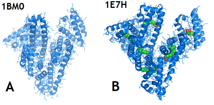
Figure 3: Structures of Human Serum Album (A; HSA) and HSA-palmitoyl complex (B).

Human serum albumin (HAS) is an abundant plasma protein. HSA is responsible for the transport of fatty acids and also binds a wide range of drug compounds. Sugio et al. in 1999 reported the crystal structure of human serum albumin. The protein structure showed that Cys34 is the only cysteine with a free sulfhydryl group that does not participate in a disulfide linkage with any external ligand. The research group identified three tentative binding sites for long-chain fatty acids located at the surface of each protein domain. Bhattacharya et al. in 2000 solved the structure of human serum albumin in complex with palmitic acids and identified a total of 11 different fatty acid binding sites.
 

Reference

Ämmälä C, Drury WJ 3rd, Knerr L, Ahlstedt I, Stillemark-Billton P, Wennberg-Huldt C, Andersson EM, Valeur E, Jansson-Löfmark R, Janzén D, Sundström L, Meuller J, Claesson J, Andersson P, Johansson C, Lee RG, Prakash TP, Seth PP, Monia BP, Andersson S. Targeted delivery of antisense oligonucleotides to pancreatic β-cells. Sci Adv. 2018 Oct 17;4(10):eaat3386. [PMC]

A A Bhattacharya 1, T Grüne, S Curry.;  Crystallographic analysis reveals common modes of binding of medium and long-chain fatty acids to human serum albumin. J Mol Biol. 2000 Nov 10;303(5):721-32.

Juliano RL. The delivery of therapeutic oligonucleotides. Nucleic Acids Res. 2016 Aug 19;44(14):6518-48. [PMC].

Prakash TP, Mullick AE, Lee RG, Yu J, Yeh ST, Low A, Chappell AE, Østergaard ME, Murray S, Gaus HJ, Swayze EE, Seth PP. Fatty acid conjugation enhances potency of antisense oligonucleotides in muscle. Nucleic Acids Res. 2019 Jul 9;47(12):6029-6044. [PMC]

Roberts, T.C., Langer, R. & Wood, M.J.A. Advances in oligonucleotide drug delivery. Nat Rev Drug Discov 19, 673–694 (2020). [Nature

S Sugio 1, A Kashima, S Mochizuki, M Noda, K Kobayashi. ; Crystal structure of human serum albumin at 2.5 A resolution. Protein Eng. 1999 Jun;12(6):439-46.  

---...---

 " Bio-Synthesis provides a full spectrum of high quality custom oligonucleotide modification services including conjugation to fatty acids, biotinylation by direct solid-phase chemical synthesis or enzyme-assisted approaches to obtain artificially modified oligonucleotides, such as mRNAs or siRNAs, containing a natural or modified backbone, as well as base, sugar and internucleotide linkages. Bio-Syntheis also provides biotinylated mRNA as well as long circular oligonucleotides".

---...---

Tumor drug penetration measurement reveals limited distribution of fluor-conjugated Herceptin antibody in a mouse breast cancer model

$
0
0
One of the major challenges for cancer therapy is the occurrence of side effects.  The problem remains unresolved for either cytostatic or cytotoxic drugs.  Whereas cytostatic drugs arrest cell cycling, cytotoxic drugs induce cell death.  While conventional therapies like the chemotherapy or radiotherapy are cytotoxic, many of the 'molecularly targeted drugs' developed in the last several decades (that inhibit proteins expressed by oncogenes) tend to be cytostatic.  Oncogenes are derived from proto-oncogenes in our genome, which are thought to promote cancer upon acquiring mutation(s) that upregulates its activity (ex. tyrosine kinase).  This is the key reason (treated cells remain viable) why targeted drugs like Gleevec (for chronic myelogenous leukemia) need to be administered perennially to avoid recurrent cancer.  Yet, even with targeted drugs, the problem of side effects persists.  For Gleevec, the side effects include rash, infection, neutropenia, anemia, edema, thrombocytopenia, etc.

Another contributing factor to side effects is that a significant fraction of intravenously injected drugs fail to reach tumors.  In the case of Taxol, nearly half of all injected drugs are eliminated after 24 h, with less than 0.5% remaining locally to treat lung cancer (Wolinsky et al., 2012).  To compensate, a greater dose of drugs may need to be administered approaching the 'maximum tolerated dose' (MTD), further exacerbating side effects. 

Several methods have been developed to address the issue.  One such approach is chemoembolization (for liver cancer), wherein the artery feeding into liver is blocked off after administering the drugs to trap them inside the liver, thus avoiding them from circulating throughout the body.  However, the utility of this strategy is limited and may not be applicable for other types of cancer.

An alternate approach involves conjugating drugs to tumor targeting delivery vectors.  One such strategy sought to utilize antibodies to guide drugs to tumors.  Tumor targeting antibodies have been increasingly used for cancer therapy.  Among them is Herceptin, a monoclonal antibody that recognizes Her2 oncogene overexpressed in a subset (~15%) of breast cancers (Figueroa-Magalhães et al., 2014).


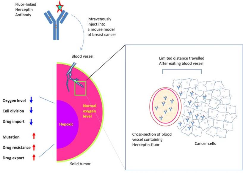
To assess its efficacy as a delivery vector, the ability of Herceptin to penetrate solid tumors was examined (solid tumors comprise >95% of human cancers).  To measure the depth of penetration, the investigators at the University of Chicago developed a novel imaging technique through which microscopic images taken at multiple depths could be integrated using computer to build the 3-dimensional 'map' of a tumor (Lee et al., 2019).  Then, by applying the principle of tomography (ex. CAT-SCAN imaging), the 3D map could be viewed at any axis (X- or Y- or Z-axis) to determine drug distribution within a solid tumor. 

To trace the injected drug, Herceptin antibody was conjugated to the fluor DyLight594.  DyLight fluors are fluorescent dyes activated at similar wavelengths as conventional fluorphores (ex. fluoresceine, Cy5, rhodamine) but may be more photostable (takes longer time to photobleach by laser). To measure the distance it travelled after leaving a blood vessel, they conjugated anti-CD31 antibody to DyLight633 fluorescent dye to locate blood vessels (for reference point).

Following the intravenous injection of both antibodies in a transgenic mouse model (overexpresses neu, a murine homologue of Her2) that spontaneously develops mammary tumors, they determined that Herceptin traveled mere 38 micrometer (several cell length) from the blood vessel boundaries after 1 hour-post-injection. 

In another report, a greater distribution of Herceptin was observed after 1 day-post-injection; however, the antibody still could not reach hypoxic regions of a solid tumor (Lee et al., 2010).  Hypoxic regions are located distant from blood vessels within a solid tumor and suffer from a low level of oxygen or nutrients, a higher mutation rate, and drug resistance (Sullivan et al., 2008; Li et al., 2017).  In this regard, delivery vectors capable of penetrating solid tumors deeply like the tumor-targeting peptides may play a greater role in reducing side effects (Joliot et al., 2004; Wright et al., 2016; Hong et al., 2000; Li et al., 2021).

The key to preventing epidemic is the ability to diagnose the infected early to preempt further propagation.  For this, Bio-Synthesis, Inc. provides primers and probes (as well as synthetic RNA control) for COVID-19 diagnosis via RT-PCR assay.  It specializes in oligonucleotide modification and provides an extensive array of chemically modified nucleoside analogues (over ~200) including bridged nucleic acid (BNA).  A number of options are available to label oligonucleotides (DNA or RNA) with fluorophoreseither terminally or internally as well as to conjugate to peptidesor antibodies.  It recently acquired a license from BNA Inc. of Osaka, Japan, for the manufacturing and distribution of BNANC, a third generation of BNA oligonucleotides.  To meet the demands of therapeutic application, its oligonucleotide products are approaching GMP grade.  Bio-Synthesis, Inc. has recently entered into collaborative agreement with Bind Therapeutics, Inc. to synthesize miR-21 blocker using BNA for triple negative breast cancer.  The BNA technology provides superior, unequalled advantages in base stacking, binding affinity, aqueous solubility and nuclease resistance.  It also improves the formation of duplexes and triplexes by reducing the repulsion between the negatively charged phosphates of the oligonucleotide backbone.  Its single-mismatch discriminating power is especially useful for diagnosis (ex. FISH using DNA probe).  For clinical application, BNA oligonucleotide exhibits lesser toxicity than other modified nucleotides. 

https://www.biosyn.com/oligo-flourescent-labeling.aspx

https://www.biosyn.com/tew/Speed-up-Identification-of-COVID19.aspx

https://www.biosyn.com/covid-19.aspx

https://www.biosyn.com/bioconjugation.aspx


References

Figueroa-Magalhães MC, et al. Treatment of HER2-positive breast cancer.  Breast. 23:128-136 (2014). PMID: 24360619

Hong FD, Clayman GL. Isolation of a peptide for targeted drug delivery into human head and neck solid tumors.  Cancer Res. 2000 Dec 1;60(23):6551-6.   PMID: 11118031

Joliot A, Prochiantz A. Transduction peptides: from technology to physiology.   Nat Cell Biol.  6:189-96 (2004).  PMID: 15039791

Lee CM, Tannock IF. The distribution of the therapeutic monoclonal antibodies cetuximab and trastuzumab within solid tumors.  BMC Cancer.  10:255 (2010).  PMID: 20525277

Lee SS, Bindokas VP, et al. Multiplex Three-Dimensional Mapping of Macromolecular Drug Distribution in the Tumor Microenvironment.  Mol Cancer Ther.  18:213-226 (2019).  PMID: 30322947

Li JQ, Wu X, Gan L, et al.   Hypoxia induces universal but differential drug resistance and impairs anticancer mechanisms of 5-fluorouracil in hepatoma cells.   Acta Pharmacol Sin. 38:1642-1654 (2017). PMID: 28713155

Li R, Wang Y, et al. Graphene oxide loaded with tumor-targeted peptide and anti-cancer drugs for cancer target therapy.  Sci Rep. 11:1725 (2021).  PMID: 33462277

Sullivan R, Paré GC, et al.  Hypoxia-induced resistance to anticancer drugs is associated with decreased senescence and requires hypoxia-inducible factor-1 activity.  Mol Cancer Ther.  7:1961-73 (2008).  PMID: 18645006

Wolinsky JB, Colson YL, et al. Local drug delivery strategies for cancer treatment: gels, nanoparticles, polymeric films, rods, and wafers.   J Control Release. 159:14-26 (2012).   PMID: 22154931

Wright CL, Pan Q, . Advancing theranostics with tumor-targeting peptides for precision otolaryngology.   World J Otorhinolaryngol Head Neck Surg.  2:98-108 (2016).  PMID: 29204554


Expanded RNA repeat diseases

$
0
0

The expansion of RNA repeats is the cause for neurodegenerative disorders. Abnormal RNA-protein interactions disrupt the physiological functions of proteins.

Many transcripts contain short tandem repeats (STRs). Mutant RNA transcripts can trigger pathological effects when a critical repeat length is reached. In some diseases, abnormal RNA-protein interactions occur, disrupting the physiological functions of bound proteins. These diseases include dominantly inherited disorders associated with long repeat expansions in the non-coding or coding regions of individual genes. 

Interaction between RNA-binding proteins and mutant RNA often results in the immobilization of these proteins in specific structures known as RNA foci. RNA foci are the pathogenic hallmarks of this type of disease.

Studies of proteins bound to CUG repeats in myotonic dystrophy type 1 (DM1) resulted in developing the "RNA gain-of-function" model. The model states that expanded repeats sequester RNA-binding proteins from their normal function.

Proteins trapped by other repeats are well studied now. The RNA repetitions list includes CUG, CAG, CGG, CCUG, AUUCA, and GGGGCC repeats.

Alternative splicing of specific pre-mRNAs is affected by abnormal RNA-protein interactions. Abnormal RNA-protein interactions alter the use of alternative polyadenylation sites of several mRNAs. Changes in nuclear transport and export affect translation, induce nucleolar stress, and dysregulate miRNA processing. Structures formed by expanded repeats appear to trigger protein recruitment.

Mutant transcripts encoded by specific nucleic acid sequences present in the expanded region can adopt stable in-vitro hairpin or G-quadruplex structures. This phenomenon is also known as "RNA-mediated toxicity" in these disorders.

However, many unanswered questions remain regarding the disease-causing molecular mechanism. Molecular probes allowing the study of proteins recruited to RNA foci may help to elucidate these mechanisms, possibly leading to new therapeutic drugs to treat these disorders.


A CTG repeat expansion causes myotonic dystrophy type I (DM1), leading to defects in developmentally regulated alternative splicing. The repeat expansion occurs within the 3’-UTR of the DMPK gene. RNA foci formed by the expanded CUG repeat in the nucleus sequester the muscle-blind-like (MBNL) protein family of splicing factors and induce upregulation of CELF1 through PKC-mediated phosphorylation and altered microRNA regulation.

In 2010, Todd & Paulson reviewed a series of proposed mechanisms by which noncoding repeat expansions give rise to nervous system degeneration and dysfunction. 

Mechanisms discussed in the review include

1. Transcriptional alterations, 

2. Generation of antisense transcripts, 

3. Sequestration of mRNA-associated protein complexes leading to aberrant mRNA splicing and processing,

4. Alterations in cellular processes, 

5. Activation of abnormal signaling cascades and failure of protein quality control pathways.

Antisense oligonucleotides are a promising therapeutic approach for diseases caused by expanded RNA repeats. In particular, antisense oligonucleotides stabilized with chemical modifications such as bridged nucleic acids (BNAs) and phosphorothioates allow the design of gapmers useful for RNA targeting therapeutics.

Table 1 : Disease causing repeats

Disease

Repeat

Gene

Normal Repeat

Disease-causing repeat

CNS Phenotype

Myotonic Dystrophy Type 1 (DM1)

CTG

3′UTR of DMPK


5–38

50~1500: Adult onset DM1
~1500+: Congenital DM1

Adult onset: Neuropsychiatric symptoms, executive dysfunction

Congenital DM1: mental retardation in>50%.

Myotonic Dystrophy Type 2 (DM2)

CCTG

Intron 1 of ZNF9


Up to 30

75–11,000

Neuropsychiatric symptoms common

No congenital onset or mental retardation

Fragile X Tremor Ataxia Syndrome (FXTAS)

CGG

5′UTR of FMR1


20–45

55–200; incomplete penetrance at all repeat lengths.

Late onset

Cerebellar ataxia, action tremor, dementia Parkinsonism.

Neuropsychiatric symptoms in females.

Spinocerebellar ataxia Type 3 (SCA 3)

CAG

Exon 10 of ATXN3


Up to 44

~45–51: reduced penetrance
~52–86: Fully penetrant

Ataxia

Parkinsonism, dystonia

dementia uncommon

Spinocerebellar Ataxia Type 8 (SCA8)

CTG

5′UTR of KLH1


15–50

71>1300; incomplete penetrance at all repeat lengths

Cerebellar Ataxia

dementia uncommon

Spinocerebellar Ataxia Type 10 (SCA10)

ATTCT

3′UTR of E46L


10–29

800–4500

Cerebellar ataxia

Seizures

cognitive decline

Spinocerebellar Ataxia Type 12 (SCA12)

CAG

5′UTR/promoter of PPP2R2B


Up to 32

51–78

Action tremor

Cerebellar ataxia

dementia uncommon

Huntington’s Disease Like 2 (HDL-2)

CTG

3′UTR of JPH-3


6–28

>41

Clinically similar to Huntington’s Disease

(Source: Todd & Paulson)

Abbreviations: ORF=Open Reading Frame; UTR=Untranslated Region; DMPK=Dystrophin Myotonica Protein Kinase; ZNF9=Zinc Finger 9; FMR1=Fragile X Mental Retardation gene 1; ATXN3=Ataxin 3; KLH1=Kelch-Like 1; PPP2R2B=protein phosphatase 2, regulatory subunit B, beta isoform.

Reference

CELF1: CUGBP Elav-like family member 1 [UniProtKB]

Ciesiolka A, Jazurek M, Drazkowska K, Krzyzosiak WJ. Structural Characteristics of Simple RNA Repeats Associated with Disease and their Deleterious Protein Interactions. Front Cell Neurosci. 2017 Apr 11;11:97. [PMC]

Magdalena Jazurek, Adam Ciesiolka, Julia Starega-Roslan, Katarzyna Bilinska and Wlodzimierz J. Krzyzosiak; SURVEY AND SUMMARY. Identifying proteins that bind to specific RNAs - focus on simple repeat expansion diseases. Nucleic Acids Research, 2016, Vol. 44, No. 19. 9050-9070.

Konieczny P, Stepniak-Konieczna E, Sobczak K. MBNL proteins and their target RNAs, interaction and splicing regulation. Nucleic Acids Res. 2014;42(17):10873-87. [PMC]

Manning KS, Rao AN, Castro M, Cooper TA. BNANC Gapmers Revert Splicing and Reduce RNA Foci with Low Toxicity in Myotonic Dystrophy Cells. ACS Chem Biol. 2017 Oct 20;12(10):2503-2509. [PMC]

Todd PK, Paulson HL. RNA-mediated neurodegeneration in repeat expansion disorders. Ann Neurol. 2010 Mar;67(3):291-300.  [PMC]

M. J. Walsh, J. Cooper-Knock, J. E. Dodd, M. J. Stopford, S. R. Mihaylov, J. Kirby, P. J. Shaw and G. M. Hautbergue;  Invited Review: Decoding the pathophysiological mechanisms that underlie RNA dysregulation in neurodegenerative disorders: a review of the current state of the art. Neuropathology and Applied Neurobiology (2015), 41, 109–134. [PMC]


---...---

 " Bio-Synthesis provides a full spectrum of high quality custom oligonucleotide modification services including back-bone modifications, conjugation to fatty acids, biotinylation by direct solid-phase chemical synthesis or enzyme-assisted approaches to obtain artificially modified oligonucleotides, such as BNA antisense oligonucleotidesmRNAs or siRNAs, containing a natural or modified backbone, as well as base, sugar and internucleotide linkages.
Bio-Synthesis also provides biotinylated mRNA as well as long circular oligonucleotides".

---...---

SARS-CoV-2 modifies the host's mRNA cap to evade an innate immunity response!

$
0
0

The new SARS coronavirus, which is the cause of the COVID-19 pandemic, hijacks the translation machinery in human host cells by converting Cap-0 to Cap-1 in the host's mRNA to avoid an innate immune response.

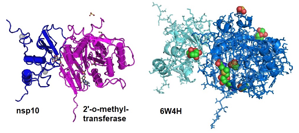
Intending to develop effective therapies for COVID-19, Viswanathan et al. studied the mechanisms permitting the virus to invade cells and evade the host's innate immune system. Viswanathan et al. in 2020 reported that the non-structural protein 16 (nsp16) methylates the 5’-end of virally encoded mRNAs to mimic cellular mRNAs. This modification hides the virus's mRNAs from the innate immune system. The research team solved the high-resolution structure of a ternary complex of full-length nsp16 and nsp10 of SARS-CoV-2 complexed to the cognate RNA substrate and a methyl donor, S-adenosyl methionine. The nsp16/nsp10 heterodimer's structural model revealed the methylation of the 2’-oxygen of the ribose sugar of the first nucleotide of SARS-CoV-2 mRNAs. Also, the structure revealed fundamental conformational changes in the RNA substrate binding site and the biochemistry of RNA Cap methylation. Furthermore, Viswanathan et al. identified a potential allosteric site as a target for developing antiviral therapies to treat SARS-CoV-2 infections.

The SARS-CoV-2 Nsp16 protein forms a complex with nsp10 to convert mRNA species from the Cap-0 (me7GopppA1) to the Cap-1 form (me7GopppA1m). The protein complex methylates the ribose 2’-OH group of the nascent mRNA's first nucleotide using S-adenosyl methionine (SAM) as the methyl donor molecule. In CoVs, the first nucleotide is usually adenosine. Cap-1 avoids induction of the innate immune response.

Figure 1: Different images of the SARS-CoV-2 nsp16/nsp10 in complex with RNA cap analog (m7GpppA) and S-adenosylmethionine (6WKS).

Figure 2: Configuration of m7GpppA as observed in the complex 6WKS.


Also, in 2020, Rosas-Lemus et al. determined the structures for nsp16-nsp10 heterodimers in complew with the methyl donor S-adenosylmethionine (SAM), the reaction product S-adenosylhomocysteine (SAH), or the SAH analog sinefungin (SFG). The research group solved structures for nsp16-nsp10 in complex with the methylated Cap-0 analog m7GpppA and either SAM or SAH. Comparing the structures with published structures for nsp16 from other beta coronaviruses revealed flexible loops in open and closed conformations at the m7GpppA-binding pocket.

Bound sulfates observed in several of the structures suggested the location of the ribonucleic acid backbone phosphates in the ribonucleotide-binding groove. The researchers also identified additional nucleotide-binding sites on the face of the protein opposite the active site.

Figure : Structures of nsp10-nsp16-SAM complex (6W4H).


Vaccinating mice with nsp16-defective SARS-CoV-1 or an immunogenic disruption of the nsp16-nsp10 interface protects mice from lethal infections. As a result, Viswanathan et al. suggested nsp16/nsp10 as an attractive drug target to treat COVID-19 patients.

Reference

Viswanathan T, Arya S, Chan SH, Qi S, Dai N, Hromas RA, Park JG, Oladunni F, Martinez-Sobrido L, Gupta YK. Structural Basis of RNA Cap Modification by SARS-CoV-2 Coronavirus. bioRxiv [Preprint]. 2020 Apr 26:2020.04.26.061705. [PMC] [6WKS]

Rosas-Lemus M, Minasov G, Shuvalova L, Inniss NL, Kiryukhina O, Brunzelle J, Satchell KJF. High-resolution structures of the SARS-CoV-2 2'-O-methyltransferase reveal strategies for structure-based inhibitor design. Sci Signal. 2020 Sep 29;13(651):eabe1202. [PubMed] [6W4H]

Teodoro Bottiglieri, S-Adenosyl-L-methionine (SAMe): from the bench to the bedside—molecular basis of a pleiotrophic molecule, The American Journal of Clinical Nutrition, Volume 76, Issue 5, November 2002, Pages 1151S–1157S. [pdf]


---...---

Coronaviruses proofread their RNA!

$
0
0

Like cellular DNA, cellular and non-cellular agents constantly damage RNA. In DNA, many lesions can block replication. The 3′ → 5′ exonuclease proofreading activity of replicative DNA polymerases reduces the misincorporation of incorrect nucleotides. Proofreading activities of replicative DNA polymerases participate in three distinct accuracy mechanisms: proofreading, mismatch repair, and Okazaki fragment maturation.

RNA virus replication has a high error rate resulting in a diverse population of genome mutants, also known as “quasispecies.” For the adaption and to thrive in host cells, RNA viruses need to maintain an intact and replication-competent genome. Coronaviruses need to achieve this with a more extensive and complex genome. A typical coronavirus genome can be as large as 32 kilobases of positive-sense RNA.

Coronavirus Replication Mechanism for SARS-CoV and SARS-CoV-2

To enter the host cell,

(1)    the coronavirus binds to the ACE2 receptor to initiate the viral entry,

(2)    internalizes the vacuole containing the virus, and

(3)    the membrane fuses with the virus to

(4)    release it into the cytoplasm of the host cell.

(5)    The genome is then translated to produce the polyproteins pp1a and pp1ab, which are cleaved by proteases to yield the 16 non-structural proteins (NSPs) that form the RNA replicase-transcriptase complex.

(6)    The Viral genome is duplicated, and mRNA encoding structural proteins are transcribed.

(7)    The subgenomic mRNAs are translated into structural proteins.

(8)    Formation of the new virion occurs on modified intracellular membranes derived from the rough endoplasmic reticulum (ER) in the perinuclear region.

(9)     Finally, the new virion is released.

Coronaviruses are known to encode a proofreading function in the non-coding protein nsp14 to minimize transcription errors and mutation rates.

Genetic inactivation of the 3’-to-5’- exonuclease (ExoN) proofreading activity in engineered coronavirus genomes resulted in increased mutation rates. The increase in mutation rates was approximately 15- to 20-fold. These findings indicated that ExoN activity is essential for the replication fidelity of the virus. Coronaviruses encode a 3’-to-5’-exoribonuclease activity (ExoN) in the nonstructural protein 14 (nsp14).

Nsp14 is a 60 kDa enzyme with an N-terminal exonuclease (ExoN) domain and a C-terminal N7-methyltransferase (N7-MTase) domain. The ExoN domain appears to be responsible for replication fidelity, and the N7-MTase domain is involved in mRNA capping. Nsp14 is also involved in several other virus life cycle processes and pathogenicity, including innate immune responses and viral genome recombination. The ExoN domain seems to correct errors made by the RNA-dependent RNA polymerase (RdRp) by removing mismatched nucleotides from the 3′ end of the growing RNA strand. In 2015, Ma et al. solved the structure of SARS-CoV nsp14–nsp10 heterodimer complexes to reveal the methyl transfer mechanism of the nsp14-mTase domain. The structure showed that methyl receptor guanosine-P3-adenosine-5’,5’-triphosphate (GpppA) binds near S-adenosyl methionine (SAM) in the complex.

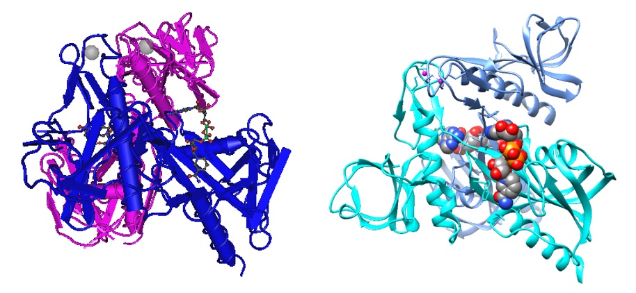
Figure 1: Two views of the nsp14–nsp10-SAH-GpppA heterodimer structure. The ligands are shown as spheres and are highlighted in color.

Ma et al. solved the structure of the nsp14–nsp10 complex by coexpressing the full-length nsp14 protein with the nsp10 protein in Escherichia coli and purifying the preformed complex. The research group refined the unliganded, SAM-bound, and S-adenosyl homocysteine (SAH)–guanosine-P3-adenosine-5′,5′-triphosphate (GpppA)–bound nsp14–nsp10 complex structures to 3.4 Å, 3.2 Å, and 3.3 Å resolutions, respectively.

Nsp14 is highly conserved within the Coronaviridae family. The nsp14-nsp10 complex excises 3’-mismatched nucleotides from double-stranded RNA. This ExoN activity of the protein complex results in a low mutation rate for the coronavirus. A DEDDh (a five catalytic amino acid) motif drives catalysis by nsp14, necessary for viral replication and transcription.

Nsp14 functions as S-adenosyl methionine (SAM)-dependent (guanine-N7) methyltransferase (N7-MTase), and the assembly of a cap1 structure at the 5′ end of viral mRNA assists in translation and evading host defense.

The formation of the cap in SARS-CoV requires four sequential reactions.

(1) First, nsp13 RNA triphosphatase (RTPase) hydrolyzes nascent RNA to yield pp-RNA.

(2) An unknown guanylyltransferase (GTase) hydrolyzes GTP, transfers the product GMP to pp-RNA, and creates Gppp-RNA.

(3) The nsp14 methylates the 5′ guanine of the Gppp-RNA at the N7 position,

(4) followed by methylation of the ribose of the first nucleotide at the 2′-O-position by nsp16.

Nsp10 is known to activate the 2′-O–MTase activity of nsp16 by stabilizing the SAM-binding pocket and extending the substrate RNA-binding groove of nsp16. Similarly, the ExoN activity of nsp14 is fully unleashed only in the presence of nsp10.

However, the exact molecular mechanism of this activation is poorly understood.

Reference

Denison MR, Graham RL, Donaldson EF, Eckerle LD, Baric RS. Coronaviruses: an RNA proofreading machine regulates replication fidelity and diversity. RNA Biol. 2011 Mar-Apr;8(2):270-9. doi: 10.4161/rna.8.2.15013. Epub 2011 Mar 1. PMID: 21593585; PMCID: PMC3127101.  https://www.ncbi.nlm.nih.gov/pmc/articles/PMC3127101/

Fazlieva R, Spittle CS, Morrissey D, Hayashi H, Yan H, Matsumoto Y. Proofreading exonuclease activity of human DNA polymerase delta and its effects on lesion-bypass DNA synthesis. Nucleic Acids Res. 2009 May;37(9):2854-66. doi: 10.1093/nar/gkp155. Epub 2009 Mar 12. PMID: 19282447; PMCID: PMC2685094. https://www.ncbi.nlm.nih.gov/pmc/articles/PMC2685094/

Ma Y, Wu L, Shaw N, Gao Y, Wang J, Sun Y, Lou Z, Yan L, Zhang R, Rao Z. Structural basis and functional analysis of the SARS coronavirus nsp14-nsp10 complex. Proc Natl Acad Sci U S A. 2015 Jul 28;112(30):9436-41. doi: 10.1073/pnas.1508686112. Epub 2015 Jul 9. PMID: 26159422; PMCID: PMC4522806. https://www.ncbi.nlm.nih.gov/pmc/articles/PMC4522806/

Robson F, Khan KS, Le TK, Paris C, Demirbag S, Barfuss P, Rocchi P, Ng WL. Coronavirus RNA Proofreading: Molecular Basis and Therapeutic Targeting. Mol Cell. 2020 Sep 3;79(5):710-727. doi: 10.1016/j.molcel.2020.07.027. Epub 2020 Aug 4. Erratum in: Mol Cell. 2020 Dec 17;80(6):1136-1138. PMID: 32853546; PMCID: PMC7402271.
https://www.ncbi.nlm.nih.gov/pmc/articles/PMC7402271/ 

---...---

 " Bio-Synthesis provides a full spectrum of high quality custom oligonucleotide modification services including back-bone modifications, conjugation to fatty acids, biotinylation by direct solid-phase chemical synthesis or enzyme-assisted approaches to obtain artificially modified oligonucleotides, such as BNA antisense oligonucleotidesmRNAs or siRNAs, containing a natural or modified backbone, as well as base, sugar and internucleotide linkages.
Bio-Synthesis also provides biotinylated mRNA and long circular oligonucleotides".

---...---

Therapeutic peptide discovery via high throughput screening of random peptide phage-display library

$
0
0
 In recent years, the method of high throughput screening has become a mainstay in both biological and pharmaceutical researches.  The ability to identify protein(s) interacting with a target protein through screening an appropriate library greatly expedites the research.  By doing so, one may bypass conventional steps entailing biochemical purification plus characterization and/or proteomic analyses. 

For pharmaceutical research, high throughput screening has been used to identify small molecule drug-like chemical compounds for treating various disorders.  Hence, to suppress the cancer-promoting property of an oncogene-encoded receptor, identifying chemical compounds capable of inhibiting specific receptor function (ex. tyrosine kinase) may be sufficient for pharmacological management.  Alternatively, identifying chemical compounds capable of inhibiting the catalytic activity of an enzyme in a biosynthetic pathway may satisfy the pharmacological objective.

Yet, the utility of screening chemical libraries has become increasingly limited with the expanding knowledge concerning the complex biochemical processes underlying human disorders.  Recent research has revealed that the downstream signaling step culminating in transcriptional regulation of target genes involves complex interaction by numerous distinct proteins. For instance, the transcriptional regulation by estrogen receptor at the estrogen-responsive elements requires the assembly of 1 to 2 megadalton-sized complex comprised of numerous transcription factors including AP2γ, RARα/γ, GATA3, STAT1, FoxA and AP1 (Liu et al., 2014). In the case of proteosome (~750 kilodalton) involved in protein degradation, the assembly of a functional complex requires the association of a number of distinct subunits and regulatory proteins (Tanaka, 2009).  

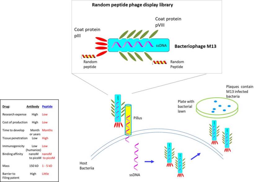
The research has further uncovered that a significant fraction of protein-to-protein interactions (~40%) are mediated through short peptides.  Though small drug-like chemicals have been effective in targeting buried hydrophobic binding pockets like the ligand-binding site of a receptor, peptides are better suited for disrupting protein-to-protein interactions that encompass a greater surface area.  Though antibodies also exhibit specificity, they suffer from poor penetration of tissues, instability (easy to denature), membrane impermeability, costly and time-consuming preparation, immunogenicity, etc.  This has inspired a greater focus by the pharmaceutical industries on improving various facets of peptides for clinical application, i.e. terminal protection, D-enantiomers, peptide cyclization, conjugation with functional groups (to improve specificity, tissue infiltration, resistance to degradation, etc.) (Lee et al., 2019).  Through these modifications, peptides exhibiting 5 times higher binding affinity than chemical compounds have been developed.


It follows that the ability to identify therapeutic peptides through a high throughput screening of a random peptide library would be highly beneficial.  To facilitate such endeavor, a biopanning system that screens random peptide library displayed on the surface of bacteriophage (bacteria infecting virus) was designed by G. Smith (Nobel prize, 2018).  The library is based on filamentous bacteriophage (ex. M13, fd, f1) containing single stranded DNA as its genome (Saw et al., 2019).  The filament-like shape helps to align and transfer its genetic material through the pilus into the cytoplasm of the host bacteria.  To construct the library, random peptides were expressed as part of the minor coat protein pIII or major coat protein pVIII.  Since its introduction, phage display library has been used extensively for multiple applications--for ex. to identify novel tumor antigens, enzyme inhibitors, antagonist/agonist of receptor, delivery vector (targeting blood vessels, tissues, cancer) or for other industrial uses.

The key to preventing epidemic is the ability to diagnose the infected early to preempt further propagation.  For this, Bio-Synthesis, Inc. provides primers and probes (as well as synthetic RNA control) for COVID-19 diagnosis via RT-PCR assay.  It specializes in oligonucleotide modification and provides an extensive array of chemically modified nucleoside analogues (over ~200) including bridged nucleic acid (BNA).  A number of options are available to label oligonucleotides (DNA or RNA) with fluorophoreseither terminally or internally as well as to conjugate to peptidesor antibodies.  It recently acquired a license from BNA Inc. of Osaka, Japan, for the manufacturing and distribution of BNANC, a third generation of BNA oligonucleotides.  To meet the demands of therapeutic application, its oligonucleotide products are approaching GMP grade.  Bio-Synthesis, Inc. has recently entered into collaborative agreement with Bind Therapeutics, Inc. to synthesize miR-21 blocker using BNA for triple negative breast cancer.  The BNA technology provides superior, unequalled advantages in base stacking, binding affinity, aqueous solubility and nuclease resistance.  It also improves the formation of duplexes and triplexes by reducing the repulsion between the negatively charged phosphates of the oligonucleotide backbone.  Its single-mismatch discriminating power is especially useful for diagnosis (ex. FISH using DNA probe).  For clinical application, BNA oligonucleotide exhibits lesser toxicity than other modified nucleotides. 


https://www.biosyn.com/oligo-flourescent-labeling.aspx

https://www.biosyn.com/tew/Speed-up-Identification-of-COVID19.aspx

https://www.biosyn.com/covid-19.aspx

https://www.biosyn.com/peptide-synthesis.aspx


References

Lee AC, Harris JL, et al. A Comprehensive Review on Current Advances in Peptide Drug Development and Design.  Int J Mol Sci.  20:2383 (2019).  PMID: 31091705

Liu Z, Merkurjev D, et al. Enhancer activation requires trans-recruitment of a mega transcription factor complex.  Cell 159:358-73 (2014).  PMID: 25303530

Saw PE, Song EW. Phage display screening of therapeutic peptide for cancer targeting and therapy.  Protein Cell  10:787-807 (2019).  PMID: 31140150

Murata S, Yashiroda H, et al.  Molecular mechanisms of proteasome assembly.  Nat Rev Mol Cell Biol. 10:104-15 (2009).  PMID: 19165213

Sugar modifications for gene silencing using siRNA

$
0
0

Modifications of the ribose unit in DNA and RNA oligonucleotides influence the biological properties and base-pairing ability of nucleic acids. In natural RNA, during RNA maturation, various enzymes can introduce chemical modifications into ribonucleotide residues. Chemical alteration can occur in the base, at the 2’-hydroxyl of the ribose, or both. Additional modifications can be introduced chemically in non-natural synthetic RNAs.

The modification of the ribofuranose in nucleic acids allows the manipulation of nucleic acid activities. The local conformation and chemical reactivity of the sugar is changes according to the modification type. Conformational restriction of the ribose units allows the increase in DNA and RNA affinity and enhances biological stability and antisense properties. Alterations in the sugar moiety conformation modify the local and potentially the global structure of nucleic acids. The properties of modified sugar units in oligonucleotides influence the recognition, binding of ligands, and enzymatic activity of proteins.

For example, 2’-modification often improves nuclease resistance, whereas the introduction of bridged nucleic acids (BNAs) significantly increases thermal stability.

Modern automated oligonucleotide synthesis methods enable technologies for the development of plasmid-based gene therapies, antisense, short interfering RNA (siRNA), short hairpin RNA (shRNA), aptamers, and other nucleic acid-based therapeutics. Since natural DNA or RNA oligonucleotides degrade easily, have low binding affinities and poor cellular uptake, chemical modifications over come many of these limitations and allow the introduction of unique properties as well.


Many chemical modifications have been made to bases, the ribose sugar and the sugar-phosphodiester backbone with the goal to improve their properties for a whole range of applications.


A


B

C


Figure 1: siRNA modification sites. A. Labeling nomenclature for ribose sugar unit and phosphate group. Sugar carbons are labeled as 1’-5’ and backbone torsion angles are described as follows:  α=O3′(i−1)−P−O5′−C5′; β=P−O5′−C5′−C4′; γ=O5′−C5′−C4′−C3′; δ=C5′−C4′−C3′−O3′; ε=C4′−C3′−O3′−P(i+1); ζ=C3′−O3′−P(i+1)−O5′(i+1) B. Possible sites of introduction of chemical modifications in the sugar moiety and phosphate linkage are marked in red. C. Possible sites for chemical modifications in nucleic acid bases.

Table 1: RNAi modifications of Sugar Units

Modification

Structure

ΔTm duplex per modification

Impact on the efficiency of RNAi

Others

2 ′ -O-methyl

(2′O-Me)

+0.5–1.5◦C

Two or more consecutive 2′O-Me inhibit RNAi.

Some siRNAs containing 75–82% 2 ′O-Me are biologically active.

Stabilizes 3′ endo ribose conformation.≥5–30% of 2′O-Me increase nuclease resistance in vitro and in vivo.

2 ′O-Me analogs of A, G and U reduce the immune response.

2 ′ -fluoro

(2′F)

+1.5–4◦C

2′F analogs in all siRNA positions only slightly reduces the activity of RNAi.

Stabilizes 3′ endo ribose conformation.

≥50% 2′F increase nuclease resistance in vitro and in vivo.

2 ′F analogs of adenine (≥7%) reduce the immune response in vitro.

>50% of the 2′F in siRNA may cause toxicity.

2 ′F-arabinonucleic acid

(2′FANA)


+1.2◦C

100% 2′FANA in the sense chain reduce the efficiency of RNAi.

 ≥30% 2 ′FANA in the antisense chain inhibit RNAi.

Stabilizes 2′ endo ribose conformation.

≥50% 2′FANA increases nuclease resistance in vitro more effectively than 2′F.

Protect siRNA from the action of exoribonucleases.

2 ′ -O-methoxyethyl

(2′O-MOE)

+0.9–1.7◦C

2′ -MOE at the flanks of the sense strand and the central part (6–11) of the antisense strand are tolerable for RNAi.Replacement of 9th or 10th nucleotides from the 5′ end to 2 ′O-MOE analogs of nucleotide increases the probability of entry in RISC.

Stabilizes 3′ endo ribose conformation.

 ≥15% 2′O-MOE at the ends of the siRNA sense chain increases nuclease resistance in vitro.


Locked nucleic acid

(LNA)

+2–8◦C

≥40%LNA in the sense chain inhibit RNAi by 5–20%.

>20% LNA in the antisense chain, or the first LNA nucleotide at the 5′ end completely inhibit RNA.

LNA can change thermal asymmetry of the duplex, increasing the efficiency of siRNA.

Reduces the conformational flexibility of nucleotides by fixing the C3′ endo conformation of the ribose.

≥10–20% LNA in siRNAs increase nuclease resistance in vitro and in vivo.

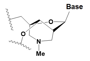
Bridged nucleic acid

(BNA)


≥40% BNA in the sense chain inhibit RNAi by 8–25%.

>20% BNA in the antisense chain, or the first BNA nucleotide at the 5′ end completely inhibit RNA.

BNA changes thermal asymmetry of the duplex, increasing the efficiency of siRNA.

Reduces the conformational flexibility of nucleotides by fixing the C3′ endo conformation of the ribose.

≥10–20% BNA in siRNAs increase nuclease resistance in vitro and in vivo.

Unlocked nucleic acid (UNA)


−5–8◦C

>15% UNA inhibit RNAi.

UNA can change thermal asymmetry of the duplex, increasing the efficiency of siRNA.

Increases conformational flexibility of nucleotides and reduces the melting point of the duplex.

UNA at the 3′ends of the duplex protect siRNA from 3′ exoribonucleases in vitro and in vivo.

4 ′ -thio-ribonucleosides (4′S)

+1 ◦C

>7–15% 4′S in the antisense strand inhibit RNAi.

>10–15% 4′S at the ends of the strands increase the nuclease resistance in vitro.

4 ′ -C-aminomethyl-2′ -O-methyl

(4’CAm2’OMe)

−1 ◦C

>2 analogs in the sense or >1 analog in the antisense strandinhibits RNAi.

≥2 modifications at the 3′ ends increase nuclease resistance in vitro.

Deoxyribonucleotide (dNMP)


−0.5◦C

>50% dNMP inhibits RNAi.

dNMP can change thermal asymmetry of the duplex, increasing the efficiency of siRNA in vitro.

Protects against exoribonucleases.

Cyclohexenyl nucleic acids (CeNA)

+1.5◦C

5% CeNA in siRNA are tolerated by RNAi.

CeNA can change thermal asymmetry of the duplex, increasing the efficiency of siRNA in vitro.

Stabilizes 3′ endo ribose conformation.

≥25% CeNA analogs increase serum nuclease resistance.


Hexitol nucleic acids (HNA)

+0.85◦C

15% HNA in siRNA are tolerated by RNAi (Fisher et al., 2009). HNA can change thermal asymmetry of the duplex, increasing the efficiency of siRNA in vitro (Herdewijn and Juliano, 2007).

Slightly increases siRNA resistance to nucleases in serum (Fisher et al., 2009).


Adapted from:  Chernikov IV, Vlassov VV, Chernolovskaya EL. Current Development of siRNA Bioconjugates: From Research to the Clinic. Front Pharmacol. 2019 Apr 26;10:444. doi: 10.3389/fphar.2019.00444. PMID: 31105570; PMCID: PMC6498891.https://www.ncbi.nlm.nih.gov/pmc/articles/PMC6498891/.

---...---

Phosphate modifications for gene silencing with RNAi

$
0
0

In RNA interference, small interfering RNAs recognize messenger RNA homologs in cells and induce their degradation.

When developing siRNA-based drugs for therapeutic use, problems encountered are their low efficiency of delivery to targeted cells and degradation by cellular nucleases. A promising approach to improve cellular delivery of siRNA is bioconjugation. Like other oligonucleotides, siRNA conjugation to lipophilic molecules, antibodies, aptamers, ligands, peptides, or polymers enhances cellular delivery and selective targeting.


Figure 1: Mechanism of systemic and transitive RNAi. Dicer cleavage of dsRNA into 21-23 nt siRNA initiates RNAi. RISC unwinds siRNA duplexes. siRNA binds to mRNA. Either the targeted mRNA is destructed or amplified via RdRP. Amplified dsRNA can transitively silence a 5’-located mRNA in a second cell {Engelke, David R.: RNA interference (RNAi). Nuts and bolts of RNAi technology. DNA Press 2003}.


Table 1: Phosphate Backbone Modifications and their Effect.

Modification

Structure

ΔTm duplex per modification

Impact on the efficiency of RNAi

Others

Phosphorothioate (PS)

−0.7°C

PS inhibits RNAi when introduced in the central part of the antisense strand.

PS protects siRNAs from the action of exoribonucleases in vitro and in vivo.

>50% PS cause toxicity in vitro and in vivo.

Dimethylethylenediamine (DMEDA)

−0.7–3.4°C

(shown only for thymidine)

10% DMEDA in the sense strand are tolerated by RNAi.

The effect on nuclease resistance of siRNA is not known.

Tert-butyl-S-acyl-2-thioethyl (tBu-SATE)

No data.

25% tBu-SATE are tolerated by RNAi.

≥20–40% tBu-SATE in siRNA increase nuclease resistance in vitro and in vivo.
Increases hydrophobicity of siRNA.

Cleaved by thioesterase in the cytoplasm of the cell giving a phosphodiester bond.

Boranophosphate (BP)

+0.4–1°C (<50% of siRNA)
−0.8–2.5°C (>50% of siRNA)

>50% PB inhibit RNAi, the central part of the antisense strand is the most sensitive to modifications.


Approximately two times more effectively protect against ribonucleases than PS, but do not cause toxicity in vitro.

Amide linker

−0.3 to +0.9°C

In some siRNA positions, a single substitution for an amide linker is tolerated by RNAi.

The introduction of two amide linkers from the 3′ ends of the duplex increases the nuclease resistance of siRNA in serum.


Adapted from:  Chernikov IV, Vlassov VV, Chernolovskaya EL. Current Development of siRNA Bioconjugates: From Research to the Clinic. Front Pharmacol. 2019 Apr 26;10:444. doi: 10.3389/fphar.2019.00444. PMID: 31105570; PMCID: PMC6498891. https://www.ncbi.nlm.nih.gov/pmc/articles/PMC6498891/.

---...--- 

 " Bio-Synthesis provides a full spectrum of high quality custom oligonucleotide modification services including back-bone modifications, conjugation to fatty acids, biotinylation by direct solid-phase chemical synthesis or enzyme-assisted approaches to obtain artificially modified oligonucleotides, such as BNA antisense oligonucleotidesmRNAs or siRNAs, containing a natural or modified backbone, as well as base, sugar and internucleotide linkages.
Bio-Synthesis also provides biotinylated mRNA and long circular oligonucleotides".

---...---


5'-Phosphate modifications for gene silencing with RNAi

$
0
0

In 2005, Pham & Sontheimer studied the molecular requirements for RNA-induced silencing (RNAi) complex (RISC) assembly in the Drosophila RNA interference pathway. The study used native gel electrophoresis to reveal components in the Drosophila RISC complex.

Component R1 forms when the Dicer-2/R2D2 heterodimer binds short interfering RNA (siRNA) duplexes. The heterodimer alone can initiate RISC assembly. Hence, other factors are dispensable for initiation. During assembly, R2 requires Argonaute 2 to convert into holo-RISC. This requirement reminds us of the RISC-loading complex, which also requires Argonaute 2 for assembly into RISC.

Pham & Sontheimer compared R2 to the RISC-loading complex and show that the two complexes are similar in their sensitivities to ATP and chemical modifications on siRNA duplexes. The researchers studied RISC formation requirements and showed that the siRNA 5'-termini are repeatedly monitored during RISC assembly. First by the Dcr-2/R2D2 heterodimer and again after R2 formation before siRNA unwinding. The 2'-position of the 5'-terminal nucleotide also affects RISC assembly because a siRNA strand bearing a 2'-deoxyribose at this position can inhibit the cognate strand from entering holo-RISC. The 2'-deoxyribose-modified strand has enhanced activity in the RNA interference pathway.


In 2007, Shah et al. observed that siRNA modified at the 5'-antisense phosphate can still cause RNAi, however not at the level affected by fully native siRNA. The researchers' result suggested an inherent tolerance of the RNAi machinery toward modification of the 5'-antisense phosphate.

In 2015, Prakash et al. reported that the electronic and spatial orientation of the 5′-phosphate analog is critical for single-stranded-siRNA activity. Chemically modified single-stranded-siRNA targeting human ApoC III mRNA showed enhanced potency for inhibiting ApoC III mRNA and protein in transgenic mice. The modified siRNAs were able to reduce the triglyceride and LDL cholesterol in transgenic mice. 

In 2017, Haraszti et al. showed that 5΄-Vinylphosphonate modification of siRNAs protects them from phosphatases, improves silencing activity, and confers novel properties to siRNAs. 

5΄-vinylphosphonate (i) increases siRNA accumulation in tissues, (ii) extends the duration of silencing in multiple organs, and (iii) protects siRNAs from 5΄-to-3΄ exonucleases.

Because 5΄-phosphate is required for loading into RNA-induced silencing complex, a 5΄-phosphate on a fully modified siRNA guide strand is expected to be beneficial. The synthetic phosphorylation of fully modified cholesterol-conjugated siRNAs increases their potency and efficacy in vitro. Unfortunately, when delivered systemically to mice, the 5΄-phosphate is removed within 2 hours.

The 5΄-phosphate mimic 5΄-(E)-vinylphosphonate stabilizes the 5΄-end of the guide strand by protecting it from phosphatases and 5΄-to-3΄-exonucleases. The improved stability significantly enhances the efficacy of cholesterol-conjugated siRNAs and the duration of silencing in vivo. 5΄-(E)-vinylphosphonate stabilizes 5΄-phosphate and enables systemic delivery to and silencing in the kidney and heart.

Table 1: RNAi modifications of the 5’-Phosphate and their Effect.


Modification

Structure

ΔTm duplex per modification

Impact on the efficiency of RNAi

Others

5′-C-methyl
(S-isomer)

−3.2°C

One (S) 5′C-methyl at the 5′-end of the antisense strand is tolerated by RNAi.

The introduction of two amide linkers from the 3′-ends of the duplex increases the nuclease resistance of siRNA in serum.

5′-(E)-vinyl-phosphonate

No data.

5′(E)-vinyl-phosphonate improves siRNA interaction efficiency with Ago2 by 2-fold.

Does not change the biological activity of siRNA in vitro.

Stabilizes 5′-phosphate. Protects from the action of phosphatases and exonucleases.
Improves the pharmacokinetics.

Enables systemic delivery to and silencing in kidney and heart.

5′-methylene-phosphonate

No data.

5′-methylene-phosphonate at the 5′-end of the antisense strand reduces the biological activity of siRNA by ~10-fold.

No data.


Reference

Chernikov IV, Vlassov VV, Chernolovskaya EL. Current Development of siRNA Bioconjugates: From Research to the Clinic. Front Pharmacol. 2019 Apr 26;10:444. [PMC]

Haraszti RA, Roux L, Coles AH, Turanov AA, Alterman JF, Echeverria D, Godinho BMDC, Aronin N, Khvorova A. 5΄-Vinylphosphonate improves tissue accumulation and efficacy of conjugated siRNAs in vivo. Nucleic Acids Res. 2017 Jul 27;45(13):7581-7592.[PMC]

Pham JW, Sontheimer EJ. Molecular requirements for RNA-induced silencing complex assembly in the Drosophila RNA interference pathway. J Biol Chem. 2005 Nov 25;280(47):39278-83. [PMC]

Shah S, Friedman SH. Tolerance of RNA interference toward modifications of the 5' antisense phosphate of small interfering RNA. Oligonucleotides. 2007 Spring;17(1):35-43. [PubMed]

Thazha P. Prakash, Walt F. Lima, Heather M. Murray, Wenyu Li, Garth A. Kinberger, Alfred E. Chappell, Hans Gaus, Punit P. Seth, Balkrishen Bhat, Stanley T. Crooke, Eric E. Swayze; Identification of metabolically stable 5′-phosphate analogs that support single-stranded siRNA activity. Nucleic Acids Research, Volume 43, Issue 6, 31 March 2015, Pages 2993–3011. [PMC]

 ---...--- 

 " Bio-Synthesis provides a full spectrum of high quality custom oligonucleotide modification services including back-bone modifications, conjugation to fatty acids, biotinylation by direct solid-phase chemical synthesis or enzyme-assisted approaches to obtain artificially modified oligonucleotides, such as BNA antisense oligonucleotidesmRNAs or siRNAs, containing a natural or modified backbone, as well as base, sugar and internucleotide linkages.
Bio-Synthesis also provides biotinylated mRNA and long circular oligonucleotides".

---...---


Nucleic Acid Base modifications for gene silencing with RNAi

$
0
0

Properly designed antisense oligonucleotides (AONs) or siRNAs allow the cleavage of specific messenger RNA (mRNA) strands.

AONs and siRNAs take advantage of endogenous cellular pathways to silence the expression of particular genes. Oligonucleotide-directed approaches target mRNAs directly before translation, eliminating the need for protein or enzyme inhibition using small molecules. However, some obstacles can prevent siRNA-based oligonucleotide therapies.

These obstacles include: 

(1) their poor extracellular and intracellular stability, 

(2) low efficiency of intracellular delivery to target cells or tissues, and 
(3) the potential for ‘‘off-target’’ gene silencing, immune stimulation, and other side effects.

The incorporation of modified nucleic acids into AONs or siRNAs may potentially allow to circumvent these obstacles. 

In 2007, Sipa et al. tested a series of siRNA duplexes containing the “rare” nucleosides, 2-thiouridine (s2U), pseudouridine (Ψ), and dihydrouridine (D), for their thermodynamic stability and gene silencing activity.

The study found that oligonucleotide duplexes with modified nucleic acid bases at terminal positions showed similar stability as a nonmodified reference duplex. The introduction of the s2U or Ψ units into the central part of the antisense strand resulted in duplexes with higher melting temperatures (Tm). However, adding D units resulted in less stable duplexes. Duplexes with s2U and Ψ units at their 3′-ends and with a D unit at their 5′-ends with respect to the guide strands were the most potent gene expression inhibitors.

Table 1: Base Modifications and their Effect.

Modification

Structure

ΔTm duplex per modification

Impact on the efficiency of RNAi

Others

2′ thiouridine
(s2U)

0–2°C

7% s2U are tolerated by RNAi.
s2U can change thermal asymmetry of the duplex and increase the efficiency of siRNA in vitro.

s2U slightly increases nuclease resistance in vitro.

Pseudouridine
(Ψ)

−1 to +1°C

One Ψ is tolerated by RNA.

Stabilizes 3′endo ribose conformation.
Reduces the PKR-induced interferon response.

Dehydrouridine

(D)

N.A.

The nonaromatic nucleobase disrupts base stacking.

D unit similarly like wobble base pair lead to less stable duplexes.

R = ribose residue.

Reference

Chernikov IV, Vlassov VV, Chernolovskaya EL. Current Development of siRNA Bioconjugates: From Research to the Clinic. Front Pharmacol. 2019 Apr 26;10:444. [PMC]

Glen F. Deleavey, Masad J. Damha; Designing Chemically Modified Oligonucleotides for Targeted Gene Silencing. Chemistry & Biology, Volume 19, Issue 8, 2012, Pages 937-954. [PubMed]

Katarzyna Sipa, Elzbieta Sochacka, Julia Kazmierczak-Baranska, Maria Maszewska, Magdalena Janicka, Genowefa Nowak, and Barbara Nawrot;  Effect of base modifications on structure, thermodynamic stability, and gene silencing activity of short interfering RNA. RNA 2007. 13: 1301-1316. [PMC]

 ---...--- 

 " Bio-Synthesis provides a full spectrum of high quality custom oligonucleotide modification services including back-bone modifications, conjugation to fatty acids, biotinylation by direct solid-phase chemical synthesis or enzyme-assisted approaches to obtain artificially modified oligonucleotides, such as BNA antisense oligonucleotidesmRNAs or siRNAs, containing a natural or modified backbone, as well as base, sugar and internucleotide linkages.
Bio-Synthesis also provides biotinylated mRNA and long circular oligonucleotides".

---...---

Increased risk for cancer in diabetic patients and innovative strategy to activate insulin receptor using a peptide independent of insulin

$
0
0
Diabetes has been described in ancient Egyptian documents as early as ca. 1500 B.C.  Yet, treatment has not been available until 1920s when insulin was first isolated (Nobel prize, 1923).  Worldwide, 425 million people are currently diagnosed with diabetes, affecting 8-9% of the global population with ~4 million deaths in 2017 (7th leading cause of mortality).  The global rate nearly doubled from 4.7% in 1980 to 8.8% in less than 30 years, and is expected to increase by another 48% in 2045.  In the U. S., the incidence of diabetes arose alarmingly from 0.9% in 1958 to 7.4% in 2015.

 Within the body, the level of glucose is regulated by insulin.  After eating, insulin is secreted by beta cells of pancreas to promote glucose uptake by the cells.  The secreted insulin binds to its receptor on the cell membrane activating its tyrosine kinase (through autophosphorylation), generating the binding site for substrates (to be kinased by the receptor).  This, in turn, activates a signaling cascade involving phosphoinositide 3-kinase (PI3K), PDK1, protein kinase B (Akt), etc., ultimately causing GLUT-4 to become embedded in the cell membrane.  GLUT-4 (encoded by SLC2A4 gene) then transports glucose into fat cells, muscle cells, etc. (Ijuin et al., 2012).

 In the event of an insulin insufficiency, the inability to store glucose by liver or muscle causes an abnormally high level of glucose in circulation, prompting its removal by the kidney through excretion.  The buildup of glucose in urine increases its osmotic pressure, causing water to be extracted from other body tissues leading to dehydration, causing further intake of fluids.  Diabetes can cause damages to the nerves, kidney, blood vessels, and may lead to dire conditions such as diabetic retinopathy (blindness) or even deaths (due to coronary artery disorder).   Currently, diabetic patients account for ~25% of kidney transplantations performed in the U.S.

The predisposition to develop type I diabetes is partly inherited and the symptoms manifest in adolescents and children (or adults).  It accounts for ~5-10% of diabetic cases and the underlying pathobiology involves the T cell mediated immunological destruction of pancreatic beta cells, causing a reduced insulin production.  In contrast, type 2 diabetes is characterized by the production of insulin by pancreas despite its inefficient usage by the body--i.e. 'insulin resistance' or reduced sensitivity to insulin.  The condition may worsen by the inability of pancreas to respond, eventually causing insulin shortage.  Specific genetic defects have not been identified though numerous DNA variations have been identified.  Type 2 diabetes accounts for 90-95% of total cases (risk increases significantly after 45 y), and lifestyles (ex. obesity, diet, lack of exercise, body fat) may contribute to the diabetic progression.

Earlier, insulin purified from animal source was used to treat diabetes.  After the determination of the amino acid sequence of insulin by F. Sanger (Nobel prize, 1958) (Stretton, 2002), other means of preparing insulin became available, ex. bovine insulin synthesized chemically.  Insulin is comprised of chain A (21 residues) and chain B (30 residues) that are held together by disulfide bonds.  In 1978, A. Riggs and K. Itakura (City of Hope National Medical Center, USA) used genetic engineering to produce synthetic human insulin in E. coli (Riggs, 2020), which was later marketed by Genentech to make it commercially available worldwide.  Currently the biosynthetically produced recombinant human insulin and/or its analogues are most widely administered.


Further advances have been made to facilitate diabetic treatment.  As insulin functions to activate the tyrosine kinase associated with insulin receptor to initiate signaling, an alternate means of activation was sought.  To this end, the investigators at Harvard Medical University (USA) discovered a 24-mer peptide derived from the transmembrane domain, which could activate the receptor in the absence of insulin (Lee et al., 2014).  Further, the peptide was able to activate insulin receptor from patients who suffer from insulin resistance--thus, bypassing the requirement for the presence of ligand-binding domain in receptor (for its activity).  Mechanistically, the peptide may disrupt the dimeric interaction of the transmembrane domains via intercalating, causing beta subunit to adopt an activate state (hence mimicking the effect of insulin binding).

Increasingly, diabetes is linked to cancer.  Multiple studies have found that diabetic patients may have an increased risk of developing cancers (liver, pancreatic, colorectal, kidney, bladder, breast, endometrial cancer but lesser risk for prostate cancer) (Abudawood, 2019; Wang et al. 2020) with diabetic women suffering from greater risk than diabetic men.  Though the precise underlying mechanism is not known, mitogenic (growth promoting) property of insulin or insulin-like growth factor (IGF), hyperglycemia (excess glucose promotes metabolism), and inflammation have been proposed.

 The key to preventing epidemic is the ability to diagnose the infected early to preempt further propagation.  For this, Bio-Synthesis, Inc. provides primers and probes (as well as synthetic RNA control) for COVID-19 diagnosis via RT-PCR assay.  It specializes in oligonucleotide modification and provides an extensive array of chemically modified nucleoside analogues (over ~200) including bridged nucleic acid (BNA).  A number of options are available to label oligonucleotides (DNA or RNA) with fluorophoreseither terminally or internally as well as to conjugate to peptidesor antibodies.  It recently acquired a license from BNA Inc. of Osaka, Japan, for the manufacturing and distribution of BNANC, a third generation of BNA oligonucleotides.  To meet the demands of therapeutic application, its oligonucleotide products are approaching GMP grade.  Bio-Synthesis, Inc. has recently entered into collaborative agreement with Bind Therapeutics, Inc. to synthesize miR-21 blocker using BNA for triple negative breast cancer.  The BNA technology provides superior, unequalled advantages in base stacking, binding affinity, aqueous solubility and nuclease resistance.  It also improves the formation of duplexes and triplexes by reducing the repulsion between the negatively charged phosphates of the oligonucleotide backbone.  Its single-mismatch discriminating power is especially useful for diagnosis (ex. FISH using DNA probe).  For clinical application, BNA oligonucleotide exhibits lesser toxicity than other modified nucleotides. 


https://www.biosyn.com/oligo-flourescent-labeling.aspx

https://www.biosyn.com/tew/Speed-up-Identification-of-COVID19.aspx

https://www.biosyn.com/covid-19.aspx

https://www.biosyn.com/peptide-synthesis.aspx


References

Abudawood M. Diabetes and cancer: A comprehensive review.   J Res Med Sci.  24:94 (2019). PMID: 31741666

Ijuin T, Takenawa T. Regulation of insulin signaling and glucose transporter 4 (GLUT4) exocytosis by phosphatidylinositol 3,4,5-trisphosphate (PIP3) phosphatase, skeletal muscle, and kidney enriched inositol polyphosphate phosphatase (SKIP).  J Biol Chem. 287:6991-9 (2012). PMID: 22247557

Lee J, Miyazaki M, et al. Insulin receptor activation with transmembrane domain ligands.   J Biol Chem. 289:19769-77 (2014).  PMID: 24867955

Riggs AD.  Making, Cloning and Expression of Human Insulin Genes in Bacteria: The Path to Humulin@.  Endocr Rev. 2020 Dec 19:bnaa029. doi: 10.1210/endrev/bnaa029.   PMID: 33340315

Stretton AO.  The first sequence.  Fred Sanger and insulin.   Genetics.  162:527-32 (2002).   PMID: 12399368

Wang M, Yang Y, Liao Z. Diabetes and cancer: Epidemiological and biological links.  World J Diabetes. 11:227-238 (2020). PMID: 32547697


Self-amplifying RNA and mRNA vaccines

$
0
0

As Pfizer-BioNTech and Moderna Inc. have recently demonstrated, synthetic RNA platforms allow for rapid, scalable, and cell-free manufacturing of vaccines based on optimized, modified mRNA. This approach utilizes in vitro transcription of antigen-encoding sequences or immunotherapies as synthetic RNA transcripts. In general, the formulation of the final mRNA vaccine products for delivery occurs in synthetic lipid nanoparticles.

The new mRNA-based vaccines from Pfizer and Moderna against the coronavirus infection are nucleoside-modified messenger RNAs (modRNAs) encoding the viral spike glycoprotein (S) from the SARS-CoV-2 coronavirus. Both vaccines do not contain a live virus and also no self-amplifying RNA. The formulation for delivery of both vaccines occurred in nanoparticles (LNP).
The Pfizer-BioNTech COVID-19 vaccine BNT162b2 is recommended for people aged 16 years and older. The Moderna COVID-19 vaccine mRNA-1273 is recommended for people 18 years and older.

Since DNA-based vaccines have performed poorly in human trials due to insufficient responses to elicit a significant clinical benefit, there is a renewed interest in RNA-based vaccines (Hobernik et al., 2018).

Currently, there are two types of synthetic vaccines in development: conventional or nonreplicating mRNA and self-amplifying RNA-based vaccines.

In vitro transcribed mRNAs encoding viral antigens have been explored as vaccines. mRNAs encoding therapeutic proteins are candidates for immunotherapy. The incorporation of chemically modified nucleotides, sequence optimization, and purification resulted in improved mRNA translation efficiency and reduced intrinsic immunogenic properties. Since antigen expression is proportional to the number of mRNA transcripts delivered during vaccination, the adequate expression of antigens for protection may need large doses or repeated administration of the vaccine.

Self-amplifying RNA vaccines are genetically engineered replicons, nucleic acid molecules replicating as units, derived from self-replicating single-stranded RNA viruses, address these limitations.


Figure 1: Example of the Semliki Forest virus-based expression systems (SFV). Schematic illustration of expression vectors. Red triangle, SP6 RNA polymerase promoter; orange triangle, Semliki Forest virus (SFV) 26S subgenomic promoter (Adapted from Lundstrom, 2020). An engineered expression vector carrying the SFV nonstructural protein genes (nsP1-4) and the gene of interest inserted downstream of the strong 26S sub-genomic promoter allows translation of in vitro transcribed RNA in cell lines and in vivo.

In 2013, Hekele et al. showed that it is possible to formulate self-amplifying RNA (saRNA) vaccines in LNPs within eight days. The so-called SAM® vaccine platform expressing seasonal influenza hemagglutinin utilizes a synthetic, self-amplifying mRNA that allowed mRNA delivery packaged in synthetic lipid nanoparticles (LNPs). 

Immunized mice showed measurable hemagglutinin inhibition and neutralizing antibody titers against the new virus within two weeks. After the second immunization, all mice had hemagglutinin inhibition titers considered as protective. These results are an indication that saRNA based vaccines are a new platform for designing vaccines useful as tools to stop newly emerging infectious viruses at the beginning of an outbreak.

Self-amplifying RNA vaccines encode 5′- and 3′- conserved sequence elements (CSEs), the nonstructural protein 1 to 4 (nsP1-4) genes, a subgenomic promoter, and the vaccine immunogen. After in situ translation, the nsP1-4 proteins form an RNA-dependent RNA polymerase (RdRP) complex that recognizes flanking CSE sequences and amplifies the vaccine-encoded transcript.

Biddlecome et al. recently reported a new gene delivery platform that illustrated the benefits of a self-amplifying (“replicon”) used in mRNA vaccines protected in a viral-protein capsid. The research group used a purified capsid protein from the plant virus Cowpea Chlorotic Mottle Virus (CCMV) for the in vitro assembly of monodispersed virus-like particles and tested it in mice. The researchers showed that immunization mice with this packaged mRNA activated dendritic cells and induced antigen-specific T-cell responses.

Single-stranded RNA viruses, such as alphaviruses, self-amplify their RNA in host cells. These characteristics make alphaviruses and flaviviruses attractive vehicles for vaccine development. Alphaviruses belonging to the family of Togaviruses such as Semliki Forest virus (SFV), Sindbis virus (SIN), and Venezuelan equine encephalitis virus (VEE) have been used for the engineering of expression vector systems. Alphaviruses are positive-sense, single-stranded RNA viruses. The 32 known alphaviruses can infect humans, rodents, fish, birds, and larger mammals. Enveloped alphavirus particles have a diameter of approximately 70 nm and a spherical 40 nm isometric nucleocapsid. The genome of alphaviruses contains two open reading frames (ORFs), one nonstructural and one structural.  All alphaviruses share antigenic sites on the capsid and at least one envelope glycoprotein. However, the viruses can be differentiated by several serological tests, particularly neutralization assays.

Reference and Links


Alphavirus:  Medical Microbiology. 4th edition.   
https://www.ncbi.nlm.nih.gov/books/NBK7633/

Alphavirus Wiki https://en.wikipedia.org/wiki/Alphavirus 

Biddlecome A, Habte HH, McGrath KM, Sambanthamoorthy S, Wurm M, Sykora MM, Knobler CM, Lorenz IC, Lasaro M, Elbers K, Gelbart WM. Delivery of self-amplifying RNA vaccines in in vitro reconstituted virus-like particles. PLoS One. 2019 Jun 4;14(6):e0215031. doi: 10.1371/journal.pone.0215031. PMID: 31163034; PMCID: PMC6548422. https://www.ncbi.nlm.nih.gov/pmc/articles/PMC6548422/

COVID-19 vaccines and pregnancy:  https://www.verywellhealth.com/who-updates-guidance-pregnancy-covid-19-vaccine-5104967

FDA and COVID-19 https://www.fda.gov/news-events/press-announcements/fda-takes-additional-action-fight-against-covid-19-issuing-emergency-use-authorization-second-covid ]

Hekele A, Bertholet S, Archer J, Gibson DG, Palladino G, Brito LA, Otten GR, Brazzoli M, Buccato S, Bonci A, Casini D, Maione D, Qi ZQ, Gill JE, Caiazza NC, Urano J, Hubby B, Gao GF, Shu Y, De Gregorio E, Mandl CW, Mason PW, Settembre EC, Ulmer JB, Craig Venter J, Dormitzer PR, Rappuoli R, Geall AJ. Rapidly produced SAM(®) vaccine against H7N9 influenza is immunogenic in mice. Emerg Microbes Infect. 2013 Aug;2(8):e52. doi: 10.1038/emi.2013.54. Epub 2013 Aug 14. PMID: 26038486; PMCID: PMC3821287. https://www.ncbi.nlm.nih.gov/pmc/articles/PMC3821287/

Hobernik D, Bros M. DNA Vaccines-How Far From Clinical Use? Int J Mol Sci. 2018 Nov 15;19(11):3605. doi: 10.3390/ijms19113605. PMID: 30445702; PMCID: PMC6274812. https://www.ncbi.nlm.nih.gov/pmc/articles/PMC6274812/ 

Lundstrom K. Self-Amplifying RNA Viruses as RNA Vaccines. Int J Mol Sci. 2020 Jul 20;21(14):5130. doi: 10.3390/ijms21145130. PMID: 32698494; PMCID: PMC7404065.   https://www.ncbi.nlm.nih.gov/pmc/articles/PMC7404065/

Lundstrom K. Application of Viral Vectors for Vaccine Development with a Special Emphasis on COVID-19. Viruses. 2020 Nov 18;12(11):1324. [PMC]

Moderna COVID-19 vaccine
mRNA-1273

Pfizer-BioNTech COVID-19 vaccine BNT162b2

---...---

NAD-RNA capping

$
0
0

The NAD-RNA cap stabilizes small regulatory RNAs in bacteria.

The 5'-terminal ends of cellular mRNAs contain a m7GpppN cap, in which N can be any nucleotide. In eukaryotes, the RNA helicase eIF4A, the scaffold protein translation initiation factor 4G (eIF4G) and the capping protein eIF4E are part of the complex that loads mRNAs onto the 40 S ribosomal subunit, together with eIF3. eIF4E has a crucial role in the regulation of translation. mRNA metabolism in the nucleus, such as capping, splicing, and polyadenylation, is mechanically linked to transcription. In addition, mRNA decay regulates mRNA metabolism whereas capping the 5’-end and polyadenylation of the 3’-end increases mRNAs' stability.

In the bacterium Escherichia coli, the nicotinamide adenine dinucleotide RNA (NAD-RNA) cap stabilizes small regulatory RNAs (
srRNAs) in vitro against nucleotide processing by RNase E and against 5’-end modification by RNA pyrophosphohydrolase RppH. RNase E is involved in RNA decay, and RppH converts 5’-triphosphate RNA into 5’-monophosphate RNA triggering endonucleolytic processing.

As recently discovered, bacterial small RNAs, yeast and human mRNAs and non-coding RNAs contain NAD cap-like structures. Bacterial RNA polymerase (RNA Pol) can also initiate transcription in vitro by accepting nucleotide metabolites capped with flavin adenine dinucleotide (FAD), uridine diphosphate glucose (UDP-Glc), and uridine diphosphate N-acetylglucosamine (UDP-GlcNAc). Capping with NAD and UDP analogs by bacterial RNA Pol is promoter-specific and stimulates promoter escape. These recent findings suggest a role for metabolite caps in regulating gene expression. As demonstrated by Wang et al., X-ray crystallography in combination with mass spectrometry allows chracterization of cap-protein interactions.


The redox factor NAD is attached to small regulatory RNAs in bacteria as a cap.

In 2016, Hoefer et al. solved the crystal structures of the nuclear migration protein NudC from Escherichia coli in complex with the substrate NAD and the cleavage product nicotinamide mononucleotide (NMN). Crystal structures of the complexes studied revealed the catalytic residues lining the binding pocket and molecular features of substrate and product recognition.


Figure 1: NudC (NADH Pyrophosphatase) in complex with NAD (5IW4).

Höfer et al.’s study revealed that NudC is a single-strand-specific RNA decapping enzyme with a strong preference for a purine as the first nucleotide. Their biochemical experiments showed that NudC preferred NAD-RNA over NAD(H) by several orders of magnitude. Hence NAD-RNA maybe its primary biological substrate. The study results suggest that NudC can bind a diverse population of cellular RNAs in an unspecific, most likely electrostatic manner.


The E. coli Nudix hydrolase NudC hydrolyzes the pyrophosphate bond and removes the NAD cap to produce nicotinamide mononucleotide (NMN) and 5’-monophosphate RNA. NudC is also known as NAD(H) pyrophosphohydrolase. As suggested by Höfer et al. it is possible that during RNA processing, NAD capping gives the bacterium additional RNA protection by using a degradation pathway that is orthogonal to the RppH-triphosphate RNA pathway.

NudC is a critical factor in mitosis, the cell division resulting in two genetically identical daughter cells. In 2015, Chen et al, used mass spectrometry to reveal that the nuclear distribution gene C (NudC or NUDC) protein, a critical factor for the progression of mitosis, is associated with the Echinoderm microtubule-associated protein (EMAP)-like 4 (EML4). The study showed that EML4 is critical for the loading of NudC onto the mitotic spindle for mitotic progression.

As reviewed by Chen et al., in mammals, NUDCL and NUDCL2 are homologs of NudC, and both proteins are thought to have specific roles in mitosis. NUDCL is phosphorylated during mitosis, and its expression is regulated during cell cycle progression. NUDCL is localized to the centrosomes and to the midbody. The depletion of this protein induces multiple mitotic defects. During mitosis, NUDCL2 is localized to the centrosome and kinetochore. Both proteins can associate with LIS1 and the dynein/dynactin complex, however, the exact mechanisms by which NUDCL and NUDCL2 accumulate to specific sites during mitosis remain unknown. 

Figure 2: Cryo-EM structure of a single dynein tail domain bound to dynactin and BICD2N (6F3A).

In 2018, Urnavicius et al. used electron microscopy and single-molecule studies to show that adaptors can recruit a second dynein to dynactin. Dynein and its cofactor dynactin form a highly processive microtubule motor in the presence of an activating adaptor, such as BICD2. 

Lissencephaly-1 homolog, LIS-1 [ uniprot/Q7KNS3 ]

NAD-capped RNA hydrolase NudC, E. coli [
uniprot/P32664 ]

NudC catalyzes the reaction of a 5'-end NAD+-phospho-ribonucleoside in mRNA + H2O to a 5'-end phospho-adenosine-phospho-ribonucleoside in mRNA + β-nicotinamide D-ribonucleotide + 2 H+.

Regulatory RNAs 

Reference

Dan Chen, Satoko Ito, Hong Yuan, Toshinori Hyodo, Kenji Kadomatsu, Michinari Hamaguchi & Takeshi Senga (2015) EML4 promotes the loading of NUDC to the spindle for mitotic progression, Cell Cycle, 14:10, 1529-1539. [tandfonline]

Höfer K, Li S, Abele F, Frindert J, Schlotthauer J, Grawenhoff J, Du J, Patel DJ, Jäschke A. Structure and function of the bacterial decapping enzyme NudC. Nat Chem Biol. 2016 Sep;12(9):730-4. [
PMC

Urnavicius L, Lau CK, Elshenawy MM, Morales-Rios E, Motz C, Yildiz A, Carter AP. Cryo-EM shows how dynactin recruits two dyneins for faster movement. Nature. 2018 Feb 7;554(7691):202-206. [
PMC

Jin Wang, Bing Liang Alvin Chew, Yong Lai, Hongping Dong, Luang Xu, Seetharamsingh Balamkundu, Weiling Maggie Cai, Liang Cui, Chuan Fa Liu, Xin-Yuan Fu, Zhenguo Lin, Pei-Yong Shi, Timothy K. Lu, Dahai Luo, Samie R. Jaffrey, Peter C. Dedon; Quantifying the RNA cap epitranscriptome reveals novel caps in cellular and viral RNA. Nucleic Acids Research, Volume 47, Issue 20, 18 November 2019, Page e130. bioRxiv 683045. [Nucleic Acids Research]

Waters LS, Storz G. Regulatory RNAs in bacteria. Cell. 2009 Feb 20;136(4):615-28. [
PMC]

 ---...--- 

 " Bio-Synthesis provides a full spectrum of high quality custom oligonucleotide modification services including back-bone modifications, conjugation to fatty acids, biotinylation by direct solid-phase chemical synthesis or enzyme-assisted approaches to obtain artificially modified oligonucleotides, such as BNA antisense oligonucleotidesmRNAs or siRNAs, containing a natural or modified backbone, as well as base, sugar and internucleotide linkages.
Bio-Synthesis also provides biotinylated and capped mRNA as well as long circular oligonucleotides".

---...---

Synthetic long single-stranded and circular DNA

$
0
0

Synthetic long single-stranded DNA (ssDNA) as well as circular DNA allow functional studies in vitro and in vivo. Improvements in DNA and RNA oligonucleotide synthesis methods enabled the production of long single-stranded DNA (ssDNA) and circular DNA, such as catenanes, chain-like molecules. Long synthetic ssDNA are valuable tools for studying their interactions with other molecules. The recombinases (RecA/Rad51), helicases, and translocases bind ssDNA to function as motor proteins. These proteins play a central role in genome maintenance. Long synthetic ssDNA may also be of use for the study of DNA repair mechanisms. Understanding the physicochemical properties of ssDNA and their exact conformations will help to elucidate their biological roles.


Single-stranded DNA (ssDNA) occurs naturally as a transient intermediate during genome maintenance processes. Genome maintenance is vital during DNA replication, repair, and recombination. Increased accumulation of ssDNA spells trouble for cells, for example, in autoimmune diseases.


Single-stranded DNA is known to occur in high incidence and concentrations in the sera of lupus patients (called systemic lupus erythematosus or SLE) at levels as high as 250 μg/ml. ssDNA is known as an immunogen for anti-ssDNA antibodies present in lupus patients. Complexes formed between ssDNA and the antibodies play a role in the pathogenesis of the acute inflammation of the kidney (glomerulonephritis) as found in lupus patients.

Scleroderma patients also have antibodies against ssDNA. Scleroderma is a disease of the connective tissues that cause scar tissue to form, usually in the skin but sometimes also in other organs.

Another example is the protein Trex1 which is the major 3’-DNA exonuclease in mammalian cells. Trex1 binds to ssDNA in mammalian cells, where it removes mismatched 3’-terminal deoxyribonucleotides at DNA strand breaks. The protein appears to play a DNA-editing role in DNA replication or gap-filling during DNA repair.

Compared to double-stranded DNA (dsDNA), the structure of ssDNA is very flexible and usually does not form well-defined secondary structures. If no internal base-pairing occurs, ssDNA is a random polymer.

Single molecules of ssDNA can be studied using biophysical methods such as single-molecule fluorescence resonance transfer (smFRET), molecular force spectroscopy, or optical tweezers. In most studies of single molecules, DNA or protein molecules are immobilized to a surface and are often mobilized with fluorophores or tethers to allow observations.

The synthesis of DNA catenanes, chain-like DNA molecules, allows the study of the secondary structure of these molecules. DNA catenanes are topoisomers of circular DNA molecules. Two or more DNA rings held together noncovalently such that one DNA circle or ring encircles the DNA strand of another to form DNA catenanes. The replication of circular DNA without the presence of topoisomerases produces DNA catenanes. A DNA gyrase also interlocks duplex DNA circles to form catenanes and resolves them as well into monomers.

When investigating DNA catenanes, Liang et al. found that secondary structures of ssDNA do form much easier than expected. Two strands of an internal loop in the folded ssDNA structure prefer to braid around instead of starting a separate circle. Also, a duplex containing only mismatched base pairs can form under physiological conditions.

Several methods are available for the formation of DNA catenanes. First, ligation of a linear oligonucleotide forms a circular one. Hybridization of another oligonucleotide on a circular one and sealing nicks using T4 DNA allows the synthesis of catenanes with specific linking numbers.The topology of these DNA products is studied using gel electrophoresis.

Reference

Circular RNA as a non-coding RNA.

Stefan Diez, Cordula Reuther, Cerasela Dinu, Ralf Seidel, Michael Mertig, Wolfgang Pompe, and Jonathon Howard; Stretching and Transporting DNA Molecules Using Motor Proteins. Nano Letters 2003 3 (9), 1251-1254. [
Nano Letters]

Extrachromosomal circular DNA in eukaryotes.

Falanga V, Medsger TA Jr, Reichlin M. Antinuclear and anti-single-stranded DNA antibodies in morphea and generalized morphea. Arch Dermatol. 1987 Mar;123(3):350-3. [PubMed]

Ha T, Kozlov AG, Lohman TM. Single Molecule Views of Protein Movement on Single Stranded DNA. Annual review of biophysics. 2012;41:295-319. [Annurev-biophys

Koffler D, Agnello V, Winchester R, Kunkel HG. The Occurrence of Single-Stranded DNA in the Serum of Patients with Systemic Lupus Erythematosus and Other Diseases. Journal of Clinical Investigation. 1973;52(1):198-204. [PMC]

Kreuzer KN, Cozzarelli NR.; Formation and resolution of DNA catenanes by DNA gyrase. Cell. 1980 May;20(1):245-54. [PubMed]

Liang X, Kuhn H, Frank-Kamenetskii MD. Monitoring Single-Stranded DNA Secondary Structure Formation by Determining the Topological State of DNA Catenanes. Biophysical Journal. 2006;90(8):2877-2889. doi:10.1529/biophysj.105.074104. [PMC

Andreas Peil, Pengfei Zhan, and Na Liu; DNA Origami Catenanes Templated by Gold Nanoparticles. Small 2020, 1905987. 1-6.

Yun-Gui Yang, Tomas Lindahl, Deborah E. Barnes; Trex1 Exonuclease Degrades ssDNA to Prevent Chronic Checkpoint Activation and Autoimmune Disease. Cell Volume 131, Issue 5, p873–886, 30 November 2007. [PubMed]

  ---...--- 

 " Bio-Synthesis provides a full spectrum of high quality custom oligonucleotide synthesis of long and circular oligonucleotides, including services of back-bone modifications, conjugation to fatty acids, biotinylation by direct solid-phase chemical synthesis or enzyme-assisted approaches to obtain artificially modified oligonucleotides, such as BNA antisense oligonucleotidesmRNAs or siRNAs, containing a natural or modified backbone, as well as base, sugar and internucleotide linkages.
Bio-Synthesis also provides biotinylated and capped mRNA".

---...---

Viewing all 591 articles
Browse latest View live