Quantcast
Channel: Latest Articles of Bio-Synthesis Inc.
Viewing all 572 articles
Browse latest View live

Protein purification methods

$
0
0

Protein purification methods


Daily thousands of laboratories or research facilities require methods or techniques for the detection and quantitation of proteins. Analytical requirements can range from standard protein detection and characterization methods to clinical diagnostics and testing even drug dosing techniques. However, multiple factors need to be considered when selecting the best approach or method for this purpouse. The following is a list of methods used for the purification and characterization of proteins from various sources. 

General methods to Purify Proteins


  • Protein Isolation using chromatography methods:

o   Ion exchange,

o   Size-exclusion chromatography or gelfiltration,

o   Affinity chromatography,

o   Liquid chromatography with perfusion columns

  • Protein Extraction and Solubilization

  • Protein Concentration Determination Methods

  • Concentrating Protein Solutions

  • Gel electrophoresis

    • Gel Electrophoresis using denaturing conditions

    • Gel Electrophoresis using non-denaturing conditions

    • 2D Gel Electrophoresis

  • Electrofocusing

Methods to analysis Protein Structures

  • X-ray crystallography

  • Protein NMR

Protein-Protein interactions

  • Yeast two-hybrid system

  • Protein-fragment complementation assay

  • Co-immunoprecipitation

  • Affinity purification and mass spectrometry

Protein-DNA interactions

  • ChIP-on-chip

  • Chip-Sequencing

  • DamID (adenine methyltransferase identification)

  • Microscale Thermophoresis

Computational methods

  • Molecular dynamics

  • Protein structure prediction

  • Protein sequence alignment (sequence comparison, incl. BLAST)

  • Protein structural alignment

  • Protein ontology, see gene ontology

Other methods

  • Hydrogen-deuterium exchange

  • Mass spectrometry

  • Protein sequencing

  • Protein synthesis

  • Proteomics

  • Peptide mass fingerprinting

  • Ligand binding assay

  • Eastern blotting

  • metabolic labeling

    • heavy isotope labeling

    • radioactive isotope labeling


IRDye® QC-1 Dark Quencher

$
0
0
Non-fluorescent quenching dyes have been used as probes in various application such as: protease activity assays, nucleic acid hybridization, and real-tie PCR. Most of these probes have been used with  visible fluorophores at wavelengths <700 nm. Higher wavelength near-infrared dyes such as IRDYe 800CW couple with IRDye QC-1 developed by Li-COR Biosciences have offer improvement which extended the benefits of fluorescent detection to Western blotting and in vivo imaging and can provide improved performance for cell-based assays, protein microarrays, microtiter plate assays, microscopy, and screening of small molecule libraries

For more information regarding higher wavelength near-infrared dyes and quencher, please click on PDF link below.

An Effective Detection Method for the EGFR Single Mutation T790M Using BNA-NC Clamping Real-time PCR

$
0
0

Abstract


Mutations on epidermal growth factor receptor (EGFR) cause a variety of cancers including breast and lung cancers. The single mutation T790M on tyrosine kinase domain of EGFR signifies the response to the popular cancer drug gefitinib, which leads to the development of resistance to gefitinib. Detecting the mutation thus guide effective therapeutical options for patients who are in need of cancer drug treatments. We sought to develop a rapid, reliable detection method for the T790M mutation using bridged nucleic acids (BNA), which has been known to enhance the hybridization affinity of oligonucleotides that contain BNA bases. Oligonucleotides containing BNA bases designed to block PCR reaction against wild-type genes, called BNA-clamp, were used to discriminate the presence of mutant genes mixed with a large number of wild-type genes. Real-time PCR in conjugation with BNA-clamping allows us to view the different levels of PCR amplifications in the degree of mixture of wild-type and mutant genes. In an effort to explore the possibility, 13-mer long clamps were prepared with various numbers of BNA bases. The clamps containing 9 BNA bases appear to be most effective in blocking the PCR reactions at an optimized concentration to distinguish the mutant from the wild-type genes. In addition to PCR results, the deferencein the Tm values for the 7 BNA bases in the 13-mer was the largest among differently designed clamps, which is consistent with the results obtained by real-time PCR. We also examined the degree of sensitivity using the clamp containing 9 BNA bases, revealing that the clamp has the ability to determine the level of mutation as low as 1% mixture of the mutant and wild-type gene. The effectiveness of blocking the PCR reaction with only 13-mers containing BNA bases allowed us to detect a single mutation, and thus, this BNA-clamping real time PCR technology may offer a promising, new avenue to detect clinically important mutations in the future.

Introduction


The receptor tyrosine kinase epidermal growth factor receptor (EGFR) plays a vital role in signal pathways including cell proliferation, migration, metastasis, evasion of apoptosis, and angiogenesis. Mutations on the EGFR, however, lead to overexpression of the receptor causing a number of cancers such as lung cancer and glioblastormamultiforme. To treat the overexpression of EGFR, gefitnib has been used as a drug. The use of gefitnib has unfortunately resulted in the emergence of resistance to gefinib by the mutation T790M located at exon-20 of EGFR (Figure 1). It suffices to say that one-half of the patients with the mutation eventually anguish with the cancer recurrence. Thus, if it would be possible to detect the T790M mutation, it would provide a better choice for cancer drugs.

BNA-NC Clamping Real-time PCR

Figure 1. A cartoon version of EGFR and the T790M location of EGFR. The T790M mutation is located at the tyrosine kinase domain, inducing activation of signaling.

To detect the mutations, highly sensitive method is required. The most promising and developing method is polymerase chain reaction (PCR) clamp technology, which has been employed as a rapid, sensitive method for the detection of gene mutations.

We have developed an easy, cost-effective, and rapid method to detect the single point mutation T790M that was synthetically generated. We have explored the possibility of a BNANC-clamp-based real-time PCR using SYBR green dye, where BNANC stands for the 3rd generation bridged nucleic acid (Figure 2). A series of BNA-clamps have been tested to understand their ability to discriminate the mutant in the large quantity of wild-type genes. The Tm values of BNA-clamps was also determined.

BNA-NC Clamping Real-time PCR

Figure 2. The structures of DNA and synthetic nucleic acids. The first one is the structure of DNA. The second one is the structure of PNA, peptide nucleic acid. The third one is the structure of LNA, locked nucleic acid. This is also known as the 1st generation BNA, 2’4’-BNA. The last one is the structure of BNANC, which is also known as the 3rd generation BNA.

Methods


BNANC-clamp real-time PCR

BNA-NC Clamping Real-time PCRBNA-NC Clamping Real-time PCRBNA-NC Clamping Real-time PCR

Results

Table 1. The BNANC-clamping oligonucleotides and Tm values of the wild-type exon-20 gene and the mutant T790M of exon-20.

NameLength# of BNATm
WTT790MDTm
BNANC-clamp-513570.965.15.8
BNANC-clamp-713775.168.66.5
BNANC-clamp-913982.172.110.0
BNANC-clamp-11131186.380.26.1
BNANC-clamp-131313N.D.*N.D.-

*N.D. represents that the Tm value cannot be determined.

BNA-NC Clamping Real-time PCR

Figure 3. Tm values of BNANC-clamp-9 against WT and T790M mutant genes. The left panel shows the data of oligonucleotide denaturation between the WT DNA and the BNANC-clamp9. The right panel shows the data of denaturation between T790M mutant and the BNANC-clamp9.

BNA-NC Clamping Real-time PCR1: 109 copies/PCR 2: 108 copies/PCR 3: 107 copies/PCR
4: 106 copies/PCR 5: 105 copies/PCR 6: 104 copies/PCR
7: 103 copies/PCR 8: 102 copies/PCR 9: 101 copies/PCR

Figure 4. Detection limit analysis. The left panel shows the real-time PCR amplification results depending on the copy number of gene. The gene used is the plasmid DNA containing WT exon-20. The right panel shows the plot of the copy numbers vs. Ct values.

BNA-NC Clamping Real-time PCR

Figure 5. Effectiveness of BNANC clamping for plasmid DNA. 104 copy number of WT plasmid DNA was used

BNA-NC Clamping Real-time PCR

Figure 6. Effectiveness of BNANC clamping for genomic DNA. 104 copy number of WT genomic DNA was used.

BNA-NC Clamping Real-time PCR

Figure 7. Sensitivity test results using 104 copy number of T790M plasmid DNA and various concentration of WT genomic DNA


Conclusions

  1. In a series of BNANC clamps listed in Table 1, we found that BNANC-clamp9 containing nine BNANCbases shows the largest difference in the Tm values between WT and the mutant T790M.
  2. The detection limit shows the possibility of amplification down to 103 copy number of gene.
  3. The BNANC-clamp9 is able to suppress the gene amplifications by the concentration of BNA clamp.
  4. In comparison between plasmid DNA and genomic DNA, no significant difference was observed in real-time PCR amplification.
  5. BNANC-clamp9 is able to determine the level of mutation as low as 1% mixture of the mutant and wild-type gene.

Bibliography

  1. Rahman, S.M.; Seki, S.; Utsuki, K.; Obika, S.; Miyashita, K.; Imanishi, T., “2',4'-BNA(NC): a novel bridged nucleic acid analogue with excellent hybridizing and nuclease resistance profiles”, Nucleosides Nucleotides Nucleic Acids., 2007, 26, 1625-1628.
  2. Rahman, S. M.; Seki, S.; Obika, S.; Yoshikawa, H.; Miyashita, K.; Imanishi, T., “Design, synthesis, and properties of 2',4'-BNA(NC): a bridged nucleic acid analogue”, J. Am. Chem. Soc., 2008, 130, 4886-4896.

Breast Cancer Genes

$
0
0
 Genes associated with breast cancer


According to the “Center of Disease Control and Prevention (CDC)” breast cancer is the most common cancer among American women. The incidence of breast cancer is rising worldwide. More recently an increase in aggressive neoplasias in young women has been observed worldwide.

A neoplasm is an abnormal growth of specific tissue cells.The term neoplasm refers to any abnormal new growth of tissue. This type of tissue grows by cellular proliferation more rapidly than normal. It continues to grow after the stimuli that initiated the new growth has passed. The newly growing tissue shows partial or complete lack of structural organization and functional coordination with the normal tissue. Often, it forms a distinct mass of tissue which can be either benign or malignant.

The cause of cancer is an abnormal mutational change in the genome. Several mutations can generate oncogenes with dominant gain of function and tumor suppressor genes with recessive loss of function. The accumulation of genetic mutations in genes involved in DNA repair and genes that control cell growth are known to cause cancer. The result is that cells carrying the mutations can grow and divide uncontrollably to form a tumor.

In breast cancer, certain cells in the breast tissue become abnormal and start multiplying without control or start forming a tumor. The most common form of this cancer begins in cells lining the milk ducts carrying milk to the nipple. This cancer type is also called ductal cancer. Other breast cancer forms can start in the milk producing glands, called lobular cancer, or even in other parts of the breast. Different variations or mutations of the BRCA1, BRCA2, CDH1, STK11, and TP53 genes are known to increase the risk of developing breast cancer. However, other genes such as AR, ATM, BARD1, CHEK2, DIRAS#, ERBB@, NBN, PALB2, RAD50, and RAD51 genes are also associated with breast cancer.


Many known risk breast cancer susceptibility alleles have now been genotyped in a large series of female BRCA1 and BRCA2 mutation carriers assembled by the Consortium of Investigators of Modifiers of BRCA1/2 (CIMBA) to evaluate their associations with risk of breast cancer for mutation carriers.


Mulligan et al. reported that of the 12 SNPs (rs2981582 in FGFR2, rs3803662 in TOX3/TNRC9, rs889312 in MAP3K1, rs13281615 at 8q24, rs381798 in LSP1, rs13387042 at 2q35, rs4973768 in SLC4A7/NEK10, rs10941679 at 5p12, rs6504950 in STXBP4/COX11, rs999737/rs10483813 in RAD51L1, rs2046210 at 6q25.1 and rs11249433 at 1p11.2) investigated so far, eight were associated with breast cancer risk for BRCA2 carriers (all but SNPs at 8q24, RAD51L1, 6q25.1 and STXBP4/COX11), whereas only three SNPs (6q25.1, TOX3/TNRC9 and 2q35) were associated with risk for BRCA1 mutation carriers.


We now know that a buildup of mutations in critical genes is the cause of cancer. Critical genes are those that control cell growth and division or the repair of damaged DNA. These mutations allow cancer cells to grow and divide uncontrollably and to form tumors. In general, genetic changes are acquired only in certain cells during a person's lifetime. These acquired genetic changes are called somatic mutations and are not inherited. In a smaller population, some inherited gene mutations increase the risk of developing cancer. However, to develop cancer additional mutations in other genes must occur. The following tables contain a list of known genes associated with breast cancer.

High-Risk Genes


#

Gene

Associated  cancers and risks

1

BRCAl


Female breast (57-84%), ovarian (24-54%), prostate (16-20%), male breast (4%), pancreatic (3%), melanoma, fallopian tube, primary peritoneal, endometrial (serous)

2

BRCA2


Female breast (41-84%), ovarian (11-27%), prostate (20-34%), pancreatic (5-7%), male breast (4-7%), melanoma, fallopian tube, primary peritoneal, endometrial (serous)

3

CDHl


Female breast (39-52%), diffuse gastric cancer (40-83%), colon


4

EPCAM

MLHl

MSH2

MSH6

PMS2

Ovarian (4-24%), colorectal (20-80%), endometrial (12-60%), stomach, pancreatic, biliary tract, urinary tract. small bowel, brain, sebaceous neoplasms


5

PTEN


Female breast (25-50%). thyroid (10%), endometrial (5-10%), colon, renal, melanoma

6

STKll


Female breast (32-54%), ovarian tumors (21%), colorectal (39%), pancreatic (11-36%), gastric (30%), lung (15%), small intestine (13%), cervical (10%), endometrial (10%), testicular tumors (9%)

7

TP53


Female breast. ovarian, soft tissue sarcoma, osteosarcoma, brain tumors, adrenocortical c'arcinoma; overall risk for cancer: nearly 100% in females, 73% in males


Moderate Risk Genes


#

Gene

Associated  cancers and risks

1

ATM

Female breast. colon, pancreatic

2

CHEK2


Female breast. male breast. colon, prostate, thyroid, renal, endometrial (serous), ovarian

3

PALB2

Female breast. male breast. pancreatic, ovarian


Newer Genes


#

Gene

Associated  cancers and risks

1

BARDl

Female breast. ovarian

2

BRIPl

Female breast. ovarian

3

FANCC

Female breast

3

NBN

Female breast. melanoma, non-Hodgkin lymphoma

4

RAD51C

Female breast. ovarian

5

RAD51D

Female breast. ovarian

6

XRCC2

Female breast. colon, pancreatic


Selected references


http://ghr.nlm.nih.gov/condition/breast-cancer


http://www.cancer.gov/


Mulligan et al., 2011; Common breast cancer susceptibility alleles are associated with tumour subtypes in BRCA1 and BRCA2 mutation carriers: results from the Consortium of Investigators of Modifiers of BRCA1/2. Mulligan et al. Breast Cancer Research 2011, 13:R110. http://breast-cancer-research.com/content/13/6/R110.


Dorothy Teegarden
Isabelle Romieu, and Sophie A. Lelièvre; Redefining the impact of nutrition on breast cancer incidence: is epigenetics involved? Nutr Res Rev. 2012 June ; 25(1): 68–95. doi:10.1017/S0954422411000199.


Niacin = Nicotininc acid = Vitamin B3

$
0
0

Niacin, Nicotinic acid, Vitamin B3


Niacin, nicotinic acid, or vitamin B3 is 
awater soluble vitamin that is a building block for the coenzymes nicotinamide adenine dinucleotide (NAD+) and nicotineamide adenine dinucleotide adenine dinucleotide (NADP+). NAD+ is a carrier of two electron equivalents for the oxidation of carbohydrates during the synthesis of adenosine triphosphate (ATP). The nicotinamide ring in NAD+ and NADP+ is capable of accepting two electrons and a hydrogen ion to produce the reduced forms of these compounds. The redox role of NAD+ is well established, however, recent evidence indicates that NAD+ is involved in the regulation of diverse pathways as well some of which appear to control life span. In general, NADH is produced by oxidative reactions such as those found in the citric acid cycle. Its reducing power is the driving force during oxidative phosphorylation to produce ATP. NADPH is primarily used in biosynthetic reactions that require reducing power. Both coenzymes, NADH and NADPH are involved in many metabolic reactions. This water soluble vitamin can be analyzed by classical high-performance liquid chromatography (HPLC) using ultra-violet (UV) detection or the more recently developed liquid chromatography-tandem mass spectrometry LC-MS(MS) techniques.

Compared to other vitamins the structure of niacin is relatively simple. It contains a pyridine ring with a single carboxylate group (see figure 1 below). The amide derivative, nicotinamide, has a similar vitamin activity.


Figure 1: Chemical formula, molecular weight and van-der-Waals model for nicotinic acid (Vitamin B3).


Food sources rich in niacin include liver and other meats, yeast, peanuts, wheat germ, and fish. The human body can also produce niacin as a byproduct of
trypthan metabolism. Approximately 2% of dietary tryptophan is metabolized by this pathway. The estimate is that 60 mg of tryptophan is equivalent to 1 mg of niacin. A balanced diet is thought to contain 600 mg of tryptophan per day. This amount contributes over half or an individual’s niacin requirement. Niacin was identified as a vitamin when the need arose to cure pellagra in the early twentieth century.

Pellagra is a disease caused by the lack of niacin or an decreased intake of tryptophan. Niacin is sometimes also called the “pellagra preventing factor”.  An excessive intake of leucine or a deficiency of the amino acid lysine can lead to a deficiency in niacin as well. If untreated, pellagra can kill within several years. Pellagra is a common disease in Africa, Indonesia, North Korea, and China. Poor, homeless, alcohol-dependent, or psychiatric patients who refuse food can also show  clinical signs of pellagra. Any human, including a vegetarian, who relies on food sources with limited amounts of niacin risks the development of pellagra.

What caused the occurrence of pellagra in eighteenth century Europe and later in the southern United States?

In 1735, a Spanish physician noticed for the first time symptoms of this strange disease. He called it mal de la rosa or the disease of the rose. The disease spread geographically with the introduction and cultivation of corn in Europe. In Italy it became known as pelleagra or rough skin, hence the term pellagra. In the first half of the 1900s the disease reached epidemic proportions in the United States producing at least 250,000 cases and 7,000 deaths per year for several decades in the southern states alone. Gradually, an association between pellagra and corn consumption was observed. Ultimately, research confirmed that a pellagra-preventative (P-P) factor, missing from corn, was necessary to prevent and cure pellagra in humans. Furthermore in 1937, it was discovered that nicotinic acid could cure black tongue in dogs. Black tongue was an early animal model for pellagra. In 1951, it was found that niacin in corn is biologically unavailable.  However, it can be released by exposure to alkaline pH. It appears that Native Americans who used corn as a dietary staple have long known about this. It is well known that many Native American societies did processes their corn with alkali before consumption.

Presently serious outbreaks still continue to occur in some developing countries. The fortification of grain products and an improved standard of living have limited the occurrence of the disease.

Niacin and cancer.

High levels of serotonin produced from tryptophan are found in carcinoid cancers. Patients with this type of cancer will be at risk for niacin deficiency if their intake of preformed niacin is low.

Carcinoid tumor starts in the hormone-producing cells of various organs and most often develop in the gastrointestinal tract, in organs such as the stomach or intestines, or in the lungs. In addition, a carcinoid tumor can also develop in the pancreas, a man’s testicles, or a woman’s ovaries and more than one carcinoid tumor can occur in the same organ.

Niacin and aging

More recent genetic research revealed additional salvage pathways for the synthesis of NAD+. Belenky et al. (2007) reported in the Journal Cell that nicotinamide riboside regulates Sir2 deacetylase activity and life span in yeast. Nicotinamide riboside is a newly discovered NAD+ precursor which is converted to nicotinamide mononucleotide by specific nicotinamide riboside kinases, Nrk1 and Nrk2. Nicotinamide riboside is an NAD+ precursor present in metabolisms from yeast to mammals. Nicotinamide riboside contains a nicotinamide ring structure connected to a ribose. It is a source of Vitamin B3. Belenky et al. discovered that exogenous nicotinamide riboside promotes Sir2-dependent repression of recombination, improves gene silencing, and extends lifespan without calorie restriction. In addition, nicotinamide riboside has been reported to increase NAD+ levels. Nicotinamide riboside acts on two pathways, the Nrk1 pathway, and the Urh1/ Pnp1/Meu1 pathway.  However, the Urh1/ Pnp1/Meu1 pathway is Nrk1 independent. Both nicotinamide riboside salvage pathways contribute to NAD+ metabolism in the absence of nicotinamide riboside supplementation. 

Nicotinamide riboside in food

Apparently nicotinamide riboside is found in milk and potentially in beer. 

References


Peter Belenky, Frances G. Racette, Katrina L. Bogan, Julie M. McClure, Jeffrey S. Smith, Charles Brenner; Nicotinamide Riboside Promotes Sir2 Silencing and Extends Lifespan via Nrk and Urh1/Pnp1/Meu1 Pathways to NAD+. Cell, Volume 129, Issue 3, 4 May 2007, Pages 473-484.

Katrina L. Bogan and Charles Brenner; Nicotinic Acid, Nicotinamide, and Nicotinamide Riboside: 

A Molecular Evaluation of NAD+ Precursor Vitamins in Human Nutrition. Annu. Rev. Nutr. 2008. 28:115–30. 

Cantó C, Houtkooper RH, Pirinen E, et al. The NAD+ precursor nicotinamide riboside enhances oxidative metabolism and protects against high-fat diet induced obesity. Cell metabolism. 2012;15(6):838-847. doi:10.1016/j.cmet.2012.04.022.


Chi, Y; Sauve, A. A. (2013). "Nicotinamide riboside, a trace nutrient in foods, is a vitamin B3 with effects on energy metabolism and neuroprotection". Current Opinion in Clinical Nutrition and Metabolic Care
16 (6): 657–61. 

John M. Denu; Vitamins and Aging: Pathways to NAD+ Synthesis. Cell. Volume 129, Issue 3, 4 May 2007, Pages 453–454.


Robert B. Rucker ... [et al.]. editors; Handbook of vitamins - 4th ed., 2007. ISBN-13: 978-0-8493-4022-2 (hardcover : alk. paper) ISBN-10: 0-8493-4022-5 (hardcover : alk. paper).


W E Schreiber; Medical aspects of biochemistry. Pp 282. Little, Brown & Co, Boston, USA. 1984. ISBN 0–316–77473–1.


Purines, Pyrimidines, and Nucleotides

$
0
0

Purines, Pyrimidines, and Nucleotides


Purines, pyrimidines, and nucleotides are ubiquitous molecules found throughout a mammalian as well as a human body. In one form or another, these molecules serve a variety of roles. Nucleotides are molecular building blocks or subunits of nucleic acids such as deoxyribonucleic acid (DNA) and ribonucleic acid (RNA). These subunits are also called monomers.  The synthesis of purines, pyrimidines, and nucleotides is an important part of mammalian metabolism. Errors in purine and pyrimidine synthesis and metabolism, inborn or acquired, often are the cause of disease or ultimately lead to disease.


Nucleotides and their structure

A nucleotide is made up of three units:

  1. A nitrogen-containing base, 
  2. A five-carbon sugar,
  3. One two three phosphate groups.

There are five common bases. Two of these bases are derivatives of purine. These are adenine and guanine. Three of the bases are pyrimidine derivatives. These are cytosine, thymine, and uracil. The addition of ribose moiety or 2-deoxyribose moiety to a base forms a nucleoside. The figure below illustrates the general structures of these molecules. Adenine, guanine, and cytosine can be found bonded to either a ribose or a 2-deoxyribose molecule. Uracil is usually found in combination with a ribose. Thymine is usually bonded to 2-deoxyribose. Ribonucleosides are called adenosine, guanosine, cytidine, and uridine. Deoxyribonucleotides are called deoxyadenosine, deoxyguanosine, deoxycytidine, and deoxythymidine.


IUPAC nucleotide codes with ambiguity

Symbol

Meaning

Origin of designation

Fw

G

G

Guanine

151.12

A

A

Adenine

135.12

T

T

Thymine

126.11

C

C

Cytosine

111.1

U

U

Uracil

112.08

M

A or C

aMino


R

G or A

puRine


W

A or T

Weak interactions
(2 H bonds)


S

C or G

Strong interaction
(3 H bonds)


Y

T or C

pYrimidine


K

G or T

Keto


V

G or C or A

not- T (not-U), V follows U


H

A or C or T

not-G, H follows G in the alphabet


D

A or G or T

not-C, D follows C


B

C or G or T

not-A, B follows A


X

G or A or T or C


N

G or A or T or C

 aNy


Reference:

Cornish-Bowden A. Nomenclature for incompletely specified bases in nucleic acid sequences: recommendations 1984. Nucleic Acids Research. 1985;13(9):3021-3030.

INTERNATIONAL UNION OF PURE AND APPLIED CHEMISTRY AND INTERNATIONAL UNION OF BIOCHEMISTRY ABBREVIATIONS AND SYMBOLS FOR NUCLEIC ACIDS, POLYNUCLEOTIDES AND THEIR CONSTITUENTS RULES APPROVED 1974 Issued by the JUPAC—IUB Commission on Biochemical Nomenclature


Discovery of Post Translational Modifications or PTMs

$
0
0
Discovery of Post-Translational Modifications (PTMs)


Many proteins, if not most, in archaea, prokaryotes and eukaryotes contain post-translational modifications (PTMs). PTMs increase the functions of proteins and define the structural plasticity of the proteome. The interplay between thousands of different molecules in cells or tissue maintains cellular integrity, morphology and regulates many important biological functions.  Post-translational modifications include covalent modifications such as phosphorylation, glycosylation, ubiquitination, nitrosylation, methylation, acetylation, lipidation, proteolysis and many others.

Post-translational modifications are now considered as a mayor regulatory mechanism in eukaryotes and influence the metabolism of normal cell biology and pathogenesis. 
In proteomic studies, the discovery of post-translational modifications is achieved with the help of advanced mass spectrometry techniques. A multitude of mass spectrometry-based proteomic strategies has been investigated and reported in the proteomic literature.

How is a PTM discovery done?


A general PTM discovery experiment entails

1.     The use of a quality control experiment or standard. For a nano-HPLC-MS/MS experiment, a standard of 125 fmol or greater is usually used. For phosphorylation studies, a phosphorylated peptide or protein or a mixture of different phosphorylated peptides or proteins may be used.


2.     Next, a proteolytic digestion is performed. For example, on proteins in gel bands resulting from SDS-PAGE experiments. Trypsin is usually used for a first pass experiment, but other proteases or mixtures of proteases can also be used.


3.   The use of nano-high performance liquid chromatography (HPLC) tandem mass spectrometry allows for the analysis of the digest. This analysis generates a dataset of fragment ions in the form of a mass list that can now be queried using search engines or bioinformatics software.

4.   Next, a database search is performed using the peptide mass finger print data.

5.   A typical search for PTMs involves the use of search engines such as X!tandem, SEQUEST, Mascot or other specialized PTM software.

6.   However, to rule out false-positives a manual verification is often necessary.


Protein PTM Identifications (PTM ID)


Proteins PTM’s are identified by digesting proteins into peptides, analyzing peptides using tandem mass spectrometry (LC-MS/MS) followed by searching for modifications using X!tandem, SEQUEST, or other specialized PTM software. The observed post-translational modifications are usually verified by hand, and a likelihood score is produced. However, results can vary depending on the specific programs used.


In general, some modifications are easier to find than others. In addition, searches or scans for specific modifications or even unknown modifications using LC-MS/MS data can be performed. Usually, modifications can be found in the unimod database.


The chances of detecting the modification a researcher is interested in are completely dependent on the stability of the modification and how that modification affects peptide fragmentation in a mass spectrometer. The stoichiometry of the modification factures in as well, as does a number of other variables. However, the PTM discovery experiment may not always work the first time around and the analysis of some specific modifications may require specialized expertise and equipment.


Proteomic Journals


International Journal of Proteomics

http://www.hindawi.com/journals/ijpro/

Journal of Proteomics

http://www.sciencedirect.com/science/journal/18743919

Journal of Proteome Research

http://pubs.acs.org/journal/jprobs

Proteomics

http://onlinelibrary.wiley.com/journal/10.1002/%28ISSN%291615-9861

Proteomics and Bioinformatics

http://omicsonline.org/proteomics-bioinformatics.php

Unimod

http://www.unimod.org/downloads.html]


Building Blocks of DNA and RNA Molecules

$
0
0

Building Blocks of DNA and RNA Molecules


The structure of DNA was solved in 1953 by Watson, a trained geneticist, and Crick, a trained physicist. This discovery is considered to be the most important discovery of the 20th century.


Numbering convention for nucleotides

$
0
0

Numbering convention for nucleotides


Nucleic acids are the building blocks for polymers of nucleotides. Oligonucleotides contain, store and transmit instructions about proteins and quantities of these a cell needs to function. This information system is called the genetic code. The chemical structures that contain and decode the code are called deoxyribonucleic acid (DNA) and ribonucleic acid (RNA). The DNA molecule stores the information and the active agent RNA decodes the stored information. Both DNA and RNA are linear polymers in their primary structures composed of monomers or single chemical units or building blocks called nucleotides. Cellular RNA molecules can range in length from leas than one hundred to thousands of monomers. However, the number of monomers in DNA can exceed a hundred million.

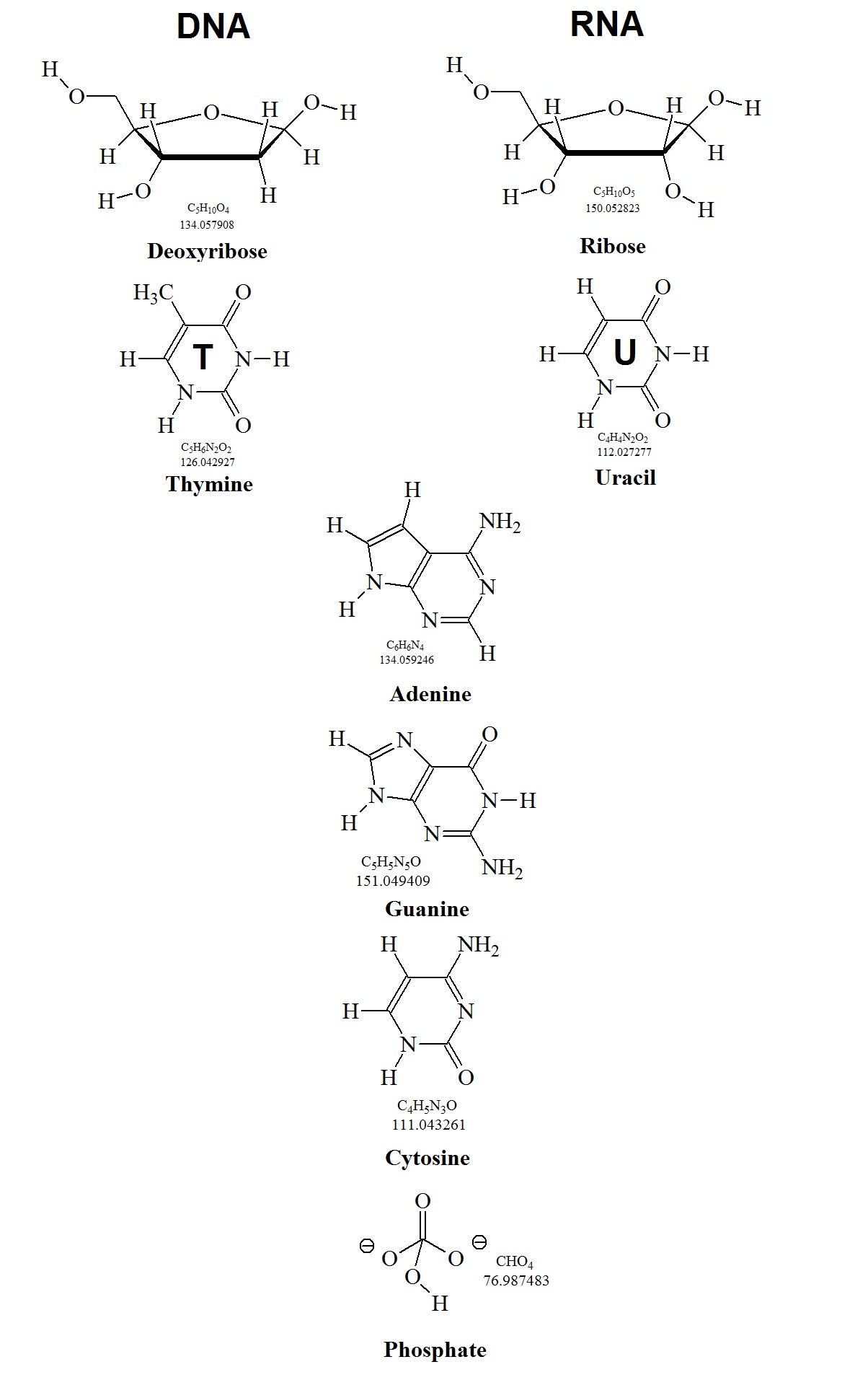
DNA and RNA are made up of only four different nucleotides. A nucleotide has three characteristic parts: (A) A nitrogenous or nitrogen-containing organic base, (B) a pentose or five-carbon sugar molecule, and (C) a phosphate group. Bases and pentoses are heterocyclic compounds. The pentose in RNA is always a ribose, however, in DNA it is a deoxyribose. In addition, one of the four nucleic acid bases differs between the two polymers.

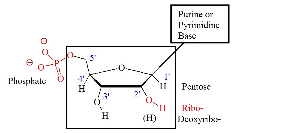
Adenine, guanine, and cytosine are present in both, DNA and RNA. Thymine is present in DNA, and uracil is only present in RNA. Abbreviations of these bases are A, G, C, T, and U, respectively. Components of the bases are either purines (A and G) or pyrimidines (C, T, or U). Figure 1 shows the general structure of nucleotides illustrating the numbering convention for the pentose ring. The ribose structure is shown in the Haworth projection.  Phosphate groups join the nucleic acid monomers together in a linear manner. Phosphate groups are attached to the 3’ and 5’ positions of the ribose sugars. Therefore, the repeating nucleic acid unit is a 3’,5’-nucleotide. The two classes of bases are often abbreviated a Y, originating from pyrimidine, and R, originating from purine. The short syllable for the phosphate group is P.


Figure 1: General structure showing the numbering convention for the pentose ring. The carbon atoms of the pentoses are numbered with primes.


Figure 2: Ribonucleic acid (RNA) fragment containing adenosine (A), guanosine (G), uridine (U), cytidine (C) linked by 3',5'-phosphodiester bonds. The chain direction from 5' to 3' is indicated by an arrow. The atom numbering scheme is indicated in the nucleotide units.


Figure 3: Atomic numbering scheme and definitions. Torsion angles for an oligonucleotide chain are shown as well. 


Figure 4: DNA and RNA building blocks. The structures and their molecular weights that make up the building block for oligonucleotides are shown.


Small concentrations of nucleosides are present in cells and extracellular fluids. The naming of the four ribonucleosides and deoxyribonucleosides is shown in table 1. Nucleotides that have phosphate groups attached are referred to as nucleoside phosphates.  

Table 1: Naming Nucleosides and Nucleotides


Bases


In

Purines

Pyrimidines


Nucleosides

RNA

Adenine, A

Guanine, G

Cytosine, C

Uracil, U

Thymine, T

DNA

Deoxyadenosine

Deoxyguanosine

Deoxycytidine

Deoxythymidine


Nucleosides

RNA

Adenylate

Guanylate

Cytidylate

Uridylate

DNA

Deoxyadenylate

Deoxyguanylate

Deoxycytidylate

Thymidylate

Nucleoside

monophosphates


AMP

GMP

CMP

UMP

Nucleoside

diphosphates


ADP

GDP

CDP

UMP

Nucleoside

diphosphates


ATP

GTP

CTP

UTP

Deoxynucleoside phosphates


dAMP

dADP

dATP

dGMP

dGDP

dGTP

dCMP

dCDP

dCTP

dUMP

dUDP

dUTP


Reference


IUPAC nomenclature: 
http://www.chem.qmul.ac.uk/iupac/misc/naabb.html


Books to review:


Genomes, 2nd edition. Terence A Brown. Department of Biomolecular Sciences, UMIST, Manchester, UK. Oxford: Wiley-Liss; 2002. ISBN-10: 0-471-25046-5

Molecular Biology of the Cell. 4th edition. Alberts B, Johnson A, Lewis J, et al. New York: Garland. Science; 2002.

Molecular Cell Biology. 4th edition. Lodish H, Berk A, Zipursky SL, et al.  New York: W. H. Freeman; 2000.

The Cell: A Molecular Approach. 2nd edition. Cooper GM. Sunderland (MA): Sinauer Associates; 2000.

Lehninger Principles of Biochemistry and Absolute Ultimate Guide Cox, Michael M., Nelson, David L.,  Lehninger, Albert L. ISBN-13: 9781429212410.  ISBN:  1429212411 Edition:  5 Pub Date:  2008. Publisher:  W. H. Freeman Company.

Principles of Nucleic Acid Structure (Springer Advanced Texts in Chemistry) Paperback – October 19, 1988 Wolfram Saenger.

Principles of Nucleic Acid Structure. Stephen Neidle. ISBN: 978-0-12-369507-9.


Guidelines for Protein Purification

$
0
0

 Guidelines for Protein Purification


Protein characterization involves the use of experimental methods that allow for the detecting and isolation of a protein and its purification. The characterization of the structure and function of a protein or gene product is usually the next step. The success of the methods and techniques used often depends on the proficiency of the person performing the purification as well as the proper selection of the purification approach. 

These general guidelines listed here are applicable to any purification process.

Define goal or objectives   

Determine purity, activity and quantity required for the final product to avoid using the wrong method.


Define properties of target protein and critical impurities 

Needed to simplify technique selection and optimization.

Develop analytical assays

Needed for fast detection of protein activity, recovery and critical contaminants.


Minimize sample handling at every stage


Important to avoid lengthy procedures which may risk losing activity or reduce recovery.

Minimize use of additives


Additives may need to be removed in an extra purification step or may interfere with activity assays.


Remove damaging contaminants early


For example, proteases that can break down the target protein may need to be removed or inactivated.


Use a different technique at each step


This takes advantage of sample characteristics that can be used for separation by combining methods that separate by size, charge, hydrophobicity,  or ligand specificity.


Minimize number of steps


Extra steps will reduce yield and increase time therefore combine steps logically.


=== Keep it simple!  ===


Apply the rule of three:


Minimize sample handling

Minimize use of additives

Remove damaging contaminants early


In addition, the requirement for how pure a protein needs to be is determined by the application it is indented to be used for.

Examples are shown below.


Purity

%

Application

Extremely high

> 99

Therapeutic use, in vivo studies

High

95 to 99

X-ray crystallography and most physico-chemical characterization methods

Moderate

< 95

Antigen for antibody production or N-terminal sequencing

-.-

Protein purification methods

$
0
0

Protein purification methods


Daily thousands of laboratories or research facilities require methods or techniques for the detection and quantitation of proteins. Analytical requirements can range from standard protein detection and characterization methods to clinical diagnostics and testing even drug dosing techniques. However, multiple factors need to be considered when selecting the best approach or method for this purpouse. The following is a list of methods used for the purification and characterization of proteins from various sources. 

General methods to Purify Proteins

  • Protein Isolation using chromatography methods:

o   Ion exchange,

o   Size-exclusion chromatography or gelfiltration,

o   Affinity chromatography,

o   Liquid chromatography with perfusion columns

  • Protein Extraction and Solubilization

  • Protein Concentration Determination Methods

  • Concentrating Protein Solutions

  • Gel electrophoresis

    • Gel Electrophoresis using denaturing conditions

    • Gel Electrophoresis using non-denaturing conditions

    • 2D Gel Electrophoresis

  • Electrofocusing

Methods to analysis Protein Structures

  • X-ray crystallography

  • Protein NMR

Protein-Protein interactions

  • Yeast two-hybrid system

  • Protein-fragment complementation assay

  • Co-immunoprecipitation

  • Affinity purification and mass spectrometry

Protein-DNA interactions

  • ChIP-on-chip

  • Chip-Sequencing

  • DamID (adenine methyltransferase identification)

  • Microscale Thermophoresis

Computational methods

  • Molecular dynamics

  • Protein structure prediction

  • Protein sequence alignment (sequence comparison, incl. BLAST)

  • Protein structural alignment

  • Protein ontology, see gene ontology

Other methods

  • Hydrogen-deuterium exchange

  • Mass spectrometry

  • Protein sequencing

  • Protein synthesis

  • Proteomics

  • Peptide mass fingerprinting

  • Ligand binding assay

  • Eastern blotting

  • metabolic labeling

    • heavy isotope labeling

    • radioactive isotope labeling

Breast Cancer Genes

$
0
0
 Genes associated with breast cancer


According to the “Center of Disease Control and Prevention (CDC)” breast cancer is the most common cancer among American women. The incidence of breast cancer is rising worldwide. More recently an increase in aggressive neoplasias in young women has been observed worldwide.

A neoplasm is an abnormal growth of specific tissue cells.The term neoplasm refers to any abnormal new growth of tissue. This type of tissue grows by cellular proliferation more rapidly than normal. It continues to grow after the stimuli that initiated the new growth has passed. The newly growing tissue shows partial or complete lack of structural organization and functional coordination with the normal tissue. Often, it forms a distinct mass of tissue which can be either benign or malignant.

The cause of cancer is an abnormal mutational change in the genome. Several mutations can generate oncogenes with dominant gain of function and tumor suppressor genes with recessive loss of function. The accumulation of genetic mutations in genes involved in DNA repair and genes that control cell growth are known to cause cancer. The result is that cells carrying the mutations can grow and divide uncontrollably to form a tumor.

In breast cancer, certain cells in the breast tissue become abnormal and start multiplying without control or start forming a tumor. The most common form of this cancer begins in cells lining the milk ducts carrying milk to the nipple. This cancer type is also called ductal cancer. Other breast cancer forms can start in the milk producing glands, called lobular cancer, or even in other parts of the breast. Different variations or mutations of the BRCA1, BRCA2, CDH1, STK11, and TP53 genes are known to increase the risk of developing breast cancer. However, other genes such as AR, ATM, BARD1, CHEK2, DIRAS#, ERBB@, NBN, PALB2, RAD50, and RAD51 genes are also associated with breast cancer.


Many known risk breast cancer susceptibility alleles have now been genotyped in a large series of female BRCA1 and BRCA2 mutation carriers assembled by the Consortium of Investigators of Modifiers of BRCA1/2 (CIMBA) to evaluate their associations with risk of breast cancer for mutation carriers.


Mulligan et al. reported that of the 12 SNPs (rs2981582 in FGFR2, rs3803662 in TOX3/TNRC9, rs889312 in MAP3K1, rs13281615 at 8q24, rs381798 in LSP1, rs13387042 at 2q35, rs4973768 in SLC4A7/NEK10, rs10941679 at 5p12, rs6504950 in STXBP4/COX11, rs999737/rs10483813 in RAD51L1, rs2046210 at 6q25.1 and rs11249433 at 1p11.2) investigated so far, eight were associated with breast cancer risk for BRCA2 carriers (all but SNPs at 8q24, RAD51L1, 6q25.1 and STXBP4/COX11), whereas only three SNPs (6q25.1, TOX3/TNRC9 and 2q35) were associated with risk for BRCA1 mutation carriers.


We now know that a buildup of mutations in critical genes is the cause of cancer. Critical genes are those that control cell growth and division or the repair of damaged DNA. These mutations allow cancer cells to grow and divide uncontrollably and to form tumors. In general, genetic changes are acquired only in certain cells during a person's lifetime. These acquired genetic changes are called somatic mutations and are not inherited. In a smaller population, some inherited gene mutations increase the risk of developing cancer. However, to develop cancer additional mutations in other genes must occur. The following tables contain a list of known genes associated with breast cancer.

High-Risk Genes


#

Gene

Associated  cancers and risks

1

BRCAl


Female breast (57-84%), ovarian (24-54%), prostate (16-20%), male breast (4%), pancreatic (3%), melanoma, fallopian tube, primary peritoneal, endometrial (serous)

2

BRCA2


Female breast (41-84%), ovarian (11-27%), prostate (20-34%), pancreatic (5-7%), male breast (4-7%), melanoma, fallopian tube, primary peritoneal, endometrial (serous)

3

CDHl


Female breast (39-52%), diffuse gastric cancer (40-83%), colon


4

EPCAM

MLHl

MSH2

MSH6

PMS2

Ovarian (4-24%), colorectal (20-80%), endometrial (12-60%), stomach, pancreatic, biliary tract, urinary tract. small bowel, brain, sebaceous neoplasms


5

PTEN


Female breast (25-50%). thyroid (10%), endometrial (5-10%), colon, renal, melanoma

6

STKll


Female breast (32-54%), ovarian tumors (21%), colorectal (39%), pancreatic (11-36%), gastric (30%), lung (15%), small intestine (13%), cervical (10%), endometrial (10%), testicular tumors (9%)

7

TP53


Female breast. ovarian, soft tissue sarcoma, osteosarcoma, brain tumors, adrenocortical c'arcinoma; overall risk for cancer: nearly 100% in females, 73% in males


Moderate Risk Genes


#

Gene

Associated  cancers and risks

1

ATM

Female breast. colon, pancreatic

2

CHEK2


Female breast. male breast. colon, prostate, thyroid, renal, endometrial (serous), ovarian

3

PALB2

Female breast. male breast. pancreatic, ovarian


Newer Genes


#

Gene

Associated  cancers and risks

1

BARDl

Female breast. ovarian

2

BRIPl

Female breast. ovarian

3

FANCC

Female breast

3

NBN

Female breast. melanoma, non-Hodgkin lymphoma

4

RAD51C

Female breast. ovarian

5

RAD51D

Female breast. ovarian

6

XRCC2

Female breast. colon, pancreatic


Selected references


http://ghr.nlm.nih.gov/condition/breast-cancer


http://www.cancer.gov/


Mulligan et al., 2011; Common breast cancer susceptibility alleles are associated with tumour subtypes in BRCA1 and BRCA2 mutation carriers: results from the Consortium of Investigators of Modifiers of BRCA1/2. Mulligan et al. Breast Cancer Research 2011, 13:R110. http://breast-cancer-research.com/content/13/6/R110.


Dorothy Teegarden
Isabelle Romieu, and Sophie A. Lelièvre; Redefining the impact of nutrition on breast cancer incidence: is epigenetics involved? Nutr Res Rev. 2012 June ; 25(1): 68–95. doi:10.1017/S0954422411000199.


Purines, Pyrimidines, and Nucleotides

$
0
0

Purines, Pyrimidines, and Nucleotides


Purines, pyrimidines, and nucleotides are ubiquitous molecules found throughout a mammalian as well as a human body. In one form or another, these molecules serve a variety of roles. Nucleotides are molecular building blocks or subunits of nucleic acids such as deoxyribonucleic acid (DNA) and ribonucleic acid (RNA). These subunits are also called monomers.  The synthesis of purines, pyrimidines, and nucleotides is an important part of mammalian metabolism. Errors in purine and pyrimidine synthesis and metabolism, inborn or acquired, often are the cause of disease or ultimately lead to disease.


Nucleotides and their structure

A nucleotide is made up of three units:

  1. A nitrogen-containing base, 
  2. A five-carbon sugar,
  3. One two three phosphate groups.

There are five common bases. Two of these bases are derivatives of purine. These are adenine and guanine. Three of the bases are pyrimidine derivatives. These are cytosine, thymine, and uracil. The addition of ribose moiety or 2-deoxyribose moiety to a base forms a nucleoside. The figure below illustrates the general structures of these molecules. Adenine, guanine, and cytosine can be found bonded to either a ribose or a 2-deoxyribose molecule. Uracil is usually found in combination with a ribose. Thymine is usually bonded to 2-deoxyribose. Ribonucleosides are called adenosine, guanosine, cytidine, and uridine. Deoxyribonucleotides are called deoxyadenosine, deoxyguanosine, deoxycytidine, and deoxythymidine.


IUPAC nucleotide codes with ambiguity

Symbol

Meaning

Origin of designation

Fw

G

G

Guanine

151.12

A

A

Adenine

135.12

T

T

Thymine

126.11

C

C

Cytosine

111.1

U

U

Uracil

112.08

M

A or C

aMino


R

G or A

puRine


W

A or T

Weak interactions
(2 H bonds)


S

C or G

Strong interaction
(3 H bonds)


Y

T or C

pYrimidine


K

G or T

Keto


V

G or C or A

not- T (not-U), V follows U


H

A or C or T

not-G, H follows G in the alphabet


D

A or G or T

not-C, D follows C


B

C or G or T

not-A, B follows A


X

G or A or T or C


N

G or A or T or C

 aNy


Reference:

Cornish-Bowden A. Nomenclature for incompletely specified bases in nucleic acid sequences: recommendations 1984. Nucleic Acids Research. 1985;13(9):3021-3030.

INTERNATIONAL UNION OF PURE AND APPLIED CHEMISTRY AND INTERNATIONAL UNION OF BIOCHEMISTRY ABBREVIATIONS AND SYMBOLS FOR NUCLEIC ACIDS, POLYNUCLEOTIDES AND THEIR CONSTITUENTS RULES APPROVED 1974 Issued by the JUPAC—IUB Commission on Biochemical Nomenclature


Discovery of Post Translational Modifications or PTMs

$
0
0
Discovery of Post-Translational Modifications (PTMs)


Many proteins, if not most, in archaea, prokaryotes and eukaryotes contain post-translational modifications (PTMs). PTMs increase the functions of proteins and define the structural plasticity of the proteome. The interplay between thousands of different molecules in cells or tissue maintains cellular integrity, morphology and regulates many important biological functions.  Post-translational modifications include covalent modifications such as phosphorylation, glycosylation, ubiquitination, nitrosylation, methylation, acetylation, lipidation, proteolysis and many others.

Post-translational modifications are now considered as a mayor regulatory mechanism in eukaryotes and influence the metabolism of normal cell biology and pathogenesis. 
In proteomic studies, the discovery of post-translational modifications is achieved with the help of advanced mass spectrometry techniques. A multitude of mass spectrometry-based proteomic strategies has been investigated and reported in the proteomic literature.

How is a PTM discovery done?


A general PTM discovery experiment entails

1.     The use of a quality control experiment or standard. For a nano-HPLC-MS/MS experiment, a standard of 125 fmol or greater is usually used. For phosphorylation studies, a phosphorylated peptide or protein or a mixture of different phosphorylated peptides or proteins may be used.


2.     Next, a proteolytic digestion is performed. For example, on proteins in gel bands resulting from SDS-PAGE experiments. Trypsin is usually used for a first pass experiment, but other proteases or mixtures of proteases can also be used.


3.   The use of nano-high performance liquid chromatography (HPLC) tandem mass spectrometry allows for the analysis of the digest. This analysis generates a dataset of fragment ions in the form of a mass list that can now be queried using search engines or bioinformatics software.

4.   Next, a database search is performed using the peptide mass finger print data.

5.   A typical search for PTMs involves the use of search engines such as X!tandem, SEQUEST, Mascot or other specialized PTM software.

6.   However, to rule out false-positives a manual verification is often necessary.


Protein PTM Identifications (PTM ID)


Proteins PTM’s are identified by digesting proteins into peptides, analyzing peptides using tandem mass spectrometry (LC-MS/MS) followed by searching for modifications using X!tandem, SEQUEST, or other specialized PTM software. The observed post-translational modifications are usually verified by hand, and a likelihood score is produced. However, results can vary depending on the specific programs used.


In general, some modifications are easier to find than others. In addition, searches or scans for specific modifications or even unknown modifications using LC-MS/MS data can be performed. Usually, modifications can be found in the unimod database.


The chances of detecting the modification a researcher is interested in are completely dependent on the stability of the modification and how that modification affects peptide fragmentation in a mass spectrometer. The stoichiometry of the modification factures in as well, as does a number of other variables. However, the PTM discovery experiment may not always work the first time around and the analysis of some specific modifications may require specialized expertise and equipment.


Proteomic Journals


International Journal of Proteomics

http://www.hindawi.com/journals/ijpro/

Journal of Proteomics

http://www.sciencedirect.com/science/journal/18743919

Journal of Proteome Research

http://pubs.acs.org/journal/jprobs

Proteomics

http://onlinelibrary.wiley.com/journal/10.1002/%28ISSN%291615-9861

Proteomics and Bioinformatics

http://omicsonline.org/proteomics-bioinformatics.php

Unimod

http://www.unimod.org/downloads.html]


Building Blocks of DNA and RNA Molecules

$
0
0

Building Blocks of DNA and RNA Molecules


The structure of DNA was solved in 1953 by Watson, a trained geneticist, and Crick, a trained physicist. This discovery is considered to be the most important discovery of the 20th century.


Numbering convention for nucleotides

$
0
0

Numbering convention for nucleotides


Nucleic acids are the building blocks for polymers of nucleotides. Oligonucleotides contain, store and transmit instructions about proteins and quantities of these a cell needs to function. This information system is called the genetic code. The chemical structures that contain and decode the code are called deoxyribonucleic acid (DNA) and ribonucleic acid (RNA). The DNA molecule stores the information and the active agent RNA decodes the stored information. Both DNA and RNA are linear polymers in their primary structures composed of monomers or single chemical units or building blocks called nucleotides. Cellular RNA molecules can range in length from leas than one hundred to thousands of monomers. However, the number of monomers in DNA can exceed a hundred million.

DNA and RNA are made up of only four different nucleotides. A nucleotide has three characteristic parts: (A) A nitrogenous or nitrogen-containing organic base, (B) a pentose or five-carbon sugar molecule, and (C) a phosphate group. Bases and pentoses are heterocyclic compounds. The pentose in RNA is always a ribose, however, in DNA it is a deoxyribose. In addition, one of the four nucleic acid bases differs between the two polymers.

Adenine, guanine, and cytosine are present in both, DNA and RNA. Thymine is present in DNA, and uracil is only present in RNA. Abbreviations of these bases are A, G, C, T, and U, respectively. Components of the bases are either purines (A and G) or pyrimidines (C, T, or U). Figure 1 shows the general structure of nucleotides illustrating the numbering convention for the pentose ring. The ribose structure is shown in the Haworth projection.  Phosphate groups join the nucleic acid monomers together in a linear manner. Phosphate groups are attached to the 3’ and 5’ positions of the ribose sugars. Therefore, the repeating nucleic acid unit is a 3’,5’-nucleotide. The two classes of bases are often abbreviated a Y, originating from pyrimidine, and R, originating from purine. The short syllable for the phosphate group is P.


Figure 1: General structure showing the numbering convention for the pentose ring. The carbon atoms of the pentoses are numbered with primes.


Figure 2: Ribonucleic acid (RNA) fragment containing adenosine (A), guanosine (G), uridine (U), cytidine (C) linked by 3',5'-phosphodiester bonds. The chain direction from 5' to 3' is indicated by an arrow. The atom numbering scheme is indicated in the nucleotide units.


Figure 3: Atomic numbering scheme and definitions. Torsion angles for an oligonucleotide chain are shown as well. 


Figure 4: DNA and RNA building blocks. The structures and their molecular weights that make up the building block for oligonucleotides are shown.


Small concentrations of nucleosides are present in cells and extracellular fluids. The naming of the four ribonucleosides and deoxyribonucleosides is shown in table 1. Nucleotides that have phosphate groups attached are referred to as nucleoside phosphates.  

Table 1: Naming Nucleosides and Nucleotides


Bases


In

Purines

Pyrimidines


Nucleosides

RNA

Adenine, A

Guanine, G

Cytosine, C

Uracil, U

Thymine, T

DNA

Deoxyadenosine

Deoxyguanosine

Deoxycytidine

Deoxythymidine


Nucleosides

RNA

Adenylate

Guanylate

Cytidylate

Uridylate

DNA

Deoxyadenylate

Deoxyguanylate

Deoxycytidylate

Thymidylate

Nucleoside

monophosphates


AMP

GMP

CMP

UMP

Nucleoside

diphosphates


ADP

GDP

CDP

UMP

Nucleoside

diphosphates


ATP

GTP

CTP

UTP

Deoxynucleoside phosphates


dAMP

dADP

dATP

dGMP

dGDP

dGTP

dCMP

dCDP

dCTP

dUMP

dUDP

dUTP


Reference


IUPAC nomenclature: 
http://www.chem.qmul.ac.uk/iupac/misc/naabb.html


Books to review:


Genomes, 2nd edition. Terence A Brown. Department of Biomolecular Sciences, UMIST, Manchester, UK. Oxford: Wiley-Liss; 2002. ISBN-10: 0-471-25046-5

Molecular Biology of the Cell. 4th edition. Alberts B, Johnson A, Lewis J, et al. New York: Garland. Science; 2002.

Molecular Cell Biology. 4th edition. Lodish H, Berk A, Zipursky SL, et al.  New York: W. H. Freeman; 2000.

The Cell: A Molecular Approach. 2nd edition. Cooper GM. Sunderland (MA): Sinauer Associates; 2000.

Lehninger Principles of Biochemistry and Absolute Ultimate Guide Cox, Michael M., Nelson, David L.,  Lehninger, Albert L. ISBN-13: 9781429212410.  ISBN:  1429212411 Edition:  5 Pub Date:  2008. Publisher:  W. H. Freeman Company.

Principles of Nucleic Acid Structure (Springer Advanced Texts in Chemistry) Paperback – October 19, 1988 Wolfram Saenger.

Principles of Nucleic Acid Structure. Stephen Neidle. ISBN: 978-0-12-369507-9.


Cancer Genomics

$
0
0
What is Cancer Genomics?


What does cancer genomics mean for a patient?

According to the NIH Publication No. 10-7556 “Cancer Genomics”, one person dies from cancer every minute in the United States. It is estimated that 1,500 people die from cancer each day. And the number of people dying per day in the future is expected to increase with the aging population. To understand, control and conquer cancer, medical researchers have to understand this disease. Since cancer starts in our DNA, many medical researchers are working hard to find cures for the many faces of cancer.

DNA or deoxyribonucleic acid carries instructions that tell cells what to do. Mistakes found in these instructions may cause the cells not to function normally. Accumulated mistakes or changes found in DNA responsible for the instructions are called mutations. Some of these mutations can be inherited, but most are acquired throughout life. Acquired mutations must be caused by environmental factors, such chemicals, or from lifestyle choices, for example, caused by smocking.


What is a genome?

The complete set of instructions in the DNA is called a genome. The term “genome” refers to the complete genetic material of an organism. In humans, it is called the “human genome.” In most cells, the genome is physically packaged into two sets of chromosomes. One set originates from the mother and one from the father. Six billion individual DNA letters or nucleotide monomers make up the human chromosomes.

The alphabet of the genome consists of four (4) letters: A, C, G, and T.  Small changes in the arrangement of these letters within the genome can result in changes of genetic instructions for a cell. Genes are parts of a genome that carry instructions to make molecules that do most work in the cell. Other parts of the genome can switch genes on and off. Depending on the function of a cell or tissue type, different genes are used by the cell. Liver cells use a set of genes needed for liver proteins whereas muscle cells use genes needed to make muscle proteins. 

What is a gene?

The term ‘gene’ refers to the functional and physical unit of heredity. This unit is passed on from parents to their offsprings or children. Most genes contain the instructions to make a specific protein. Germline DNA contains inherited mutations.

What is germline DNA?

Germline DNA is found in germ cells. Eggs and sperm cells are germline cells that join to form an embryo. This type of DNA is the source for all other cells in the body and is also called constitutional DNA.

What is somatic DNA?

Somatic DNA is the DNA found in body cells but not in the germ cells. Therefore, somatic mutations are alterations or changes in the DNA that occur after conception. Somatic mutations are not present in germ line DNA and are therefore not passed on to children. Many somatic mutations can cause diseases such as cancer. However, not all somatic mutations cause diseases.

What are the effects of changes or mutation in DNA?

Changes or mutation in DNA can cause cells to make the wrong amount of one protein or change the shape of a protein so that it does no longer work as it should. Because proteins control the behavior of cells, mutations may lead to health problems. In cancer, these changes can cause cells to grow out of control. As a result, these cells can damage the surrounding tissues.

What is the Cancer Genome Atlas?

The Cancer Genome Atlas (TCGA) Data Portal provides a platform for researchers to search, download and analyze data sets. These data sets were generated by TCGA. The National Cancer Institute at the National Institute of Health (NCI/) established the atlas. The goal of the TCGA is to help researchers understand what turns a normal cell into a cancer cell.

The comparison of DNA from normal and cancer tissue has already shown that

•    Certain areas of the genome are commonly affected in several types of cancer. Affected genes are often genes that control signaling pathways that cause cells to divide and survive when they normally would die.

•    Specific changes or cell signatures often also called biomarkers allow researchers to tell what type of cancer is present. These signatures may help medical doctors to identify specific cancer types. Medical researchers already know that different types of cancer respond differently to various treatments or have a different prognosis. 


Reference

http://cancergenome.nih.gov/

An Effective Detection Method for the EGFR Single Mustation T790M Using BNA-NC Clamping Real-time PCR

$
0
0
Abstract:

Mutations on epidermal growth factor receptor (EGFR) cause a variety of cancers including breast and lung cancers. The single mutation T790M on tyrosine kinase domain of EGFR signifies the response to the popular cancer drug gefitinib, which leads to the development of resistance to gefitinib. Detecting the mutation thus guide effective therapeutical options for patients who are in need of cancer drug treatments. We sought to develop a rapid, reliable detection method for the T790M mutation using bridged nucleic acids (BNA), which has been known to enhance the hybridization affinity of oligonucleotides that contain BNA bases. Oligonucleotides containing BNA bases designed to block PCR reaction against wild-type genes, called BNAclamp, were used to discriminate the presence of mutant genes mixed with a large number of wild-type genes. Real-time PCR in conjugation with BNAclamping allows us to view the different levels of PCR amplifications in the degree of mixture of wild-type and mutant genes. In an effort to explore the possibility, 13-mer long clamps were prepared with various numbers of BNA bases. The clamps containing 9 BNA bases appear to be most effective in blocking the PCR reactions at an optimized concentration to distinguish the mutant from the wild-type genes. In addition to PCR results, the deference in the Tm values for the 7 BNA bases in the 13-mer was the largest among differently designed clamps, which is consistent with the results obtained by real-time PCR. We also examined the degree of sensitivity using the clamp containing 9 BNA bases, revealing that the clamp has the ability to determine the level of mutation as low as 1% mixture oft he mutant and wild-type gene. The effectiveness of blocking the PCR reaction with only 13-mers containing BNA bases allowed us to detect a single mutation, and thus, this BNAclamping real time PCR technology may offer a promising, new avenue to detect clinically importantmutations in the future.

Niacin = Nicotininc acid = Vitamin B3

$
0
0

Niacin, Nicotinic acid, Vitamin B3


Niacin, nicotinic acid, or vitamin B3 is a water soluble vitamin that is a building block for the coenzymes nicotinamide adenine dinucleotide (NAD+) and nicotinamide adenine dinucleotide adenine dinucleotide (NADP+). NAD+ is a carrier of two electron equivalents for the oxidation of carbohydrates during the synthesis of adenosine triphosphate (ATP). The nicotinamide ring in NAD+ and NADP+ is capable of accepting two electrons and a hydrogen ion to produce the reduced forms of these compounds. The redox role of NAD+ is well established, however, recent evidence indicates that NAD+ is involved in the regulation of diverse pathways as well some of which appear to control life span. In general, NADH is produced by oxidative reactions such as those found in the citric acid cycle. It's reducing power is the driving force during oxidative phosphorylation to produce ATP. NADPH is primarily used in biosynthetic reactions that require reducing power. Both coenzymes, NADH and NADPH are involved in many metabolic reactions. This water soluble vitamin can be analyzed by classical high-performance liquid chromatography (HPLC) using ultra-violet (UV) detection or the more recently developed liquid chromatography-tandem mass spectrometry LC-MS(MS) techniques.

Compared to other vitamins the structure of niacin is relatively simple. It contains a pyridine ring with a single carboxylate group (see figure 1 below). The amide derivative, nicotinamide, has a similar vitamin activity.


Figure 1: Chemical formula, molecular weight and van-der-Waals model for nicotinic acid (Vitamin B3).


Food sources rich in niacin include liver and other meats, yeast, peanuts, wheat germ, and fish. The human body can also produce niacin as a byproduct of tryptophan metabolism. Approximately 2% of dietary tryptophan is metabolized by this pathway. The estimate is that 60 mg of tryptophan is equivalent to 1 mg of niacin. A balanced diet is thought to contain 600 mg of tryptophan per day. This amount contributes over half or an individual’s niacin requirement. Niacin was identified as a vitamin when the need arose to cure pellagra in the early twentieth century.

Pellagra is a disease caused by the lack of niacin or a decreased intake of tryptophan. Niacin is sometimes also called the “pellagra preventing factor”.  An excessive intake of leucine or a deficiency of the amino acid lysine can lead to a deficiency in niacin as well. If untreated, pellagra can kill within several years. Pellagra is a common disease in Africa, Indonesia, North Korea, and China. Poor, homeless, alcohol-dependent, or psychiatric patients who refuse food can also show  clinical signs of pellagra. Any human, including a vegetarian, who relies on food sources with limited amounts of niacin risks the development of pellagra.

What caused the occurrence of pellagra in eighteenth century Europe and later in the southern United States?

In 1735, a Spanish physician noticed for the first time symptoms of this strange disease. He called it mal de la rosa or the disease of the rose. The disease spread geographically with the introduction and cultivation of corn in Europe. In Italy it became known as pelleagra or rough skin, hence the term pellagra. In the first half of the 1900s the disease reached epidemic proportions in the United States producing at least 250,000 cases and 7,000 deaths per year for several decades in the southern states alone. Gradually, an association between pellagra and corn consumption was observed. Ultimately, research confirmed that a pellagra-preventative (P-P) factor, missing from corn, was necessary to prevent and cure pellagra in humans. Furthermore in 1937, it was discovered that nicotinic acid could cure black tongue in dogs. Black tongue was an early animal model for pellagra. In 1951, it was found that niacin in corn is biologically unavailable.  However, it can be released by exposure to alkaline pH. It appears that Native Americans who used corn as a dietary staple have long known about this. It is well known that many Native American societies did processes their corn with alkali before consumption.

Presently serious outbreaks still continue to occur in some developing countries. The fortification of grain products and an improved standard of living have limited the occurrence of the disease.

Niacin and cancer.

High levels of serotonin produced from tryptophan are found in carcinoid cancers. Patients with this type of cancer will be at risk for niacin deficiency if their intake of preformed niacin is low.

Carcinoid tumor starts in the hormone-producing cells of various organs and most often develop in the gastrointestinal tract, in organs such as the stomach or intestines, or in the lungs. In addition, a carcinoid tumor can also develop in the pancreas, a man’s testicles, or a woman’s ovaries and more than one carcinoid tumor can occur in the same organ.

Niacin and aging

More recent genetic research revealed additional salvage pathways for the synthesis of NAD+. Belenky et al. (2007) reported in the Journal Cell that nicotinamide riboside regulates Sir2 deacetylase activity and life span in yeast. Nicotinamide riboside is a newly discovered NAD+ precursor which is converted to nicotinamide mononucleotide by specific nicotinamide riboside kinases, Nrk1 and Nrk2. Nicotinamide riboside is an NAD+ precursor present in metabolisms from yeast to mammals. Nicotinamide riboside contains a nicotinamide ring structure connected to a ribose. It is a source of Vitamin B3. Belenky et al. discovered that exogenous nicotinamide riboside promotes Sir2-dependent repression of recombination, improves gene silencing, and extends lifespan without calorie restriction. In addition, nicotinamide riboside has been reported to increase NAD+ levels. Nicotinamide riboside acts on two pathways, the Nrk1 pathway, and the Urh1/ Pnp1/Meu1 pathway.  However, the Urh1/ Pnp1/Meu1 pathway is Nrk1 independent. Both nicotinamide riboside salvage pathways contribute to NAD+ metabolism in the absence of nicotinamide riboside supplementation. 

Nicotinamide riboside in food

Apparently nicotinamide riboside is found in milk and potentially in beer. 


References


Peter Belenky, Frances G. Racette, Katrina L. Bogan, Julie M. McClure, Jeffrey S. Smith, Charles Brenner; Nicotinamide Riboside Promotes Sir2 Silencing and Extends Lifespan via Nrk and Urh1/Pnp1/Meu1 Pathways to NAD+. Cell, Volume 129, Issue 3, 4 May 2007, Pages 473-484.

Katrina L. Bogan and Charles Brenner; Nicotinic Acid, Nicotinamide, and Nicotinamide Riboside: A Molecular Evaluation of NAD+ Precursor Vitamins in Human Nutrition. Annu. Rev. Nutr. 2008. 28:115–30. 

Cantó C, Houtkooper RH, Pirinen E, et al. The NAD+ precursor nicotinamide riboside enhances oxidative metabolism and protects against high-fat diet induced obesity. Cell metabolism. 2012;15(6):838-847. doi:10.1016/j.cmet.2012.04.022.

Chi, Y; Sauve, A. A. (2013). "Nicotinamide riboside, a trace nutrient in foods, is a vitamin B3 with effects on energy metabolism and neuroprotection". Current Opinion in Clinical Nutrition and Metabolic Care 16 (6): 657–61. 

John M. Denu; Vitamins and Aging: Pathways to NAD+ Synthesis. Cell. Volume 129, Issue 3, 4 May 2007, Pages 453–454.


Robert B. Rucker ... [et al.]. editors; Handbook of vitamins - 4th ed., 2007. ISBN-13: 978-0-8493-4022-2 (hardcover : alk. paper) ISBN-10: 0-8493-4022-5 (hardcover : alk. paper).


W E Schreiber; Medical aspects of biochemistry. Pp 282. Little, Brown & Co, Boston, USA. 1984. ISBN 0–316–77473–1.


Cancer Genomics

$
0
0
What is Cancer Genomics?


What does cancer genomics mean for a patient?

According to the NIH Publication No. 10-7556 “Cancer Genomics”, one person dies from cancer every minute in the United States. It is estimated that 1,500 people die from cancer each day. And the number of people dying per day in the future is expected to increase with the aging population. To understand, control and conquer cancer, medical researchers have to understand this disease. Since cancer starts in our DNA, many medical researchers are working hard to find cures for the many faces of cancer.

DNA or deoxyribonucleic acid carries instructions that tell cells what to do. Mistakes found in these instructions may cause the cells not to function normally. Accumulated mistakes or changes found in DNA responsible for the instructions are called mutations. Some of these mutations can be inherited, but most are acquired throughout life. Acquired mutations must be caused by environmental factors, such chemicals, or from lifestyle choices, for example, caused by smocking.


What is a genome?

The complete set of instructions in the DNA is called a genome. The term “genome” refers to the complete genetic material of an organism. In humans, it is called the “human genome.” In most cells, the genome is physically packaged into two sets of chromosomes. One set originates from the mother and one from the father. Six billion individual DNA letters or nucleotide monomers make up the human chromosomes.

The alphabet of the genome consists of four (4) letters: A, C, G, and T.  Small changes in the arrangement of these letters within the genome can result in changes of genetic instructions for a cell. Genes are parts of a genome that carry instructions to make molecules that do most work in the cell. Other parts of the genome can switch genes on and off. Depending on the function of a cell or tissue type, different genes are used by the cell. Liver cells use a set of genes needed for liver proteins whereas muscle cells use genes needed to make muscle proteins. 

What is a gene?

The term ‘gene’ refers to the functional and physical unit of heredity. This unit is passed on from parents to their offsprings or children. Most genes contain the instructions to make a specific protein. Germline DNA contains inherited mutations.

What is germline DNA?

Germline DNA is found in germ cells. Eggs and sperm cells are germline cells that join to form an embryo. This type of DNA is the source for all other cells in the body and is also called constitutional DNA.

What is somatic DNA?

Somatic DNA is the DNA found in body cells but not in the germ cells. Therefore, somatic mutations are alterations or changes in the DNA that occur after conception. Somatic mutations are not present in germ line DNA and are therefore not passed on to children. Many somatic mutations can cause diseases such as cancer. However, not all somatic mutations cause diseases.

What are the effects of changes or mutation in DNA?

Changes or mutation in DNA can cause cells to make the wrong amount of one protein or change the shape of a protein so that it does no longer work as it should. Because proteins control the behavior of cells, mutations may lead to health problems. In cancer, these changes can cause cells to grow out of control. As a result, these cells can damage the surrounding tissues.

What is the Cancer Genome Atlas?

The Cancer Genome Atlas (TCGA) Data Portal provides a platform for researchers to search, download and analyze data sets. These data sets were generated by TCGA. The National Cancer Institute at the National Institute of Health (NCI/) established the atlas. The goal of the TCGA is to help researchers understand what turns a normal cell into a cancer cell.

The comparison of DNA from normal and cancer tissue has already shown that

•    Certain areas of the genome are commonly affected in several types of cancer. Affected genes are often genes that control signaling pathways that cause cells to divide and survive when they normally would die.

•    Specific changes or cell signatures often also called biomarkers allow researchers to tell what type of cancer is present. These signatures may help medical doctors to identify specific cancer types. Medical researchers already know that different types of cancer respond differently to various treatments or have a different prognosis. 


Reference

http://cancergenome.nih.gov/

Viewing all 572 articles
Browse latest View live


Latest Images